Monday, November 6, 2023

Stiff Person Syndrome Mimicking as Atypical Parkinson’s Disease - A Diagnostic Dilemma - Juniper Publishers

Neurology & Neurosurgery - Juniper Publishers

Abstract

Stiff person syndrome (SPS) is a rare neurological disorder characterized by stiffness and spasm of the truncal muscles and subsequent postural deformities. In this report, we present a case of a woman in her early 60s who presented to us with muscle stiffness, back pain, and frequent falls. On examination, she was found to have increased muscle tone and mild tremors. Given her age and presentation, a provisional diagnosis of Atypical Parkinson’s was made. Patient was treated with Diazepam. Because of presence of autoimmune co-morbid conditions, anti-glutamic acid decarboxylase (GAD) antibodies were tested, which were positive, and thus, diagnosis of SPS was made. Treatment with Diazepam was continued, and her symptoms significantly improved. Even though SPS is a rare disorder, our case report highlights the need to keep it in differential diagnoses when an elderly patient presents with stiffness and postural instability, as treatment for SPS and atypical Parkinson’s vary considerably.

Keywords: Stiff person syndrome; Atypical Parkinson’s; Anti-GAD antibodies; Rare disorder

Abbreviations: SPS: Stiff Person Syndrome; PD: Parkinson’s Disease; GAD: Glutamic Acid Decarboxylase

Introduction

Stiff person syndrome is a progressive neurological disease even with treatment and results in significant disability in patients affected [1]. In this case report, we will go over our patient’s presentation, the variants of SPS and the presentation of these variants. We will also discuss Atypical Parkinson’s disease and why this diagnosis was considered in our patient.

The main pathogenesis of SPS is thought to be blockade of glutamic acid decarboxylase (GAD) enzyme which is responsible for maintaining inhibitory pathways in the central nervous system. GAD blockade results in reduced levels of gamma butyric acid (GABA) producing loss of neural inhibition. GAD blockade mainly results from anti-GAD antibodies that are associated with multiple autoimmune disorders, the most commonly associated disease being type 1 diabetes mellitus [2].

There are several variants of the disease [3]. The classic variant is associated with other autoimmune disorders while the partial SPS is characterized by stiffness limited to only to one limb. Progressive encephalomyelitis with rigidity and myoclonus is another uncommon variant of the disease characterized by transient oculomotor disturbance. Another rare variant of this disease is the paraneoplastic SPS which is suspected in patients who are anti-GAD negative. These patients have antiamphiphysin antibodies and have associated underlying malignancy of the breast and lung.

Classic SPS is the most common variant of the disease and is characterised by the presence of anti-GAD antibodies and presence of autoimmune diseases. The disease presents with muscle stiffness involving the axial group of muscles, lumbar group, and the proximal limb muscles. The disease is progressive and leads to deformities. As the stiffness and rigidity in the axial muscle group progresses, patients also develop a wide based gait and tend to have frequent falls, thus predisposing them to fractures.

Atypical Parkinson’s on the other hand, are a group of neurodegenerative disorders which have features resembling Parkinson’s disease (PD) with additional features that differentiate them from PD. These include Dementia with Lewy bodies characterized by the presence of visual hallucinations and REM sleep behaviour disorder, Multi system atrophy that presents with postural instability and cerebellar findings like gait ataxia. Progressive supranuclear palsy is another subtype characterized by vertical gaze palsy, akinesia, and progressive aphasia. The last type, called corticobasilar degeneration, is relatively rare and presents with myoclonus and dystonia [4].

A common denominator among SPS and Atypical PD is the presence of rigidity and gait instability producing frequent falls. Due to the similar features in both diseases, diagnosing them clinically can be challenging, particularly in older patients. Not all individuals with Atypical Parkinson’s Disease will exhibit resting tremors, or bradykinesia during their initial presentation; some may only display muscle rigidity and postural instability.

Case Report

A 60-years-old female presented with complaints of backache and stiffness of 4 months duration which was gradual in onset and progressive in nature. She also complained of worsening of stiffness and tightness in her back since the last 2 months and also gave history of 5 episodes of falls. She also developed difficulty in getting up from squatting position. Her past medical history was significant for diabetes mellitus and hypothyroidism for which she was on regular medications. On examination, her vitals were stable. Neurological examination findings revealed increased muscle tone in all four limbs and mild tremors of the hands were noted. Deep tendon reflexes were exaggerated. Cerebellar examination did not reveal any findings. In view of progressive muscular rigidity, tremors and postural instability in an elderly patient, a provisional diagnosis of Atypical Parkinson’s was made while keeping Stiff person syndrome in the differential because of presence of autoimmune conditions diabetes mellitus and hypothyroidism. Magnetic resonance imaging (MRI) of the brain and spine were normal. A nerve conduction study was done which was normal and electromyography was inconclusive.

Serum panel to detect auto antibodies was sent to the lab. The reports came back positive for anti-GAD antibodies and thus the conclusive diagnosis of SPS was established.

During her stay in the hospital, patient was treated with tablet Diazepam 10mg three times daily, which alleviated her symptoms. Her blood sugars were controlled, and thyroid function tests were also done, which were normal. Her symptoms significantly improved following treatment with diazepam, and she was discharged in stable condition. She will be regularly followed up on out-patient basis.

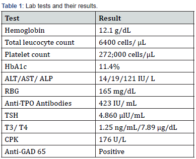
A summary of her lab reports can be found in Table 1.

HbA1c : Glycated Haemoglobin; ALT : Alanine Transaminase; AST : Aspartate Transaminase; ALP : Alkaline Phosphatase; RBG: Random Blood Glucose; Anti-TPO Antibodies: Anti-Thyroid Peroxidase Antibodies; TSH: Thyroid Stimulating Hormone; T3/T4: Triiodothyronine/ Thyroxine; CPK: Creatinine Phosphokinase; Anti: GAD Antibodies: Anti-Glutamic Acid Decarboxylase Autoantibodies.

Discussion

The prevalence of Stiff Person Syndrome is only 1 – 2 people per million. Though the pathophysiology of the disease is unclear, neurologists find autoimmunity to be the main culprit, as majority of the cases are associated with anti-GAD positive antibodies. While their presence supports the diagnosis, their absence does not rule out the disease. The disease mainly affects people in their 2nd to 5th decade of life and affects women more than men [5].

The clinical course is progressive stiffness slowly leading to deformities and functional impairment. Intellect and memory are spared. There is no confirmatory test, although anti-GAD antibody test is widely accepted as the diagnostic test. Imaging is noncontributory, neurological testing may reveal continuous firing of action potential. Treatment is usually symptomatic and managing any co-morbid conditions. Death can result from diaphragmatic spasm [6].

Even though the disease is rare, because of its similarity in presentation with that of Atypical Parkinson’s, this differential must be kept in mind, especially in elderly patients.

Hence in elderly patients presenting with gait instability, frequent falls and muscle rigidity, physicians need to consider both SPS and atypical PD and ruling out one of them based on the past medical history and lab findings.

Conclusion

Stiff person syndrome being a rare disorder can easily be missed. It’s prognosis is guarded, and thus early recognition of the disease and initiation of treatment can slow down the functional decline in these patients. As the diseases progresses, patients lose the ability to ambulate and require walking assistance device [7]. They may also require assistance for activities of daily living, and this can have a psychological effect on these patients. Providing psychological counselling is equally crucial to ensure the wellbeing of individuals, a fact that might be disregarded by physicians primarily focused on treating the physical aspect of the disease.


To Know more about Open Access Journal of Neurology & Neurosurgery
Click here: https://juniperpublishers.com/oajnn/index.php

To Know more about our Juniper Publishers
Click here: https://juniperpublishers.com/index.php


Thursday, November 2, 2023

Primary Multiple Metachronous Cervical Cancer: Difficulties in Differential Diagnosis - Juniper Publishers

Journal of Case Studies - Juniper Publishers


Abstract

This observation shows a combination of primary multiple tumors, where one of the tumor localizations is the cervix, the other is the mammary gland, the third is the lung.

For differential diagnosis in cases of primary multiple tumors, the most informative is CT of the lungs, PET-CT, ultrasound of the pelvic organs and abdominal cavity, histological examination, immunohistochemical examination. In case of difficulties in differential diagnosis, it is possible to perform extended genetic testing of frequent somatic mutations in the TP53, GATA3, EGFR, KRAS, NRAS, ESR, PIK3CA, AKT genes, which allows you to determine the source of metastasis, and in this regard, select the necessary antitumor therapy.

Despite the progression of cervical cancer (metastases to the supraclavicular lymph nodes), the patient was not denied specialized treatment for lung cancer. The multidisciplinary approach adopted in the clinic in the diagnosis and treatment of patients made it possible to detect a malignant lung tumor at the preclinical stage and perform a radical operation, which allows us to hope for a good long-term result. At the same time, the metastatic lesion was not confirmed, the data of pathomorphological examination and immunohistochemical studies established an independent primary nature of the tumor.

Keywords: Metachronous cervical cancer; Cervical cancer; Recurrence

Introduction

Cervical cancer (CC) is the second most common cancer among women under 65 years of age and is the most common cause of death from gynecological cancer worldwide. In terms of morbidity and mortality, cervical cancer ranks fourth among all cancers. In 2020, there were 604,127 new cases of cervical cancer and 341,831 deaths worldwide. In the structure of mortality from malignant neoplasms in women under 45 years of age, cervical cancer ranks 1st. Tumors with localization in the cervix, mammary gland, lungs and other organs are not casuistry, but represent an extremely complex and not fully resolved problem of oncology. In the available literature, we did not find a combination of primary multiple cancers of the breast, lung and cervix. The frequency of metachronous tumors in women reaches 4.5%. We present to your attention an extremely rare case - the presence of primary multiple metachronous cervical cancer.

Patient S., 60 years old, applied to the Novosibirsk Regional Clinical Oncological Center in 2014 with a diagnosis of breast cancer III st T2N1M0. - radical left-sided mastectomy with lymphadenectomy.

Histological examination No. 26854-69 dated August 27, 2014. Conclusion: infiltrating ductal cancer of the left breast, maligna II with growth in the vessels, secondary changes in two lymph nodes.

Immunohistochemical study dated 09/04/2014. - ER 8, PR 6, Her2neo 1+ Ki67 - 25%. Adjuvant radiation therapy was prescribed - a dose of 46-50Gy for 23-25 fractions 5 times a week for the operated mammary gland, supra-/subclavian, ipsilateral parasternal and part of the axillary zone (as a zone of special risk) ROD 2Gy, SOD 45-50Gy.

Polychemotherapy courses - according to the FAC scheme: Cyclophosphamide 800mg, Doxorubicin 80mg, 5 fluorouracil 800mg 4 courses, hormone therapy with Tamoxifen 20mg. The patient was registered with an oncologist and regularly underwent examinations.

In 2015, cervical cancer II st T2bN0M0 was diagnosed, cervical biopsy No. 30867-79 dated 10/14/2015. - squamous cell nonkeratinizing cancer.

Courses of chemoradiation therapy, remote beam therapy in a total dose of 45-50Gy, simultaneously with chemotherapy (3 cycles of cisplatin 50mg each week) followed by brachytherapy, EBRT - 2Gy per fraction, in the irradiation mode 5 times a week, were carried out. The total dose to the focus from combined radiation therapy was 75Gy, to the lymphatic outflow path - 58Gy.

In 2017, according to the results of a histological examination dated February 21, 2017. - Metastasis of squamous cell carcinoma of the cervix to the supraclavicular lymph nodes on the left, courses of polychemotherapy Paclitaxel 175 mg/m2 IV 3 hours on the 1st day + Doxorubicin - 8 courses, completed in September 2017, palliative course of radiation therapy to the region of the supraclavicular lymph nodes on right. According to the control studies - stabilization of the process.

In February 2020, according to the results of bone scintigraphy - metastases in the bones of the spine Th8-Th10, reactive strontium therapy was prescribed, Fulvestrant 500mg 1 time in 28 days/m, continued growth of Th8-Th10, Palbociclib was added from 02.2020.

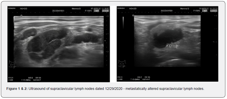
Ultrasound of the supraclavicular lymph nodes from 12/29/2020 - on the left: multiple (9) hypoechoic with clear and fuzzy contours ranging in size from 3 to 12mm (previously from 3 to 10mm) are located (Figure 1 & 2).

01/13/2021 - excisional biopsy of supraclavicular lymph nodes on the left.

01/20/2021 Histological examination of surgical material: No. 442-5/21: the histological picture and immunophenotype of tumor cells correspond to metastases of squamous cell carcinoma, taking into account the expression to p16 (Clone CINtec, Ventana) - carcinoma of the cervix.

MRI of the pelvis from 01/19/2021 - no pathology was detected.

Ultrasound of the abdominal organs from 01/29/2021 - echo signs of a liver cyst, a cyst of the left kidney, diffuse changes in the liver parenchyma, pancreas.

02/08/2021 - concilium - chemotherapy is indicated according to the scheme: Paclitaxel 175mg/m2 IV drip + Cisplatin 75mg/m2 IV drip on day 1 Cycle 1 time in 21 days, up to 6 courses with an assessment of the effect after 3 and a decision issue of radiation therapy 6 cycles of chemotherapy were performed (treatment was completed on 05/25/2021) MRI of the pelvis from 12/09/2021 - MRI signs: - abdominal infiltrate of the uterine bladder tissue with inhomogeneous contents and air with fistulas in the bladder and vagina, - post-radiation changes in the pelvis .

To clarify the process, PET-CT of the whole body was performed: - In the parenchyma of the right lung, nodular metabolically active seals in S2 - 1.2 * 0.8cm, in S6 - 0.9 * 0.8cm. Infiltration of the small pelvis.

According to PET-CT of the whole body dated 02.11.2021. - zone of tissue resorption in the region of the alveolar process of the upper jaw.

Consultation of the maxillofacial surgeon from 02.11.2021 - More evidence for osteonecrosis with bisphosphonates.

Due to difficulties in diagnosing the zone of tissue resorption in the region of the alveolar process of the upper jaw, an extended genetic testing of frequent somatic mutations in the genes TP53, GATA3, EGFR, KRAS, NRAS, ESR, PIK3CA, AKT was performed in the laboratory of pharmacogenomics of the Institute of Chemical Biology and Fundamental Medicine SB RAS: in block 26854-56, a mutation was found in the GATA3 gene (chromosome 10, position 8115971 in the assembly of the human genome No. 19) with a content of 30.4% with a coverage of 171 reads; in block 30867, a mutation was found in the TP53 gene (chromosome 17, position 757754) in the assembly of the human genome 19 with a content of 1.15% with a coverage of 783 reads, a mutation in the TP53 gene (chromosome 17, position 7577541 in the assembly of the human genome No. 19) was found in plasma with a content of 1.86% with a coverage of 22872, respectively.

Conclusion: tumor DNA corresponding to sample 30867 (cervical cancer) circulates in blood plasma Concilium at the Novosibirsk Regional Clinical Oncological Dispensary dated January 12, 2022 - given the progression of the disease, continued tumor growth is indicated for monochemotherapy: Karboplatin AUC 5 IV 1 hour on the 1st day every 3 weeks, up to 6 courses in total (with an assessment of the effect after 3 and 6 courses - CT of the chest + MRI of the small pelvis) MSCT of the chest on March 18, 2022 - no dynamics.

MRI of the pelvic organs on March 18, 2022 - no dynamics.

6 courses of monochemotherapy with Carboplatin AUC 5 IV 1 hour on the 1st day every 3 weeks were carried out, treatment was completed in May 2022.

Since August 2021, a vesico-vaginal fistula was diagnosed (Figure 3), which significantly reduced the quality of life of the patient.

Examined by a urologist in May 2022, cystoscopy was performed: - Formations: on the right side wall with a transition to the posterior multiple small bullous-solid formations - a biopsy was taken, urine was taken for atypical cells, cytological examination - no atypia. Histological examination - Moderately expressed active cystitis. Tumor growth was not detected in the preparation.

In July 2022, a set of examinations was carried out: pelvic ultrasound, abdominal ultrasound, CT of the lungs, skeletal bone scan, PET-CT, and stabilization of the process was established.

In this regard, on July 20, 2022, fistuloplasty was performed at the Avicenna clinic (Figure 4-6), the postoperative period was uneventful, without complications, and she was discharged on the fifth day. The patient notes a significant improvement in the quality of life.

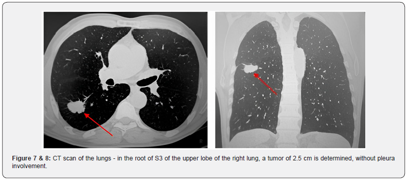
In August 2022 according to the results of follow-up examinations on CT scan of the lungs, there are signs of metastatic lesions of the upper and lower lobe of the right lung, videoassisted thoracoscopic upper lobectomy on the right with atypical resection of the lower lobe is recommended (Figure 7 & 8).

At the Institute of Oncology and Neurosurgery of the Federal State Budgetary Institution “N.N. ak. E.N. Meshalkin” 30.08.2022 - videothoracoscopic upper lobectomy on the right with atypical resection of the lower lobe. Intraoperatively: in the root of S3 of the upper lobe of the right lung, a tumor of 2.5cm was determined, without pleura involvement. In the lower lobe on the right S6, S8, S9, 3 subpleural foci are determined. Performed upper lobectomy with atypical resection of the lower lobe.

According to the pathomorphological study dated 09/02/2022. No. 10147-10168 - taking into account the morphology of the formation - the presence of a pronounced exophytic component, the tumor stem, the solidary nature of the neoplasm, the most likely primary highly differentiated keratinizing squamous cell carcinoma S3 of the bronchus of the upper lobe of the right lung, pT1cN0M0.

A revision of the pathomorphological report was performed at the Tomsk National Research Medical Center dated 09/08/2022. No. 10147-10168, where the diagnosis was also confirmed - primary highly differentiated keratinizing squamous cell carcinoma S3 of the bronchus of the upper lobe of the right lung, pT1cN0M0.

According to the ultrasound of the small pelvis, ultrasound of the abdominal cavity dated December 14, 2022. - no negative dynamics, signs of post-radiation changes.

According to CT of the chest organs dated November 22, 2022. - condition after surgical treatment of the right lung. There are no new foci, lung metastases without dynamics, metastases of Th8- Th10 bodies - without dynamics.

According to colposcopy dated 12/17/2022. - no local recurrence, post-radiation changes (Figure 9).

Conclusion

This observation shows a combination of primary multiple tumors, where one of the tumor localizations is the cervix, the other is the mammary gland, the third is the lung. Figure 7 & 8 - CT scan of the lungs - in the root of S3 of the upper lobe of the right lung, a tumor of 2.5 cm is determined, without pleura involvement.

1. For differential diagnosis in cases of primary multiple tumors, the most informative is CT of the lungs, PET-CT, ultrasound of the pelvic organs and abdominal cavity, histological examination, immunohistochemical examination.

In case of difficulties in differential diagnosis, it is possible to perform extended genetic testing of frequent somatic mutations in the TP53, GATA3, EGFR, KRAS, NRAS, ESR, PIK3CA, AKT genes, which allows you to determine the source of metastasis, and in this regard, select the necessary antitumor therapy.

2. Despite the progression of cervical cancer (metastases to the supraclavicular lymph nodes), the patient was not denied specialized treatment for lung cancer. The multidisciplinary approach adopted in the clinic in the diagnosis and treatment of patients made it possible to detect a malignant lung tumor at the preclinical stage and perform a radical operation, which allows us to hope for a good long-term result. At the same time, the metastatic lesion was not confirmed, the data of pathomorphological examination and immunohistochemical studies established an independent primary nature of the tumor.

3. In the treatment of primary multiple tumors, a multidisciplinary approach is required with the involvement of a thoracic surgeon, an oncogynecologist, an oncomammologist, a molecular oncologist, and a pathologist. The final conclusions on this issue can be obtained by accumulating a sufficient number of observations.

Therefore, each case of a primary multiple metachronous lesions should be especially carefully studied and presented in international medical literature.


To Know more about our Juniper Publishers

Wednesday, November 1, 2023

Review on Bovine Viral Diarrhea - Juniper Publishers

 Dairy & Veterinary Sciences - Juniper Publishers


Abstract

Bovine viral diarrhea virus is a pest virus that can cause severe clinical signs; Major economic losses continue to result from reproductive losses and exacerbation of concurrent bacterial or viral infections. Of less clinical importance is severe clinical disease due to BVDV alone, either as result of acute infection by highly virulent isolate or by development of mucosal disease in persistently infected calves. Disease due to BVDV can arise at any stage of life and is often dependent on the virulence of infecting isolate. Survival of BVDV virus in population depended up on the characteristics of persistently infecting its host. Persistent infection arises by the unique ability of the BVDV to survive by immune tolerance in bovine fetus through evasion of both innate and acquired immunity in utero. Adaptive immunity is avoided by immune tolerance through infection of fetus prior to development and maturation of adaptive immunity. The pathogenesis of BVDV infection is complex, with infection pre- and post-gestation leading to different outcomes. Infection of the dam during gestation results in fetal infection, which may lead to embryonic death, teratogenic effects, or the birth of persistently infected, (PI) calves. PI animals shed BVDV in their excretions and secretions throughout life and are the primary route of transmission of the virus. These animals can usually be readily detected by virus or viral antigen detection assays (RT-PCR, ELISA), except in the immediate post-natal period where colostral antibodies may mask virus presence. Acute infection with BVDV results in transient viraemia prior to seroconversion and can lead to reproductive dysfunction and immunosuppression leading to an increased incidence of secondary disease. Antibody assays readily detect virus exposure at the individual level and can also be used in pooled samples (serum and milk) to determine herd exposure or immunity. Diagnostic tests can be used to diagnose clinical cases, establish disease prevalence in groups and detect apparently normal but persistently infected animals.

Keywords:Bovine viral diarrhea; Mucosal disease; Persistent infection; Virus

Abbreviations:BVD: Bovine Viral Diarrheal; BVDV1: Bovine Viral Diarrhea Virus type1; BVDV2: Bovine Viral Diarrhea Virus type2; BVDV: Bovine Viral Diarrheal Virus; Cp: Cytopathogenic; CSFV: Classical Swine Fever Virus; ELISA: Enzyme Linked Immune Sorbent Assay; FCS: Fetal Calf Serum; FCS-SM: Fetal Calf Serum Supplemented Media; IHC: Immune Histocompatibility; MD: Mucosal Diseases ; MLV: Modified Live Vaccine NCP: Non cytopathogenic; OIE: Office of International Epizootics; PI: Persistently Infected; RNA: Ribonucleic Acid; RT-PCR: Reverse Transcriptase Polymerase Chain Reaction; VI: Virus Isolation; VN: Virus Neutralizations

Introduction

Bovine viral diarrhea virus (BVDV) is a Pest virus of the family Flaviviridae capable of causing serious clinical diseases in cattle [1]. The virus is divided into two genomes (BVDV-1 and BVDV-2) because of antigenic and genetic differences [2]. And Infection with BVDV is also known to have significant financial impact stemming primarily from the reproductive and immunosuppressive effects of acute infection [3]. As such, control and eradication programs are becoming increasingly common across much of the cattle-producing world including legislated, regional schemes and voluntary, herd-based schemes [4]. While healthy, immune-competent cattle (or late-term, immune-competent fetuses) may suffer from acute BVDV infection resulting in sero-conversion; the disease is primarily spread and maintained in cattle populations by persistently infected (PI) individuals that Persistency of infection arises from fetal infection in early gestation Following acute infection in the dam [5]. Most control programs aim to eliminate PI cattle and hence the source of continuing infection [4].

This requires the efficient and effective use of accurate diagnostic tests, and a wide range is readily available for the detection of BVDV virus, antigen (Ag) or specific antibodies (Ab) that the tests will return varying results depending on the current or historical BVDV infection status and Animals that have never been exposed to the virus will test negative for Ab, Ag, and virus with the broad nature of disease, transmittance, and lack of treatment made BVD global pandemic and one of the most significant cattle diseases in world Coupled with vast clinical symptom and associated death loss, gravity of BVD makes it a disease no longer overlooked [6]. The overall effects of BVD are difficult to examine, due to the breadth of the disease. Research and data highlighting effects of the disease is very limited, simply since too many variables are present for accurate measurement. Furthermore, if considered the worldwide prevalence of BVD, it is easily understood why BVD is an important cattle disease from an economic standpoint [7].

The areas affected by BVD, specifically reproduction, production and immunosuppression, are what make it such an expensive disease; Reproductive disorders & syndromes associated with BVDV include: abortion, early embryonic loss, fetal defects, conception failure, normal to prolonged returns to estrous, increase in return to service numbers, spread out calving pattern, slow to grow and weak calves, birth defects such as blind calves or calves with neurological problems, poor weaning rates due to ill thrift and death and A good pregnancy rate does not indicate a BVDV free herd as reproductive losses is associated with the timing of infection [4].

Production effects span those regarding decline in feed to gain efficiency, lower carcass quality, and decreased milk production and because of the vague and diverse clinical signs observed with the disease, it is very difficult to clinically diagnose BVD infected cattle. The difficulty of clinically diagnosing BVD positive cattle impedes the studying of these cattle in a clinical setting. Regardless, understanding the nature of viral infections in animals it should be noted that the side effects are not usually contained to one specific area of the body. A virus such as BVD has been noted to have potential complications with every body system in cattle. Although rare, BVD viruses have manifested in many ways as rare as causing skeletal deformities to severe neurological damage (Houe, 1994). Finally, immunosuppression may also lead to secondary disorders, along with a whole host of other [8].

With above background, the objectives of this paper are:
 To provide a brief review on the economic importance of BVD in production and reproduction.
 To know the management systems of BVD infection during any outbreak time and infectious period.
 To understand the nature and characteristics of BVD.
 To provide information on the epidemiology of BVD.
 To emphasize diagnostic, prevention and control methods of BVD.

General Overview

Etiology

BVDV is a member of the pest virus genus caused by a virus called Bovine Viral Diarrhea Virus (BVDV) that there are several ways in which BVDV infection can manifest in a herd and cause economic loss of that the most important craffied one’s are the immunosuppressive effects, reproductive losses and losses associated with persistently infected animals (Becher et al., 2011).

Viral taxonomy

There are four recognized species within the pest virus genus. These species are BVDV-1, BVDV-2, border disease virus of sheep and classical swine fever virus, previously known as hog cholera virus [9]. (See box below)

Epidemiology The occurrence of BVD infection

Acute infection is the result of horizontal transmission to a susceptible animal following contact either with an acutely infected animal that is shedding BVDV, or with a PI animal that sheds BVDV continuously that it may also be acquired through several routes of transmission such as inhalation, ingestion and transplacental [10].

The spread of BVD

The greatest period of spread is during yarding, particularly when cattle from different groups or herds are mixed or penned in adjacent pens [11]. And the main source of infection is persistently infected (PI) animals that shed many viral particles in all body fluids and excretions (milk, urine, feces, saliva). Infection in susceptible animals tends to occur from direct animal to animal contact that may occur during yarding, mustering, agricultural shows and sales and the most common ways that BVDV is introduced to a herd through the introduction of replacement breeding animals, new bulls and occasionally over the fence contact with infected animals [3].

Persistence of BVD

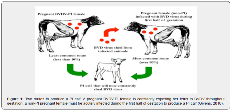
Persistently infected (PI) calves are created when a fetus is exposed to BVDV during the first half of gestation that on this time, the fetal immune system is not developed enough to respond to a BVDV infection so the fetus might be aborted, but if the fetus survives it will likely develop into a PI calf. Some PI calves grow poorly while others may look healthy and grow very well, making it impossible to consistently detect PI animals visually. Most PI animals die by 2 years of age, but some will survive for several years and constantly shed BVDV throughout their life (Givens et al, 2010).

PI cattle are much more efficient at transmitting the disease due to the higher amount of systemically circulating virus present within the animal. With higher amounts of circulating virus these animals will secrete higher levels of the virus. Additionally, PI cattle will secrete the virus in various forms from inoculation of the virus until their death. The potency of transmission is of major concern, and it has been shown that horizontal transmission from a PI animal to a seronegative animal can be detected within 20 hours after direct contact [9] (Figure 1).
Transmission and source of infections Transmission

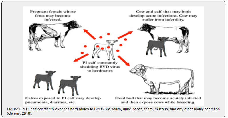
Bovine pest virus is spread by close contact (nose-to-nose) between cattle. The greatest period of spread is during yarding, particularly when cattle from different groups or herds are mixed or penned in adjacent pens [11]. BVDV does not usually survive in the environment very long (less than 3 weeks), so direct transmission between animals is the most common route of transmission that Acutely infected animals are a temporary source of BVDV transmission, but PI animals shed millions of viral particles every day; therefore, serve as a constant source of BVDV exposure in a herd because they continuously shed virus in saliva, mucous, tears, milk, feces, urine, and any other bodily secretion and its transmission may occur vertically (i.e., before birth), leading to congenital infection of the fetus, or the transmission may occur horizontally (i.e., after birth) that Congenital infections may cause resorption, abortion, still birth, or live-birth and Congenitally infected fetuses that survive in utero infection (i.e., the live-births) may be born as BVDV-infected calves [12] (Figure 2).
Source of infection

Persistently infected carrier calves play a key role in spreading the virus as they can shed viruses throughout their life, acting as a source of infection for other animals. This most commonly occurs when these cattle are between 6–24 months of age. However, some PI’s have been known to survive and enter the breeding herd. They may die from a range of diseases caused by the immunosuppressive effects of BVDV infection and occasionally they may develop Mucosal Disease [13].

Risk factors and host range Risk factors for BVDV infections.

The risk factors for BVDV infections are often a reflection of the risk of direct or indirect contact with PI cattle. The suspected risk factors include livestock trade, pasturing animals, use of common pasture for infected and susceptible cattle, cattle density, number of infected neighboring herds, broken fences, animal contact between herds (e.g., livestock exhibitions), weak hygiene, and presence of other susceptible ruminants, such as sheep and wildlife [14].

Host range

Other than cattle carrying the BVD virus, domesticated farm animals have been shown to have the potential of becoming infected with BVD as well. So, the virus has been found in sheep, pigs, and wildlife, although the chance of transmission to or from cattle has not been fully established but transmission between sheep and cattle has been experimentally proven [15]. The BVD virus has been found in pigs as classical swine fever virus (CSFV), but once again the transmission to cattle is considered doubtful. Initially this was thought to be a possible mode of transmission to and from cattle, and some even thought that the disease may have originated from wildlife populations such as serological evidence exists that deer can become infected with BVD [16].

Pathogenesis

BVDV infection has a wide range of clinical presentations and unique diagnostic challenges; so, to select an appropriate diagnostic test, it is important first to understand the pathogenesis of the disease. BVDV is divided into noncytopathogenic (ncp), and cytopathogenic (cp) biotypes based on effects on cultured cells rather than in the infected host. Cp biotypes induce apoptosis in cultured cells while ncp biotypes do not [17]. Non-cytopathogenic BVDV, however, appears to be the cause of acute infections and can be transmitted in a wide range of body fluids, including nasal discharge, urine, milk, semen, saliva, tears, and fetal fluids [18]. Also, Cp BVDV has been shown to be capable of inducing acute infection under experimental conditions [19].

Clinical Signs and symptoms Clinical signs

Clinical signs of infected cattle cover a wide spectrum from showing no signs to death. The varying clinical signs depend on what form of the virus the animal is carrying along with the extent of the infection. Cytopathic strains of BVD will result in more severe reproductive effects including embryonic loss, stillbirths, and congenital defects. Additionally, it forms of the virus contracted in older cattle will cause these animals to present themselves critically in a clinical setting causing fever, depression, lack of appetite, labored breathing, oral ulcerations, and diarrhea to be observed also in the most severe cases Bleeding of internal organs and death has been observed. Supportive care such as fluids and anti-inflammatory drugs may be used, but the likelihood of losing the animal is still relatively high. Noncytopathic strains of the virus will cause infected cattle to show mild clinical signs if any at all. So, this type of virus will create PI calves if they are infected in utero and will cause transiently affected animals if contracted by older animals. In general, a noncytopathic form of the virus will have less detrimental effects than a cytopathic strain of the virus [20].

Secondary Diseases

Other than the consequences of reproductive disorders, Bovine Respiratory Disease (BRD) is a major disease that is often coupled with BVD. It is much like BVD, an umbrella term for a wide range of bacterial and viral caused respiratory diseases. So, those that contained high levels of BVD seropositive cattle were at a higher risk of Bovine Respiratory Syncytial Virus (BRSV) [14]. Viruses are the principle pathogenic instigators of BRD, making prevention of BVD key to keeping BRD herd levels at a minimum and should be noted that immunosuppression is not the only explanation for BRD being a secondary disease to BVD (Hansen et al, 1996).

Clinical syndromes associated with BVD.

There is a wide spectrum of outcomes associated with BVDV that its Infections may be subclinical (no outward signs of disease), clinical or chronic. This depends on the immune status of the animal, the stage of gestation of female animals and the amount or strain of virus that the animal is exposed to. Common outcomes of exposure to BVDV in cattle include one or more of the following Reproductive Syndromes such as embryonic loss, stillbirths, and congenital defects the most common once; conception failure, normal to prolonged returns to estrous, increase in return to service numbers, spread out calving pattern, slow to grow and weak calves; birth defects such as blind calves or calves with neurological problems, poor weaning rates due to ill thrift and death [14].

Pathology

Pathologically, PI cattle are viremic, yet clinically they often show little or no signs of being infected. The classical PI animal is the one that is never shows signs clinically and may seem fine but is the silent culprit for regenerating the disease; so, these reasons that PI animals are of greatest concern to producers. The common pathology of the disease is cerebral hypoplasia, ulcerative esopagitis, hemorrhagic necrosis of peyers patchs on ileum, colitis and crypt dilatation unilateral corneal opacity are good examples of BVD [21] (Figure 3(a,b,c)).

Diagnostics

In general, diagnostic assays for detection of BVDV infections can be divided into Serological methods, i.e. methods aimed at detection of virus-specific antibodies as an indication of exposure to the pathogen, and methods aimed at detection of infectious virus or viral components (antigen or nucleic acids), as an indication of current infection [22].

Serological methods

In the context of BVDV control, the main objectives of serological methods are to Differentiate between exposed and non-exposed herds, to monitor progress and Drawbacks within an ongoing control programme, and to investigate immune status in individual animals in infected herds, to identify possible PI animals. A PI animal is, as previously mentioned, generally seronegative, unless it has persisting maternal antibodies, or has been exposed to and infected with a heterologous strain. Serological methods can also be used to diagnose acute infection by detection of seroconversion in paired samples [8]. Virus neutralization (VN): is the reference test for antibody detection. VN is sensitive and specific, but cell culture dependent and labour demanding and will typically take 5-6 days to perform. Thus, it is mostly used as a reference test for back-up and calibration purposes [8].

Antibody ELISA: For testing of large series of samples, enzyme-linked immunosorbent assays (ELISAs) have many advantages over VN. They are rapid, relatively inexpensive both to establish and run, and are suitable for automation. Two principally different ELISA formats are common in use for antibody detection: indirect or blocking (competitive) assays. In the indirect format, specific antibodies are trapped by immobilized viral antigen, and subsequently detected using enzyme conjugated [22]. Virus isolation (VI): in bovine cell cultures, followed by identification of the viral isolate by immunoperoxidase or immunofluorescence staining is considered the standard reference test for detection of infectious virus. Although VI demands time, resources, and skill, it is a reliable and widely used method. The presence of antibodies, or cell toxic substances, or both in the sample, however, can yield a false negative result. Furthermore, because BVDV is a common contaminant of biological by-products of bovine origin such as fetal calf serum (FCS) and, consequently, bovine cell cultures grown in FCS-supplemented media, the cell culture system must be monitored to ensure freedom from contamination with virus or antibodies [23].

Detection of antigen

Assays for detection of viral antigen rely on existing BVDVencoded antigens in the sample material, i.e., there is no amplification of the target, as is the case for VI and RNA detection protocols. This minimizes the risk of cross contamination of Samples, and flavors detection of PI over acutely infected animals [22]. Several formats of ELISAs are commercially available for detection of viral antigens. The basic principle consists of the use of virus-specific monoclonal antibodies to capture viral antigens, followed by detection of antigen-antibody complex with enzyme-conjugated antibodies. Antigen ELISAs are widely used for identification of PI animals and can be used for detection of viruses in serum, Buffy coat cells or skin biopsies (e.g., ear notch samples). Like VI, antigen ELISAs may yield false negative results if antibodies are present in the sample. This should be considered when testing young animals that might have persisting maternal antibodies [8].

Immunohistochemical detection of antigen in skin biopsies, particularly ear notch samples, is another method that is used for identification of PI animals [24]. By use of separate sets of primers and probes, BVDV-1 and -2 can also be discriminated between in the same assay [25]. Genotyping: The genetic typing of BVDV has most frequently been based on sequence comparison of partial sequences of the 5’ NCR, Npro or E2 regions [26]. Analysis of the 5’ NCR, a highly conserved region of the genome, has shown to be a reliable and reproducible method for genetic characterization of BVDV isolates even though the genetic resolution obtained is not as high as that obtained with more variable regions as Npro or E2 [27]. Furthermore, it is the target region for most PCR-based diagnostics, and as such a suitable target for direct sequencing from the PCR product [28].

Differential diagnosis of BVD

BVD is differentially diagnosed with several diseases like: Malignant catarrhal fever bilateral corneal opacity(edema) or uveitis, dermatitis are typical; Rinderpest- tiger or zebra striping lesion is prominent in large intestine on chronic case; Bluetongue- Hemorrhage on the base of pulmonary artery and aorta with large quantity of plasma like fluid in thorax and pericardial sac; Salmonellosis-no oral lesion, diarrhea but have is zoonotic bluereddish discoloration of extremities, cyanosis; Johne’s disease corrugated appearance of intestine & also has no oral lesion and bottle jaw are typical and prominent mucosal folds [21].

Treatment Reducing Clinical Effects

It is common for producers to treat BVD animals with antibiotics, thinking it will help the cattle “get over the hump” of whatever they are experiencing. In animals that are showing clinical effects of BVD, the only true treatment available is supportive. Optimizing the animal’s ability to neutralize the virus may consist of reducing fever, countering any nutritional or metabolic disorders, and providing basic husbandry practices that one would apply to a diseased animal. Use of antibiotics may be an option to prevent secondary diseases such as BRD and in a form may help reduce clinical effects of BVD. An animal with an acute form of BRD will cause the immune system to weaken, allowing a BVD virus to overcome the animal’s immune system, causing an acute form of BVD. The use of antibiotics has been proven to be effective at fighting respiratory infections in cattle. Broad spectrum agents such as Nuflor, Excenel, LA-200, and Micotil, can reduce the severity and length of a bacterial infection. In the likely case that an animal is fighting BRD along with BVD, by reducing the extent of the respiratory infection it will reduce the likelihood that the animal shows clinical signs of BVD [29,30].

No True Cure

Due to the nature of viral infections, there is no treatment to fully cure an animal of a viral infection. As we have discussed there is a limited amount of treatment that may be performed with BVD animals; the key lies in prevention of disease that are: All purchased animals should be tested for PI status and quarantined to give time for acute infections to clear; Any animal leaving the farm (shows, leased bulls, etc.) should be quarantined for 30 days before re-entering the herd. Purchased pregnant animals should not only be tested themselves for PI status, but also their calves should be tested at birth to make sure they are not a PI [31].

Prevention and control Prevention

The global nature of the disease, along with the ease of transfer between carriers, makes prevention of BVD a formidable challenge. The challenge of eradicating, or even just controlling BVD, can be seen by the state-wide and national attempts made by groups of producers to control the disease. When the time comes for a producer to implement a prevention program, one must keep in mind that the program must be specifically attuned to the type of operation (cow-calf, stocker, feedlot, dry lot dairy, pastured dairy cattle, dairy calf ranch, etc.). There is no “one size fits all” prevention program. It is crucial that the Producer design and continuously adapt a program that specifically fits their operation [22]. Animal Husbandry: The start of any effective prevention program begins with basic animal husbandry practices. Scoring of the body condition may be done with relative ease, especially with the help of a veterinarian and records should be maintained to compare past results. The importance of not underestimating the body condition of the animals is a crucial step in preventing BVD within a herd. Animals under nutritional deficiency may become immunocompromised, hindering the immune response time [32].

Passive Immunity: Adequate quantity and quality of colostrum within the desired 3-hour post parturition window. The wellknown benefits of adequate transfer of immunoglobulins from dam to offspring can be summed up simply by the formation of a competent and responsive immune system. The rewards of immunoglobulin absorption of offspring therefore are linked directly to the protection of clinical diseases including prevention of BVD [28,29]. Active Immunity and Vaccination: Although removal of a PI animal is the primary form of halting the shedding of BVD, it has been shown that transmission of the disease will be hindered by an adequate vaccination program [7]. Wide ranges of vaccine-induced neutralizing titers were formed in calves with a two-series BVD specific vaccine of either an inactivated or modified live vaccine [7]. Other important aspects of the study were that the study population was of colostrum-deprived calves; although the importance of colostrum and passive immunity should not be underestimated, this shows that regardless of degree of passive transfer, vaccines are still effective at stimulating titers in immunoglobulin naive calves [28,29]. It is apparent that vaccination and passive immunity both offer degrees of protection against BVD but, when coupled together; the combination conferred a potent efficacy against shielding viruses [28, 29].

Testing: Testing cattle for BVD is the most important aspect of prevention for the disease in white blood samples will be necessary to detect BVD in some PI animals. Different test methods used by laboratories, veterinarians and producers will be highlighted in the following discussion. Immunohistochemistry (IHC) is the most practical approach to testing a herd for BVD. Ear notch samples are the most common tissue sample used for this test. This requires little labor in obtaining samples from large numbers of cattle with the use of an instrument such as ear notch pliers and the samples are also suitable for transport. This test is very accurate at identifying PI animals but cannot provide accurate results for transient animals [33]. Also, Polymerase Chain Reaction (PCR) tests used to detect BVD have been shown to be timelier compared to virus isolation tests. Additionally, PCR has the potential to detect BVD in its antigen-antibody complex. Therefore, unlike IHC, this test can be performed on calves under 6 weeks of age, because it makes no difference if colostral antibodies are bound with the antigen [24].

Culling: Within any herd attempting to prevent BVD a strict “test and cull” policy must be practiced. Regardless of other factors including the worth of animals, market prices, labor involved, these animals must be removed from the herd. Every day a transient or PI animal is with its herdmates, the potential for the disease to spread will not decrease [34]. Closed Herd: Once the herd has been tested and is clean of BVD, the next challenge in preventing the disease is maintaining a closed herd. Simply put, any animal being introduced to the herd should be properly quarantined until tested as negative for BVD. At this time, it is then safe for the animal to be turned out with other animals. Replacement heifers and bulls raised on a dairy or ranch are obviously exempt from this protocol, because of their origination from the herd. Maintaining a closed herd is a difficult task for producers to accomplish due to the transition of the beef and dairy industry to fewer and larger farms. This means that a larger number of animals are being moved in and out of farms in a shorter period [35].

Control

For long, attempts to control BVDV infections were limited to prophylactic treatment practices by using modified live vaccine and killed virus vaccine, implemented primarily to reduce, or prevent clinical disease on a herd basis. Today, however, it is well established that the benefit of preventing clinical disease in transiently infected animals is negligible when considering the overall prevention and control of the disease [27].

Bovine Viral Diarrhea Infection in Ethiopia

In Africa, few studies conducted on seroprevalence of the disease have indicated that the prevalence’s were varied from 70–83% [36]. Although Seroepidemiology of BVD were reported in European and some African countries (OIE, 2008), there is no research or published information available as to these authors knowledge on the investigation of BVDV infection in Ethiopia. But there was one study had done to detect the presence of BVDV infection and to determine the Seroepidemiology and associated factors in three agroecological zones in Ethiopia such as Jimma, Southwest Shoa, and West Shoa where BVD is more prevalent where due to the following factors tropical climate and humidity of the area with bimodal heavy annual rainfall ranging and traditional mixed crop-livestock agriculture of areas makes the infection more prevalence on these areas as a study [37].

Economic Significance Of BVD

BVDV adversely affects both health and productivity. The losses due to infection are diarrhea, decreased milk production, reproductive disorders, increased occurrence of other diseases, and death. The losses from fetal infection include abortions; congenital defects; weak and abnormally small calves; unthrifty, persistently infected (PI) animals; and death among PI animals. [6]. As we have introduced, the effects of BVD are wide-ranging that vague and diverse clinical signs observed with the disease, it is very difficult to clinically diagnose BVD infected cattle. The difficulty of clinically diagnosing BVD positive cattle impedes the studying of these cattle in a clinical setting. Regardless, understanding the nature of viral infections in animals it should be noted that the side effects are not usually contained to one specific area of the body. So, a virus such as BVD has been noted to have potential complications with every body system in cattle. Although rare, BVD viruses have manifested in ways as rare as causing skeletal deformities to severe neurological damage [34- 46].

Conclusions and Recommendations

An understanding of the pathology and pathogenesis of BVDV infection will guide and optimize diagnostic approaches. As the age of the animals and stage of infection varies, different tests and testing protocols need to be utilized. Highly sophisticated, but relatively cheap diagnostic tests, such as a combination of RTPCR and Ab monitoring on pooled material allow classification of the BVDV infection status of individuals and herds. A molecular epidemiological approach may be used to trace routes of BVDV transmission, and to identify and prevent risky behaviour within a BVDV control programme.

A single introduction of BVD into a susceptible population may result in widespread infection, and an introduced strain may circulate for many years and remain genetically unaltered. The significance of a single animal being ill may be overlooked, as often occurs with animals that are persistently infected. It is important to realize that this single case could be an indicator of the potential for greater losses, due to the reproductive, productive, and other effects of the pestivirus. Awareness of the presence of pestivirus, followed by appropriate management strategies, will minimize losses. Finally, the immune suppression, Persistent infection, production loss, reproduction defect and neurological defects all these make BVD as one the most important economical disease in veterinary world.

Based on the above conclusion the following recommendations are forwarded.

  • It should be maintained on closed herd (not always practical).
  • Don does not purchase replacements from auction.
  • Quarantine new arrivals and show stock for 30 days and test new arrivals for BVD- PI.
  • Test purchased pregnant animals and their calf at birth. Keep records (reproductions and health).

To Know more about Journal of Dairy & Veterinary Sciences
Click here: https://juniperpublishers.com/jdvs/index.php

To Know more about our Juniper Publishers
Click here: https://juniperpublishers.com/index.php

Tuesday, October 31, 2023

Surgical Management for Meckel’s Diverticulum: A Case Series and Literature Review - Juniper Publishers

Journal of Surgery - Juniper Publishers

Abstract

Introduction: Meckel’s diverticulum (MD) is the most common developmental anomaly of the gastrointestinal tract [1]. MD is typically asymptomatic, however it can cause complications such as bleeding in children and small bowel obstruction in adults [2].

Cases: Two adult patients presented to Tullamore Hospital with features of small bowel obstruction (ileus). Both patients underwent CT scan, subsequent diagnostic laparoscopy and diverticulectomy of inflamed MD. The third case is a 12yr old boy who was presented to Khartoum Teaching Hospital with PR bleeding, then after two days developed abdominal distension and tenderness, with significant drop on hemoglobin. The patient underwent exploratory laparotomy which demonstrated MD with two perforated peptic ulcers at the ileum. Wedge resection was performed for the MD and primary repair of the ulcers after biopsy and edge refashioning. The post-operative period passed uneventfully for all three patients, with outpatient clinic follow up.

Discussion: The appropriate operation for symptomatic MD can be aided using a recommended HDR as cutoff 2.0 [3]. Basal ligation and frozen biopsy offer a novel and effective operative technique, as it can significantly shorten operative time [4]. For hemorrhagic ulcers and perforation, segmental and wedge resection for MD is recommended [5].

Conclusion:The laparoscopic approach was found to be effective as a diagnostic and therapeutic modality in patients with symptomatic MD. The foremost therapeutic objective is to achieve complete resection of MD along with ectopic mucosa. The HDR aids in determining whether diverticulectomy or wedge resection is the most appropriate MD resection technique

Keywords: Meckel’s diverticulum; CT-Abdomen & Pelvis; Height-Diameter Ratio; Heterotopic Gastric Mucosa; Per-rectum; Chest X-Ray

Abbreviations: MD: Meckel’s diverticulum; CT-AP: CT-Abdomen & Pelvis; HDR; Height-Diameter Ratio; HGM: Heterotopic Gastric Mucosa; PR: Per-rectum; CXR: Chest X-Ray.

Introduction

Meckel’s diverticulum is the most common developmental anomaly of the gastrointestinal tract with a prevalence of 2.0%. MD is reported to be up to four times more frequent in men [6]. This anomaly is understood to result from incomplete obliteration of the omphalomesenteric duct [1]. MD is a true diverticulum found at the antimesenteric border of the ileum, two feet proximal from ileocecal valve, two heterotopic mucosae found on MD, and two inch in length (The role of two) [5]. Usually MD is an asymptomatic, incidental finding however it can often cause complications such as small bowel obstruction due to inflamed Meckel’s or forming a loops of small bowel may become entangled around a fibrous cord. Meckel’s diverticulum can also lead to intussusception, volvulus, adhesions, stricture, and ileus or may become incarcerated within a hernia sac as in the litter hernia [2]. The aim of surgical management is to respect the diverticulum and all associated ectopic mucosa [7].

Case Presentation (1)

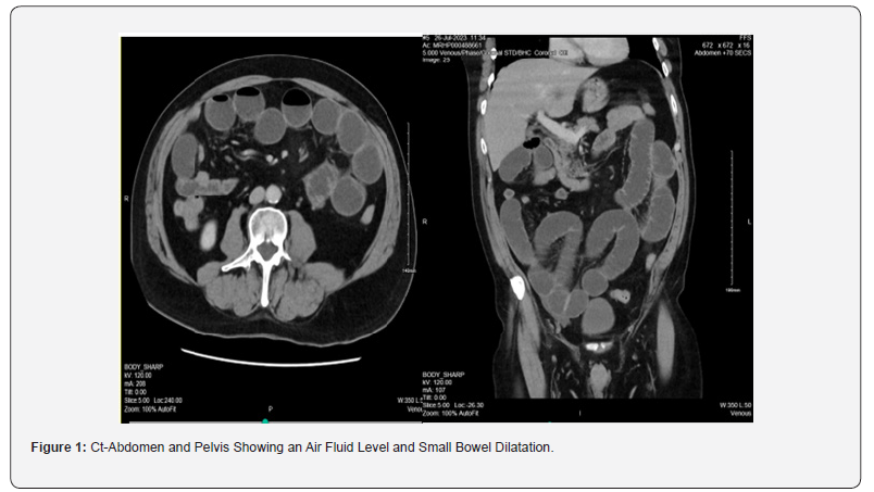
A 65-year-old male presenting to Tullamore Regional Hospital, complaining of abdominal pain, vomiting and absolute constipation for 3 days prior to admission. The patient’s abdomen was distended with generalised tenderness, however no surgical scars were observed. The patient’s admitted and laboratory results were as follows: Hb 13.0 units/dL, WCC 12.13 units/dL, CRP 15.5 units/dL, K+ 3.9 units/dL. A working diagnosis of small bowel obstruction was made, and the patient received I.V. fluids with nil per mouth. A CT-AP Figure 1 was ordered which confirmed a small bowel obstruction and the patient was consented for an emergency diagnostic laparoscopy.

Radiological Findings (Case 1) (Figure 1)

Procedure (Case 1)

Intra-operative laparoscopic exploration revealed an inflamed MD, and the entire small bowel was dilated sparing only last 60cm of distal ileum. Complete diverticulectomy was performed for inflamed MD with a linear stapler Figure 2. Subsequent histopathological analysis reported only an inflamed MD with no ectopic mucosa or neoplasia. The post-operative period passed uneventfully. The patient was discharged home in good condition on day four post-op. He was reviewed in the Outpatient’s Clinic six weeks later. The patient reported no post-operative complaints and the wounds well-healed.

Intra operative images (Case 1): (Figure 2).

Case Presentation (2)

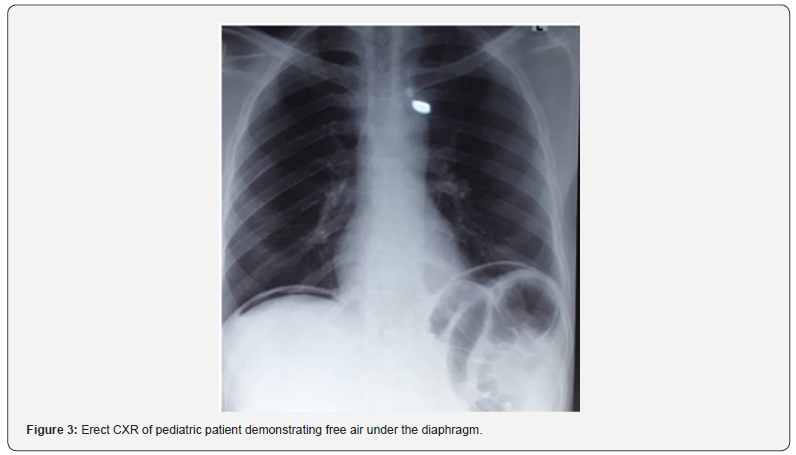
A 12-year-old boy was admitted to the Paediatric Surgical Unit in Khartoum Teaching Hospital with abdominal pain and bleeding per rectum. On examination the patient’s abdomen was soft, with right sided tenderness. Digital rectal exam was normal with no haemorrhoids or anal fissure noted. Two days postadmission the patient developed generalized abdominal pain, vomiting and there was abdominal distension and peritonitis. Routine haematological investigations revealed the patient’s serum haemoglobin had dropped from 14grams to 11grams. The patient therefore received a 50ml/kg transfusion of packed red blood cells, as well as IV antibiotics and I.V. fluids. An erect chest x-ray Figure 3 showed air under diaphragm. In discussion with the patient’s family, the consent was made for a laparotomy.

Radiological Imaging (Case 2): (Figure 3)

Procedure (Case 2): A right lower transverse laparotomy incision was made to explore the bowel. Surgical exploration revealed a short Meckle’s Diverticulum with a wide base at the distal ileum with two perforated peptic ulcers. The MD was removed via wedge resection. The perforated ulcer edges were then biopsied and refashioned before closure via primary repair. Postoperatively, intravenous antibiotics and PPI infusion were given, and postoperative period passed uneventfully, the patient tolerated oral intake. The patient was discharged on day seven post-op with follow up in the outpatient clinic in three months. Histology afterwards confirmed a completely resected MD with ectopic gastric mucosa.

Case Presentation (3)

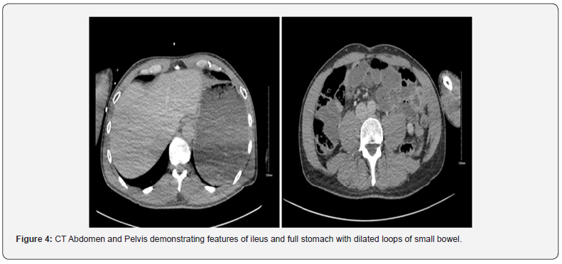
A 60-year-old male presented to the emergency department of Tullamore Regional Hospital with signs and symptoms of small bowel obstruction beginning 2 days prior to admission. Examination revealed a distended abdomen with generalised tenderness. Routine investigations: WCC 8.23 units/dL, CRP 31.9 units/dL, K+ 4.2 units/dL. The patient was admitted for nasogastric tube suction, I.V. fluids and analgesia. A CT-AP showed partial small bowel ileus with dilated loops of small bowel Figure 4. The patient was consented for an emergency diagnostic laparoscopy.


Radiological Imaging (Case 3): (Figure 4)

Procedure (Case 3)

Laparoscopic exploration detected an inflamed MD at the distal ileum with cord adhesion to the umbilicus Figure 5. Dilated small bowel was observed proximal to the adhesions. The inflamed MD was resected at the base with a linear stapler (diverticulectomy). The histopathology report showed an inflamed MD with ectopic gastric mucosa. The post-operative period passed unremarkably. The patient was discharged day three post-op and reviewed at OPD at 4 weeks with well healed wounds and no post-operative complaints.

Intra operative images (Case 3)

(Figure 5)


Discussion

Symptomatic Meckel’s diverticulum typically occurs in the first two decades of life and almost exclusively before the fifth decade [6]. The adult cases outlined above both contradict this pattern given both patients were above 60 years of age. In adults, MD is usually an asymptomatic condition with only about 4% of those affected developing complications [8]. The reported mechanism of intestinal obstruction in MD includes intussusception, adhesion, and volvulus. Furthermore, inflammation of the MD can cause small bowel ileus as seen in our two adult cases [1]. While obstruction is the most common complication in adult patients, gastrointestinal bleeding is the most common presenting symptom of MD in children [2]. This aligns well with the presenting complaints of the three cases in this report. Moreover, another study reported haemorrhage as the most common presentation in children and is reported in over 50% of their symptomatic cases [7]. Other complications of MD include perforation, umbilical fistula, cyst, fibrous cord with umbilicus, while double Meckel’s and carcinoid tumour are also reported in the literature [8]. 90% of bleeding diverticula contain heterotrophic mucosa, most often gastric mucosa (71%), contains acid-secreting parietal cells which can lead to ulceration and haemorrhage as observed in our paediatric case. Histopathological analysis demonstrates ectopic pancreatic tissue in 12.0% of resected MD [6].

The diagnosis of MD may be challenging as the condition often remains asymptomatic or may mimic various diseases which obscures the clinical picture. For example, MD often mimics the presentation of acute appendicitis in children, as 11% of complicated MD were initially misdiagnosed as acute appendicitis [8]. The probability of onset of complications decreases with age, therefore the diagnosis of MD in adults is often incidental [9]. Exploratory laparoscopy is an important diagnostic tool, especially when perforation has occurred [9]. In the case of hemorrhage, as often seen in pediatric MD, mesenteric angiography and embolization are reported as diagnostic and therapeutic tools [5, 10]. The advent of technetium (Tc) 99m radionuclide scanning has greatly facilitated the diagnosis of MD especially when the anomaly contains heterotopic mucosa [5]. Since Meckel’s diverticulum (MD) is rarely diagnosed in adults, there is no consensus on the optimum operative technique for asymptomatic MD. Indeed, there is disagreement as to whether an incidentally discovered MD should indeed be respected at all [11]. However, research suggests up to 57% of resected MD were found incidentally which contradicts the view that routine resection is not indicated in incidentally discovered cases [3].

Resection of incidental MD is explicitly not recommended in patients with peritonitis, major abdominal trauma, ascites, or in the context of older age, malignancy, or immunosuppression. However, research does suggest resection of incidental MD may be appropriate in the context of risk factors which may predict future complications. Risk factors for MD complications include male gender, age younger than 40 years, diverticulum longer than two centimeters, narrow neck of the diverticulum and the presence of macroscopically mucosal abnormalities noted at surgery [11]. On the other hand, some researchers disagree with the view that the external appearance of the MD can reliably predict the presence of HGM, and therefore argue macroscopic appearance cannot reliably aid resection decisions in the context of an incidentally discovered MD [3]. Nevertheless, the definitive treatment of symptomatic MD is laparoscopic or open diverticulectomy via wedge or segmental resection. The type of procedure depends on; (a) the integrity of diverticulum base; (b) the presence and location of ectopic tissue within MD [8]. Location of ectopic mucosa can be predicted based on height-to-diameter ratio. Long diverticula (height-todiameter ratio >2) have ectopic tissue located at the body and tip, whereas short diverticula have wide distribution of ectopic tissue surrounding the diverticulum base. A greater HDR has also been found to be an independent risk factor for perforation [11].

When indication of surgery is simple diverticulitis, diverticulectomy should be performed for long and wedge resection for short MD [11]. Simple transverse resection is not recommended for short MD, as resection followed by anastomosis seems preferable to wedge resection or tangential mechanical stapling, in order reduce the risk of leaving behind abnormal heterotopic mucosa [4]. New operative methods for MD include basal ligation combined with intraoperative frozen section. Using intraoperative histological feedback on the existence of residual ectopic mucosa, surgery is either terminated or further wedge intestinal resection or bowel resection was performed as required. Basal ligation is a safe and effective operation method as it can significantly shorten operation times and postoperative fasting time. For hemorrhagic ulcer and perforation, segmental and wedge resection are indicated as performed in our pediatric case [5,12-15].

Conclusion

The laparoscopic approach was found to be effective as a diagnostic and therapeutic modality in patients with symptomatic MD. Once MD has been diagnosed, the fundamental therapeutic aim is to achieve complete resection of the diverticulum along with ectopic epithelium. The HDR can assist in determining the most appropriate resection technique. Longer MD should be resected with diverticulectomy whereas wedge resection is the optimum technique for short MD.


To Know more about Open Access Journal of Surgery
Click here: https://juniperpublishers.com/oajs/index.php

To Know more about our Juniper Publishers
Click here: https://juniperpublishers.com/index.php
Monday, October 30, 2023

Violence Against the Older Adults: A Brazilian Tragedy - Juniper Publishers

 Forensic Sciences & Criminal Investigation - Juniper Publishers


Abstract

Context and Objective: The occurrence of suspected cases of abuse against older adults is a global phenomenon; health professionals must pay close attention to the possibility of abuse against this population. This study aimed to describe the profile of notifications of abuse against older adults reported in SINAN (the official database of the Brazilian Ministry of Health) between 2009 and July 2014.

Design: Cross-sectional study.

Setting: The manuscript was produced in the Department of Legal Medicine of the Medical School of Universidade de São Paulo and ABC Faculty of Medicine.

Methods: Data on domestic, sexual, and other forms of violence were obtained from the Ministry of Health’s official website (Sistema de Informação e Agravos de Notification – SINAN) between 2009 and July 2014 (http://dtr2004.saude.gov.br/ sinanweb/index.php). The selected subjects were elderly aged 60 years or older of both sexes.

Results: The number of notifications showed a proportional increase between 2009 and mid-July 2014; most of the attacks occurred in the victims’ homes, and the most frequent aggressors were sons or daughters, followed by unknown people and acquaintances. Women were the most attacked. Many aggressions were recurrent and in multiple forms, but physical aggression and negligence were the most reported.

Conclusion: This study’s great importance is exposing the quantitative data of this violent scenario so that health professionals who care for older adults remain alert to the possibilities of mistreatment and that they notify suspected or proven cases, as determined by Brazilian law.

Keywords: Older adults; Abuse; Neglect; Physical signs; Incidence; Violence

Introduction

With the global growth of the elderly population, specific issues related to this group of individuals should be widely discussed. Among these issues is violence, which is essential because mistreatment of older people is associated with distress and increased mortality [1]. Therefore, it is a serious public health problem.

Abuse is defined as violating an individual’s human and civil rights. The abuse might consist of a single or repeated act, often affecting vulnerable people and occurring in any relationship. The following six types of abuse have been described: physical, sexual, psychological, financial, or material, neglect or act of omission, and discriminatory [2]. According to the Brazilian Institute of Geography and Statistics (IBGE) [3], in 1950, older adults comprised 4% of the country’s total population; in 1991, they represented 7.3%; 8.6% in 2000 (14.5 million) [4]; and 10% in 2010. Thus, according to the World Health Organization (WHO), Brazil is experiencing a rapid increase in the number and percentage of older people, often within a single generation, and needs to adjust to this new reality [5]. Brazilian laws are strict regarding the notification of confirmed or suspected cases of abuse against older people. Law nº. 12.461 (2011), amended Article 19 of the Statute of the Elderly (Law nº. 10.741/2003), is more rigorous than the previous law. It mandates that the notification of abuse against older adults be compulsory, even in suspected cases, and should be performed by health authorities through private and public health services and any official agency (Police authority; Public Ministry; Municipal Council for the Elderly, the State Council of the Elderly, and the National Council for the Elderly) [5,6].

Concerning the sub notification, Cooper et al. [1] in a systematic review published in 2008, concluded that 6% of older people reported significant abuse in the last month, 5.6% of couples reported physical violence in their relationship in the last year, and one in four vulnerable elders are at risk of abuse; however, only a tiny proportion of this abuse is currently detected. In Brazil, according to the Information System of Aggravation of Notification (SINAN Net), which is associated with the Brazilian Ministry of Health, there were significant fluctuations in the total numbers of specific notifications of abuse and violence-related deaths among people over age 60 between 2009 and 2011 [7]. In 2009, there were 1748 notifications of abuse, 2.5% of which resulted in death; 3298 notifications of abuse, with 1.17% of these resulting in death, occurred in 2010, and 5307 notifications of abuse, with 4.6% of these cases resulting in death, occurred in 2011 [6,7]. Mascarenhas et al. [7] evaluated many aspects of the reports of violence against older adults in the Brazilian Health Services [7,8]. These authors described the victims’ profile as Caucasian males, between 60 and 69 years of age, single or widowed, with low education levels, and with no physical disabilities or mental or behavioral disorders. Physical violence was prevalent among men, whereas psychological violence, sexual violence, and neglect were more prevalent among older women [7,8]. Another Brazilian study showed that women over 75 years of age, single or widowed, with low education levels and chronic diseases, are primary abuse victims [8,9]. This data will facilitate the assessment of the primary at-risk groups suffering from violence and provide information for establishing public policies to reduce the number of abused elderly individuals. Thus, medical physicians and forensic examiners should recognize these signs to identify cases of abuse against older adults and avoid recurrences in non-fatal violence and vengeful perpetrators.

Objectives

This study aimed to describe the profile of notifications of abuse against older adults reported in SINAN (the official database of the Health Ministry of Brazil) from 2009 to July 2014.

Methods

The results of this cross-sectional study were conducted through analysis of data published on the official website of the Brazilian Ministry of Health: SINAN Net. (http://dtr2004.saude. gov.br/sinanweb/). The information in this database included notifications of violence (domestic, sexual, and other types of violence against the older adults), which were included in the list of compulsory notifications of violence according to the Ordinance GM/MS Nº 104, January 25, 2011. We analyzed the notifications from 2009 to July 2014. The study parameters included the sex and age of the victim, the victim’s relationship to the perpetrator, the place of the aggression, and the type and form of the aggression. The results were compared with data from the literature.

Results

Total numbers

In the period from January 2009 to July 2014, the SINAN (official database of the Brazilian Ministry of Health) received 655,720 compulsory notifications of violence, and 35,155 of these cases involved the older adults population (over 60 years of age), representing 5.36% of the total.

Setting of the aggression, sex, and ethnicity of the victims

68% of these cases (24,167) occurred in the victim’s residence, and were reported 12,314 cases recurrent violence, which resulted in 1111 deaths. The women were the most frequent victims, accounting for 54% of total aggressions (maintaining the ratio above 50% for all study years), ethnicity was Caucasian (Graph 1), and 33,3% had low levels of education.

In the analyzed period, the average percentage of elderly victims of total violence remained stable, at around 5.14%, compared to the total violent notifications (all ages), as shown in Table 1.

The physical violence and deaths

However, from 2009 to July 2014, notification of physical violence against older people increases in Brazil (Graph 2). The partial number of notifications until July exceeded the total number in 2011. In 2010 there was an increase of 103% compared to the previous year; in 2011, the increase compared to 2010 was 58,7%, and 68,44% in 2011 compared to 2012. However, there was a slowdown in 2013 when the increase in notification was 14,69% concerning 2012. The total of notifications of physical abuse was 23,542 in the period (2009 to July 2014), with 1111 deaths (Graph 3); 14,000 cases were caused by beating.

Physical abuse was more frequent in men (51,5%) than women, but the difference was irrelevant. The perpetrators were sons or daughters in 4,305 notifications. A stranger was the aggressor in 3,304 cases; friends were 2,696, and husband or ex-husband, the number of notifications, was 2,478. In the 2,558 aggressions, the instrument used was the knife.

Neglect, abandonment, and sexual violence

Neglect and abandonment were the second most frequent kind of violence against older people. In the period of this study, 9,296 notifications were performed, with 129 deaths. Women were the preferred victims, 4,872 cases (52,4%). Table 2 shows the notifications according to race. There were 9,241 cases of sexual and psychomoral violence reported during the period, of which 6,595 were women (71.36%). Considering only reports of isolated sexual violence, there were 782 cases, 94.91% of which involved women.

Discussion

Our study showed that 5.36% of the total notifications from January 2009 to July 2014 were related to older people.

A Portuguese study applied between January and June 2013 demonstrated that 23.5% of older adults have suffered some abuse, emotional and neglect especially; these results were obtained through a questionnaire [10], which may have facilitated the complaint. In Brazil, in 2013, notifications of violence against the older population were 5.7%. According to the WHO, 36% of American nursing home staff reported witnessing abuse in the past year, with 10% witnessing physical abuse and 40% witnessing psychological abuse.9,10 These data do not show the true scope of this type of crime, as most abusers are close relatives (i.e., sons and daughters). Another study (a systematic review of the prevalence of abused elderly individuals) showed that the prevalence of abused elderly individuals varies according to culture and the definition and measure of violence. According to these authors, 25% of older adults are at risk of abuse, although few cases have been identified1. Many forms of violence are imposed against older people, as these victims are vulnerable individuals who are often unable to react to attacks or depend on the abuser. In some situations, an old individual has difficulty understanding the violent act or does not want to report the abuser due to their relationship. The term “abuse against the elderly” has often been associated with physical aggression; however, neglect and emotional abuse (psychological) play essential roles [9,11]. Few studies consider neglect as a separate form of abuse. The definition of abuse against the elderly is complex, involves multiples phenomena, and varies considerably among researchers and between the laws of different countries, even among the states of a country, as in the U.S. Concerning determining the age limit of elderly individuals, some studies suggest ages above 60 years. In contrast, others only consider ages over 65 years. This varying definition is a problem that affects epidemiological studies of the incidence of these events. According to Akaza et al. (2010), other confounding factors involve the different classifications of the types of maltreatment (violence). For some authors, violence is classified into four types (physical, emotional, financial, and neglect). In contrast, others add self-neglect, sexual abuse, and other miscellaneous forms of abuse [11,12]. The prevalence of elderly abuse is difficult to determine, as some studies define older adults as over 60. In contrast, other studies consider people over 65 years. Although all people can be battered, the most frequent victims are vulnerable individuals, such as children, teenagers, women, homosexuals, people with disabilities or mental disorders, and the elderly [8,11]. The Ministry of Health warns that falling accidents among the elderly and their consequences have become an epidemic in Brazil, with more than 20,000 hospitalizations in 2009. The Brazilian government recommends that all physicians who evaluate elderly individuals suffering from falls (accidental or not) ask these individuals whether there were any other injuries or falls in the last six months [12,13].

In 2007, according to the Ministry of Health, 18,946 older adults died from external causes, representing the seventh leading cause of death, and 125,000 hospitalizations were recorded that same year. In 2010, the WHO reported that 41 elderly individuals die each day worldwide from external causes, representing this group’s sixth leading cause of death, emphasizing that these events remain incredibly underreported [13,14]. In our study, relatives, mainly daughters, and sons, were the significant perpetrators of abuse against older people, and the aggression occurred inside the home. These data were similar to those observed in medical literature. Alcoholism among the offenders was a factor in many reports. Other similarities between the Brazilian results and the descriptions in the literature showed that women were the principal victims. The differences between the gender data obtained in SINAN were mild, although women were the most victimized every year [7,14]. These results were similar to those of Souza et al. [15], perhaps reflecting the greater longevity of women [15].

Although abuse against the elderly is not recent, only from the late 1980s, it has become a topic of interest. Indeed, the notifications recorded in the databases do not represent the actual number of deaths or injuries suffered within this population. Under notification might partially reflect that the clinical manifestations of abuse are often mistaken for the manifestations of diseases inherent to aging [15,16] (e.g, bruises). Some studies show that the presence of bruises greater than five centimeters in size on the lateral arm or right shoulder, neck, head, thoracolumbar region, buttocks, and soles of the feet are not often observed in accidental injuries and should be considered to result from an act of violence [17].

Multiple wounds in different stages of cicatrization; bruises at different stages of evolution (purple, brown, green, yellow, and finally brownish yellow); injuries in the shape of any instrument, marks of containment on the wrists, ankles, and heels; traumatic alopecia, edema of the scalp, and fractured teeth or nose or burns are other warning signs. Radiological images showed that misaligned fractures in different stages of consolidation could also be signs of physical aggression [17-21]. On the other hand, pressure sores, localized areas of tissue damage, or necrosis are common in the older population. However, Santos et al. [20] state simple care can prevent pressure sores. Therefore, the presence of these lesions might be an indicator of inadequate care. Is important to point out that, as Mosqueda et al. [22] say, “innocent bruises frequently occur on the extremities”. Although recognizing this abuse is difficult due to physiological aging, it should always be considered, particularly when the clinical history contradicts the findings of physical or laboratory examinations.

Another explanation for the low rate of notification of abuse against the elderly is that health professionals are not attentive to this possibility and that older people frequently are silent victims. The violence perpetrated through negligence principally occurs in a domestic environment and frequently involves the family, which makes diagnosis difficult. In many instances, the victim does not report the violence because they do not understand the incident as a form of violence or are afraid to report their close relatives as the perpetrators. According to Minayo [13], deaths, injuries, and trauma from traffic accidents and falls could also suggest negligent acts committed by the authorities or people involved with the accessibility of the elderly individual. Finally, we have to remember that signs suggestive of sexual violence injuries are also frequent, and include itching, anal or vaginal bleeding and pain, sexually transmitted diseases, and spotting or bleeding in the underwear21. Physicians, including coroners, should pay attention to elderly individuals with these lesions during their examinations, as there are many types of abuse against the elderly, and the diagnosis is difficult.

Final Considerations

The importance of our study is to show the large number of cases of violence that occur in our country, totaling 23,542 cases in the period studied, with an unspeakable tragedy represented by the 1,111 deaths that occurred in this period. Notably, our study also showed that assistance from health professionals facilitates the identification and confirmation of cases of maltreatment against the elderly and reporting, which is required by Brazilian law. Worrisome, this study showed that the number of abuse notifications against the elderly has grown in Brazil. Men and women are victims of physical, sexual, and neglect abuse, often recurrent and fatal. With the increasing life expectancy of the world population, issues involving older individuals are becoming common in medical practice. Among these issues, violence against older adults must be analyzed carefully, as many forms of violence leave no visible marks (neglect, psychological and economic violence), and many injuries to the elderly result from advanced age (fractures from falls and bruises). The information provided to physicians does not always represent the truth. Thus, physicians need to be alert to the possibility of violence against older adults, as the recurrence of maltreatment, and to report suspected or confirmed cases.


To Know more about Journal of Forensic Sciences & Criminal Investigation
Click here: https://juniperpublishers.com/jfsci/index.php

To Know more about our Juniper Publishers
Click here: https://juniperpublishers.com/index.php 

Understanding The Acute Interventions for Complicated Pleural Effusions - Juniper Publishers

  Pulmonary & Respiratory Sciences - Juniper Publishers Abstract Pleural effusion affects approximately 1.5 million individuals in the U...