Showing posts with label Lung Cancer. Show all posts

Wednesday, July 14, 2021

RNF168 is a Potential Therapeutic Target in Non-Small Cell Lung Cancer - Juniper Publishers

 Cancer Therapy & Oncology - Juniper Publishers 

Abstract

Prostate cancer is somewhat unusual when compared with other types of cancer. This is because many prostate tumors do not spread quickly to other parts of the body. Some prostate cancers grow very slowly and may not cause symptoms or problems for years or ever. Even when prostate cancer has spread to other parts of the body, it can be managed for a long time, allowing men even with advanced prostate cancer to live with good health and quality of life for many years. Vitamin D is a steroid hormone that is thought to play a role in the etiology and progression of prostate cancer. Hormone activity requires binding to the vitamin D receptor (VDR), which contains several genetic polymorphisms that have been associated with risk of prostate cancer.

Introduction

Lung cancer is one of the most prevalent cancers in humans. In 2018 alone, lung cancer took the lives of 1.6 million people worldwide [1]. There are two main types of lung cancer, non-small-cell lung cancer (NSCLC) and small-cell lung cancer (SCLC), with NSCLC comprising ~85% of the cases [2]. A histologic sub-category of NSCLC is lung adenocarcinoma, accounting for ~40% of all lung cancer cases. The other main histologic sub-category is squamous cell carcinoma. Intra-tumor heterogeneity as well as high mortality among patients with NSCLC emphasize the unmet need to identify reliable prognostic markers that are unique to each subtype. Further, despite recent advances in treatment options for NSCLC, such as targeting of driver mutations, immunotherapy, radiotherapy, and platinum-based chemotherapy [3], inherent or acquired resistance to treatment is still inevitable. Thus, how to overcome treatment resistance and identify molecular biomarkers that can be used to discriminate between responders and non-responders remains a major research focus.

Insufficient DNA damage response (DDR), especially against DNA double-strand breaks (DSBs), results in genetic instability that is a root cause of most cancers. Nevertheless, cancer cells are addicted to existing DDR pathways to maintain infinite replication, making these pathways an obvious central target in anticancer therapy [4] RING (Really Interesting New Gene) domain-containing E3 ubiquitin ligases are a large family of enzymes that associate with an E2 ubiquitin-conjugating enzyme to ubiquitinate their substrates. The RING E3 ubiquitin ligase RNF168 -dependent ubiquitination signaling plays an essential role in the DDR [5]. RNF168 localizes at DNA repair sites and promotes ubiquitination of key DDR proteins to drive DNA DSB repair [6]. Previous studies have shown that RNF168 is a factor in chemotherapy resistance in various cancers [7]. In esophageal cancer, over-expression of RNF168 has been found to correlate with poor prognosis [7]. RNF168 is understudied in NSCLC, and nothing is known concerning whether it may have a role in NSCLC subtypes as a potential prognostic biomarker.

Here we investigated the role of RNF168 as a potential prognostic marker in NSCLC using popular online databases such as CBioPortal, Oncomine, KMPlotter, ProteinAtlas, and CellMiner CDB. We found that high mRNA levels of RNF168 correlated with poor patient prognosis in lung adenocarcinoma. Furthermore, we found that this gene was overexpressed in NSCLC, in both adenocarcinoma and squamous cell carcinoma. Finally, we identified RNF168 to predict sensitivity to docetaxel in lung adenocarcinoma.

Results

RNF168 is over-expressed in non-small-cell lung cancer

We used CBioPortal, to study the genetic alteration of RNF168 in NSCLC. We found the gene to be altered 16.78% of the time in a total of 1,144 cases of NSCLC, with amplification being the most common alteration, present in 15.12% of cases (Figure 1A). We then delved into NSCLC subtypes and found RNF168 to be altered in 4.78% of adenocarcinoma cases (Figure 1B). RNF168 alteration was greatest in squamous cell carcinoma, with 46.07% of the cases showing gene amplification (Figure 1C). We used Oncomine to compare the expression of RNF168 in normal lung cells to that of lung adenocarcinoma cells and found greater expression in adenocarcinoma cells (Figure 1D). As expected, RNF168 mRNA expression was higher in squamous cell carcinoma than in normal pulmonary cells (Figure 1E). Thus, RNF168 overexpression was present in NSCLC subtypes.

High expression of RNF168 is associated with lower patient survival in NSCLC overall and lung adenocarcinoma

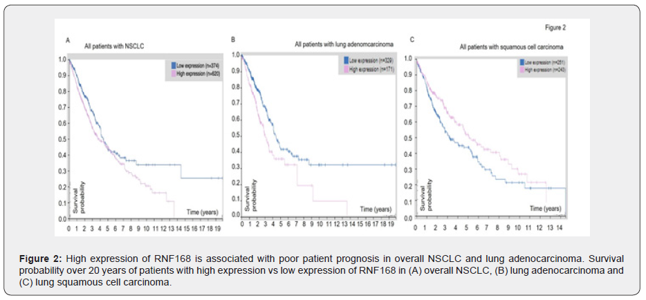
Next, we looked at the patient prognosis using the Human Protein Atlas database to determine whether there was a correlation between gene expression and patient survival. We found that higher RNF168 expression was associated with significantly (P <0.007) decreased survival for all NSCLC patients, as shown in Figure 2A. This trend of poor patient prognosis when RNF168 level was high remained true for lung adenocarcinoma (P = 0.001) Moreover, the 5-year survival for patients with high levels of RNF168 was 36%, whereas of those who had low levels, 43% were alive at 5 years (Figure 2B). In squamous cell carcinoma, however, this trend was reversed. High levels of RNF168 consistently indicated better probability of survival (P = 0.02) (Figure 2C). Overall, our analysis of patient prognosis showed that high levels of RNF168 was detrimental to patients in overall NSCLC, and especially adenocarcinoma, whereas low levels of RNF168 indicated better patient prognosis for overall NSCLC and adenocarcinoma. In contrast, for squamous cell carcinoma, high expression of RNF168 was associated with better patient prognosis, and low expression of RNF168 was associated with poorer prognosis.
Docetaxel can be used to treat patients with high levels of RNF168
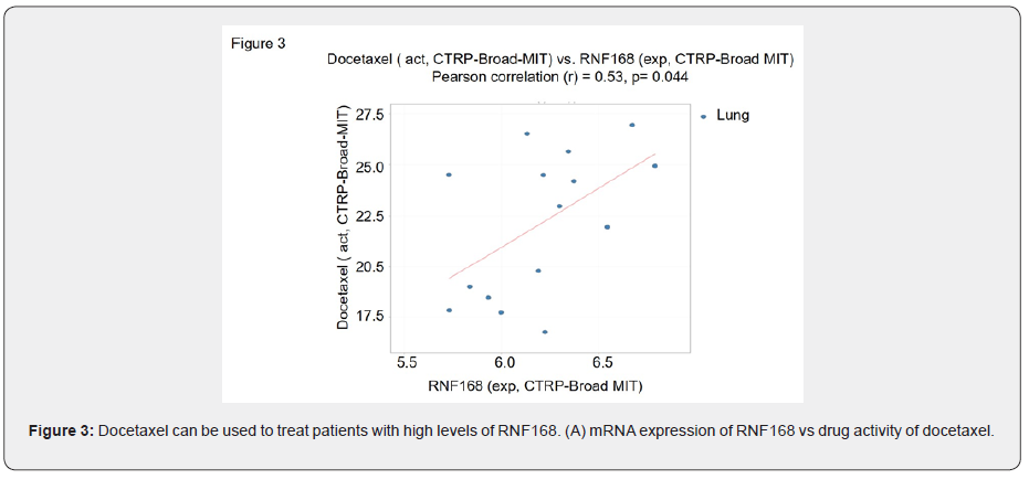
Our analysis of data from the Human Protein Atlas clearly showed that high expression of RNF168 suggested a poorer prognosis for patients with lung adenocarcinoma., Therefore, we used CellMiner CDB to look for drugs that exhibited a high efficacy in lung cancer cells with high RNF168 mRNA expression. We identified docetaxel as a drug that can potentially be used to treat patients with high levels of RNF168 (r = 0.53, r2 = 0.28, and P = 0.044; Figure 3).

Methods

CBioPortal

Through CBioPortal (https://www.cbioportal.org/), we identified RNF168 alteration frequencies in various types of lung cancer such as NSCLC, LUAD, and Lung Squamous Cell Carcinoma (LUSC). More specifically, we looked at the frequency of mutations, alterations, deep deletions, and multiple alterations in each histologic sub-category. We analyzed at the MSK, MSKCC, TracerX, University of Turin, and TCGA study datasets for NSCLC and LUAD to determine that RNF168 is over-expressed in NSCLC, LUSC, and LUAD.

Oncomine

Oncomine (https://www.oncomine.org) was used to analyze the over-expression of RNF168 between different types of lung cancer cells and normal cells, given by log2 median-centered intensity and box and whisker plots indicating median intensities. The Hou Lung dataset indicated RNF168 over-expression between patients with LUSC and normal patients, while the Okayama Lung dataset showed the relationship between RNF168 expression in LUAD patients and normal patients. Using Oncomine, we could verify the over-expression of RNF168 in LUAD by comparing the expression of the gene in LUAD with LUSC and normal lung without cancer.

Human Protein Atlas

Using Protein Atlas (https://www.proteinatlas.org), we obtained prognostic data for RNF168 in overall NSCLC, as well as adenocarcinoma and squamous cell carcinoma. We used the prognostic information to analyze the effect of RNF168 on survival in NSCLC, LUAD, and LUSC, indicated by survival probability over 20 years. Patient data was taken from TCGA data sets.

CellMiner CDB

We used CellMiner CDB (https://discover.nci.nih.gov/cellminercdb/) to identify potential drugs that can target high RNF168 expression in adenocarcinoma. Univariate analyses allowed us to compare the patterns between mRNA expression (Z-score) and DNA copy number for RNF168 and drug activity for a variety of drugs. To determine the effectiveness of the identified drugs, the correlation between drug activity and RNF168 expression was analyzed using Pearson’s correlation, and we looked at cell lines from NCI-60, CCLE-Broad-MIT, GDSC-MGH-Sanger, and CTRP-Broad-MIT.

Discussion

We have found that high RNF168 levels were associated with a decreased probability of survival in patients with NSCLC patients, and particularly so for those with lung adenocarcinoma. Moreover, although high levels RNF168 indicates poorer survival for patients with adenocarcinoma, it has a better prognosis of patients with lung squamous cell carcinoma. Ultimately, we find that docetaxel is a potential treatment to patients with high RNF168 expression. Ubiquitin ligases (E3) have been of much interest to scientists because of their role in the degradation of proteins. RNF168 is no exception, and its potential to facilitate cancer progression as an oncogene has previously been identified. For example, RNF168 has been found to be over-expressed in esophageal cancer samples and correlated with poor patient prognosis. This study also showed that RNF168 promoted JAK-STAT signaling, which is involved in many different types of cancers [7]. Not only has RNF168 been shown to be involved in esophageal cancer, but many other cancers as well. Others have reported that overexpression of RNF168 can lead to mutations in the BRCA1 and BRCA2 genes, which are both tumor-suppressors [8]. BRCA mutation could lead to increased carcinogenicity and the investigators theorized that RNF168 can increase the risk of BRCA-mutated cancers. The impact of RNF168 on lung cancer has not been previously investigated. Our study adds to existing literature for RNF168 by giving further evidence of its potential as an oncogene. Our research also proof of yet another cancer that RNF168 contributes to, and thus underscores its role as a potential target in cancer. RNF168 is currently understudied, however, given its tumorigenic potential in adenocarcinoma, its role and regulation should be investigated in greater detail in the future studies.

Surprisingly, we found that high expression of RNF168 was associated with a better survival rate for squamous cell carcinoma patients. Most smokers who develop lung cancer suffer from squamous cell carcinoma, whereas adenocarcinoma is more likely to develop in non-smokers [9]. One of the primary causes of lung adenocarcinoma is genetic mutations, whereas squamous cell carcinoma is caused by the inhalation of carcinogens, damaging the cells that line the lungs [10]. The overexpression of RNF168 has previously been linked to the development of mutations, which could explain why high expression is detrimental in adenocarcinoma [7].

Conclusion

In conclusion, using data from public databases our study identifies a novel role of RNF168 as a potential biomarker in NSCLC [11-13]. While we find a positive correlation between overall survival in lung adenocarcinoma and negative correlation in case of lung squamous cell carcinoma, further research is needed, e.g. using RNF168 knockdown experiments in cell lines or mouse models to clarify a more deterministic role of RNF168 and mechanisms underlying the potential relationships between RNF168 expression and lung adenocarcinoma and between RNF168 and squamous cell carcinoma.

To Know more about Cancer Therapy & Oncology   


To Know more about our Juniper Publishers

Click here: https://juniperpublishers.com/index.php    


Wednesday, November 27, 2019

Airway Microbiota and Allergic Diseases: Clinical Implications-Juniper Publishers

INTERNATIONAL JOURNAL OF PULMONARY & RESPIRATORY SCIENCES

Introduction

Bronchial Asthma is an airway disease with variable degrees of bronchial mucosal inflammation and intermittent episodes of airway obstruction and bronchial hyperesponsivness. That asthma is a syndrome consisting of different phenotypes has been recognized for a long time by clinicians [1]. New evidence indicates that the composition of airway microbiota differs in states of health and disease. Different chronic airway diseases had been related to changes in microbiota due to various factors which could affect severity of symptoms and even response to treatment [2]. Micro biome may be one of the protective factors against asthma in early life [3].


What is Airway Microbiota
It a complex variety of microbes present intrachea and different generations of the bronchi either on the mucus layer or the epithelial surfaces or even both. These microbes include bacteria, yeasts, viruses and bacteriophages. The bacterial part of microbiomeis the most prevalent component with various genera: Prevotella, Sphingomonas, Pseudomonas, Acinetobacter, Fusobacterium, Megasphaera, Veillonella, Staphylococcus, and Streptococcus. The bronchial tree for instance contains a mean of 2000 bacterial genomes per cm2 surface [4]. The mucosal surfaces in the human body are the home of 10-100 trillion microbes with a diversity of greater than 1,000 species [5]. The highest concentration of microbes is found in the GI tract, compared to those found in the lower airways. Healthy human lungs are not sterile, as previously believed, but it is unknown whether the microbes in the lungs form a stable community or are a series of transient colonizers [6].

However, various theories about the origin of lower airway microbiota in healthy individuals had been suggested. As it may represent true colonization of the lower generations of bronchi, or it is the result of turnover of the microbial community or it is just contamination of oropharynx during lower airway sampling or even linked potentially to those who are incorrectly categorized as truly healthy [7].

Importance of microbiota

The commensal bacteria are nonpathogenic and defend our airways against the pathogens. There are several possible mechanisms:
  1. Commensals are the native competitors of pathogenic bacteria, because they occupy the same niche inside the human airways.
  2. They are able to produce antibacterial substances called bacteriocins which inhibit the growth of pathogens. Genera Bacillus, Lactobacillus, Lactococcus, Staphylococcus, Streptococcus, and Streptomyces are the main producers of bacteriocins in respiratory tract.
  3. Commensals are good inducers of anti allergic Th1 cascade with anti-inflammatory interleukin (IL)-10, FOXP3, and secretory immunoglobulin A (sIgA) production [7].

Airway epithelial cell and microbiota interaction

The airway epithelium together with alveolar macrophages and dendritic cells collectively can recognize of bacterial products trapped into the lower airways with the inhaled air. Some of these products are can potentiate pro inflammatory stimuli. So it is a challenging issue to distinguish between pathogens and commensals to avoid development of constant or persistent inflammation and help to develop tolerance against harmless microbiota [8].

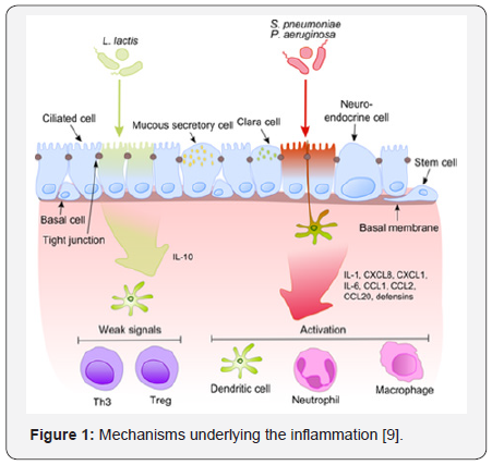
Once pathogenic bacterium (e.g., S. pneumoniae, P.aeruginosa) has been attached to activated pattern recognition receptors located on/in bronchial epithelial cells, the proinflammatory cytokines pathways are predominant via release of IL-1, IL-6 and IL-8 which induce neutrophils, dendritic cells and macrophages chemotaxis to target cells (e.g., neutrophils, dendritic cells and macrophages. Standard microbiota fail to induce strong signaling, thus aborting inflammation. (Figure 1) [9].
This process becomes much more intriguing when taking into account that commensals often share their surface molecules with pathogens. Epithelial cells are equipped with very sensitive recognition tools - toll like receptors (TLRs), NOD like receptors (NLRs) and retinoic acid-inducible gene (RIG)-I-like receptors (RLRs) which determine presence of non commensal bacteria which activate cellular components of the adaptive and innate immunity and recruit them to the infection site [7].

NF-κB is the principal regulators of different response to harmful microbiota as it is become activated by a number of stimuli as bacterial cell walls or inflammatory cytokines. This results in its translocation from the cytoplasm into the nucleus to activate epithelial cells pro-inflammatory genes. These specific genes can recognize a particular nucleotide sequence (5’-GGG ACT TTC T-3’) in upstream region of response genes. [10]. Inspite of expressing express the same microbe-associated molecular patterns (MAMPs), harmless bacteria fails to translocate NF-κB into the nucleus thus preventing the inflammation. The balance between pathogens and commensals is extremely important in the maintenance of homeostasis in the respiratory tract [9].

Pediatric acterial airway microbiota in early life

A neonatal mouse exposed to a broad-spectrum antibiotic has been shown to increase allergen-induced airway inflammation susceptibility [4]. Germ-free mice also exhibit enhanced airway inflammation upon allergen exposure [3], while colonizing OF germ free mice with microbiota from conventional mice decreased accumulation of natural killer T (NKT) cells in their airways .This was only observed in neonates not in adult mice. This highlights the importance of early life as a critical period for intervention [11].

Absence of airway colonization during this critical neonatal window resulted in sustained susceptibility to allergicinflammation through adulthood. This ensure long-term control of allergic airway inflammation via controlling commensal bacteria communities early in early life [12].

Microbiota and climax community

Climax community is defined as a microbial community that has reached a final or “climax” steady state best adapted for growth at that specific niche along the mucosa. However, this climax community is dynamic and still exhibits both resistance and resilience [13]. Evidence is now accumulating that longterm dietary pressures , repeated antibiotic use, GI illnesses or medications such as antacids, proton pump inhibitors, and nonsteroidal anti-inflammatory drugs can break both the resistance and resilience of a community and result in it re-assembling into another climax community, although this may be accompanied by detrimental changes in host mucosal immuno biology and physiology. One mechanism underlying the activity of probiotic microbes and prebiotic nutrients may be the ability to restructure a climax community to improve host mucosal immuno biology and physiology [14].

Microbiota (microflora) hypothesis

Several theories had been suggested to explain the increase in the incidence of asthma and other allergic diseases over the past 30 years and the discrepancy between the higher rates of allergic disease among industrialized relative to developing countries. One rising assumption is a lack of early microbial stimulation which results in aberrant immune responses to innocuous antigens later in life “hygiene hypothesis” [15]. Life style modifications and over use of broad spectrum antibiotics raise the concept of disturbance of mechanisms of mucosal immunologic tolerance due to changing diversity of gastrointestinal (GI) microbiota composition in westernized areas [16].

Epidemiologic and clinical data supporting this interpretation include

  1. a positive correlation between increasing risk for asthma/allergies and increasing use antibiotics in industrialized countries,
  2. Altered fecal microbiota composition had been correlated to different atopic diseases
  3. Oral probiotics orsignificant dietary changes lead to some successful prevention/reduction of severity of allergic diseases.
Experimental data in mice compared that immune response generation and normal ones which showed numerous defects in immune response [17]. Altogether, these experimental, epidemiologic, and clinical observations support the hypothesis that even minor changes in the quality or quantity of airway microbiota can be one of the predisposing factors for allergic disease [10].

Cross-talk between the gut and the lung

The existence of the gut–lung axis and its implications for airway disease provide a portal for potential therapeutic intervention in prevention or management of asthma [18]. Oral supplementation with probiotic strain of Bifidobacterium and prebiotic non-digestible oligosaccharides reduced airway IL6 and IL4 levels and protected against HDM-induced airway inflammation. This suggest that some intestinal bacteria have the capacity to suppress inflammation at a distal mucosal site [19].

Oral tolerance and airway tolerance

Oral tolerance is defined as the propensity of ingested antigens to abort subsequent systemic immune responses. Gastrointestinal tract may be also involved in tolerance to inhaled and ingested antigensvia CD4+ regulatory T cells (Tregs) that produce immunosuppressive cytokines, IL-10 and TGFβ, in what is termed “bystander suppression.” [19,20]. Mucosal signals, such as those from the microbiota, keep resident dendritic cells in an immature or non-inflammatory state [15].

Airway microbiata diversity in asthma

In asthmatic patients, certain airway microbial composition was associated with airway eosinophilia and AHR to mannitol but not airway neutrophilia. Comparing eosinophilic and noneosinophilic asthmaas regards airway microbiome revealed that Asthmatic patients with the lowest levels of eosinophils had an altered bacterial microbial profile, with more Neisseria, Bacteroides, and Rothia species and less Sphingomonas, Halomonas, and Aeribacillus species compared with asthmatic patients with high eosinophilia. This may invite furtherresearch on effect of modulating diversity of microbiota to modulate various asthma phenotypes [21].

Airway microbiota dysbiosis in asthma

Airway dysbiosis in patients with severe asthma appears to differ from that observed in those with milder asthma. Specific Bacterial communities as Proteobacteria were associated with worsening ACQ scores and sputum total leukocyte values in severe and poorly controlled asthma. Actinobacteria had been associated with stable or even improving ACQ scores and can predict steroid responsiveness [22].

Airway microbiota and asthma heterogeneity

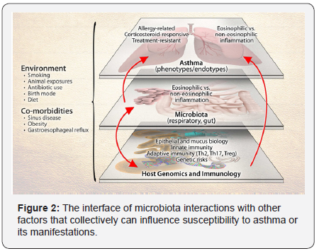
Dissecting the role of the microbiome in asthma is challenged by the heterogeneity of the disease at multiple levels (Figure 2). These levels include asthma’s clinical and inflammatory heterogeneity, genetic factors that contribute to asthma risk, and the multiplicity of immune pathways involved in asthma. The potential effects of environmental exposures on gene function, immune responses, as well as microbiota composition add further complexity. As with genetics, mechanistic consequences of the altered microbiome may explain certain aspects or phenotypes of asthma as the development of allergic or non-allergic asthma,and treatment-resistant asthma) [22] Components of the depicted system-host genetics and immunology, microbiota, environmental exposures, and the disease of asthma- are themselves heterogeneous entities, presenting challenges to more precisely dissect the role(s) of the microbiome in asthma.

Upper airway microbiota and asthma

Bisgaard et al. [23] demonstrated that the nasopharyngeal microbiome composition was influenced by the early life exposures, including attending day care, having siblings, and taking antibiotics. Haemophilus, Streptococcus, Moraxella had been previously associated with airway disease and increased risk for asthma exacerbations. Early colonization with either Moraxella, or Streptococcus was strongly associated with acute lower respiratory viral infections. This colonization can be predictor for asthma development later in life.

Thus, probiotic intervention studies of animals provide encouraging evidence for intentional manipulation of the intestinal microbiota as a strategy for asthma prevention and management. A meta-analysis of a large number of randomized trials of probiotic supplementation, on atopic sensitization and asthma in children, however, shows that the success of these interventions in mice does not translate easily to disease prevention in humans. At a minimum, this highlights that different probiotics may have distinct interactions with the host microbiome and that some strains might be more specific for modulating atopic inflammation but many other considerations, such as diet, age of intervention, coincident environmental exposures, length of supplementation period, and other as yet unknown factors, are likely important [24].

Airway microbiota and severity of asthma

Relationships between the airway microbiome and disease features have also been examined in patients with in severe asthma. Different clinical phenotypes of severe asthma have been described, suggesting the possible involvement of alternate mechanistic pathways, as has been surmised for asthma in general. A preliminary analysis of the bronchial microbiome in these subjects, poorly controlled despite high-dose ICS therapy, noted significant relationships between different bacterial community profiles and features such as body-mass index and measures of asthma control [25]. A similar study of sputum bacterial composition in 28 treatment-resistant asthmatics found that the relative abundance of M. catarrhalis, Haemophilus, or Streptococcus spp. correlated with worse lung function and higher sputum neutrophil counts and IL-8 concentrations [19].

Microbiota and therapy of allergic disease

The composition of the microbiota can be manipulated by combinations of antibiotics, probiotics, and dietary components which may have direct growth promoting or inhibiting activity for specific microbes. [26]. Certain types of fatty acids, phenolic compounds, and carbohydrates may modulate these microbiota. However, a single type of probiotic or dietary component will not be efficacious in all individuals. This likely due to differences in the types of microbial communities in different individuals. The objective of the international Human Microbiome Project is to characterize and define the human microbiome in states of health and disease [10]. The challenge for future research is to use this information to optimize probiotic/dietary therapy to improve human health and prevent microbiota-associated diseases, such as allergies .They are likely to include short chain fatty acids and ionic polysaccharides [27] .

Microbiota and prevention of allergic disease

Probiotic intervention studies of animals provide encouraging evidence for intentional manipulation of the intestinal microbiota as a strategy for asthma prevention and management. However, A large number of randomized trials on the value of probiotic supplementation, on asthma incidence and severity in children, could not show the same success of these interventions as in mice [28-30]. This may be due to many other considerations, such as diet, age of intervention, coincident environmental exposures, length of supplementation period, and other as yet unknown factors, are likely important [24,31- 34].

To Know More About International journal of pulmonary & Respiratory Sciences Please click on: 

To Know More About Open Access Journals Please click on: ttps://juniperpublishers.com/index.php

Artificial Intelligence System for Value Added Tax Collection via Self Organizing Map (SOM)- Juniper Publishers

  Forensic Sciences & Criminal Investigation - Juniper Publishers Abstract Findings:  Based on our experiments, our approach is an effec...