Showing posts with label Magnetic Resonance Imaging. Show all posts

Wednesday, March 9, 2022

A Rare Case of Metastatic Malignant Paraganglioma : Following with F-18 FDG PET/MRI - Juniper Publishers

 Juniper Online Journal of Case Studies - Juniper Publishers

Abstract

Paragangliomas are rare tumors arising from undifferentiated cells of the primitive neural crest. We present a 43-year-old male patient with a glomus tumor in the left carotid space and a retroperitoneal tumor with bone metastases, with invasion of the left renal vein in the left paraaortic area posterior to the third part of the duodenum. Serum epinephrine and norepinephrine levels were high. A CT-guided trucut biopsy was performed from this lesion, and histopathological examination and immunohistochemical analyzes concluded the diagnosis of retroperitoneal paraganglioma. The tumor was judged to be secretive and inoperable; therefore, it was decided to treat it with radiotherapy and systemic chemotherapy.

Keywords: Paragangliomas; Neuroendocrine tumors; Computed tomography; Magnetic resonance imaging; PET/MRI

Introduction

Paragangliomas (PGLs) are rare neuroendocrine tumors originating from the neural crest that originate from chromaffin sympathetic tissues or parasympathetic paraganglia and can occur in a wide localization from the skull base to the pelvic floor [1]. Retroperitoneal paragangliomas are rare tumors. As only a minority of cases secrete catecholamines, they are usually non-functional tumors [2]. About 10% of paragangliomas arise outside the adrenal gland. Few cases of metastatic retroperitoneal paragangliomas have been reported in the literature [3].

Precise localization of primary and metastatic lesions is important because of the presence of critical vascular and neural structures in the vicinity of the mass. It can change the management strategy from surgery to radiation, radionuclide therapy or observation.

PET/MRI imaging systems have proven to be comparable to PET/CT in oncological imaging, and the use of MRI instead of the CT component has the theoretical advantages of lower ionizing radiation exposure, as well as improved soft tissue contrast and local tumor elaboration [4].

In this article, F-18 FDG PET/MRI findings and treatment response of a case of secretory retroperitoneal metastatic paraganglioma treated with radiotherapy and systemic chemotherapy are presented.

Case Presentation

Here, we present a rare case of retroperitoneal paraganglioma with bone metastases in a 43-year-old male patient.

The patient with a diagnosis of left glomus tumor known for 10 years was admitted to our hospital with the development of hypertension attacks. The blood pressure values were found to be 220/110mmHg in the mean measurements. In the clinical examination, our patient had a complaint of pain in the lumbar region.The patient had no smoking history or family history of paraganglioma or other neuroendocrine tumor. In the laboratory tests of the case; Urine normetanephrine value was 3358 (68-444) microgram, Vanillyl mandelic acid (VMA) was 17.1 (0-8), CrgA value was 451ng/ml and it was found above normal measurements. Urinary metanephrine value was within the normal range. Another blood tests, including liver and renal biochemistry were normal.

On PET MRI performed in January 2021, a 2x2.5cm FDG (+) mass from a glomus tumor was observed at the level of the left internal-external carotid artery bifurcation. (suvmax : 6.8) A mass lesion of approximately 3x5.5x7.5cm in size with irregular lobulated contours with FDG uptake (SUVmax: 32.47) and evaluated in favor of hypervascular retroperitoneal paraganglioma was observed in the left paraaortic area inferior to the third part of the duodenum. The defined mass seemed to invade the posterior adjacent left renal vein and the opening of the renal vein to the inferior vena cava, and surrounded the left renal artery more than 180°.

Metastatic lesions were observed in all vertebrae with FDG (+) 3.5cm in diameter, the largest of which was located on the right peduncle of the L4 vertebra (SUVmax 21.60). In addition, multiple FDG (+) bone metastases were observed in the manibrium sterni, on the right lower ribs, and bilaterally proximal to the femur (Figure 1).
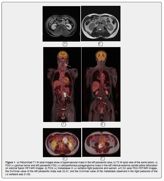
A percutaneous biopsy of the L4 vertebra under CT guidance confirmed that the lesion was a paraganglioma metastasis.The patient was diagnosed with metastatic paraganglioma and underwent palliative chemotherapy.

Radiotherapy was applied to the metastases at the level of L4 vertebra with 35Gy and for the mass in the paraaortic area 45Gy. Simultaneous 12 cycles of systemic chemotherapy were also given. In chemotherapy, cyclophosphamide, vincristine and dacarbazine (CVD) were applied. Zoledronic acid was also given. Our patient received radiotherapy between January 8 and February 11. At the end of radiotherapy, our patient's blood pressure was within normal limits. (125/80mmHg)

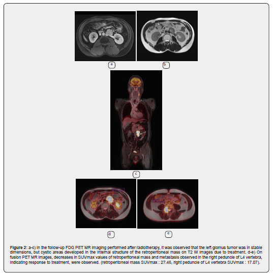
In the control PET MRI examination of the patient, which was performed in August 2021, the mass showing significant FDG uptake (SUVmax 27.46) in the retroperitoneum in the left paraaortic area, although there is no significant difference in size, shows metabolic minimal regression. In addition, there was metabolic regression (SUVmax 17.07) in the metastasis observed in the L4 vertebra. Mass lesion consistent with paraganglioma observed at the level of the left internal-external carotid artery bifurcation were stable (Figure 2).
Discussion

Paraganglioma is a rare tumor that usually occurs in the 3rd to 4th decades. 40% of paragangliomas produce high catecholamine levels, resulting in symptoms such as headache, palpitations, profuse sweating, and elevated urinary metanephrine or vanillylmandelic acid levels. The most common site of involvement for paraganglioma in the retroperitoneum is the organs of Zuckerkandl located anterior to the aorta at the origin of the inferior mesenteric artery [5].

Paragangliomas frequently occur with recurrence or metastasis, as 30% to 40% are hereditary and the other 15-20% carry somatic mutations in one of the more than 20 well-characterized PGL susceptibility genes [1].

Computed tomography (CT) and magnetic resonance imaging (MRI) are effective non-invasive imaging modalities for the detection and evaluation of paragangliomas. MRI has advantages over CT in better soft tissue contrast, higher intrinsic flow sensitivity, and non-ionizer [8]. Magnetic resonance imaging has proven superior to other imaging techniques in demonstrating a more accurate assessment of lesion margins and invasion of adjacent structures [6].

As it is known, Paragangliomas are hypervascular tumors with imaging features of soft tissue mass with intense contrast enhancement and “salt and pepper” appearance on MRI in conventional spin-echo sequences [7].

Diffusion-weighted imaging and ADC value measurement, which are functional imaging methods taken simultaneously in PET-MRI, are used as diagnostic or differential diagnosis tools in oncology [9].

Recently, [68Ga]-DOTATATE PET/CT has emerged as a more sensitive modality for identifying occult PGLs and metastasis by targeting somatostatin receptor 2 (SSTR2) overexpression at PGLs with greater affinity than other somatostatin receptor analogues. A new modality, PET/MR imaging systems have proven to be comparable to PET/CT in oncological imaging. The use of MRI instead of the CT component has the theoretical advantages of lower exposure to ionizing radiation, as well as improved soft tissue contrast and identification of the local tumor and surrounding structures. Some case studies showed much greater soft tissue contrast with [68Ga]-DOTATATE PET/MR compared to PET/CT in PGL patients, allowing for more accurate tumor identification resulting in changes in preoperative planning [10-12]. The prospective analysis of 8 PGL patients with [18F]-FDG PET/CT and [18F]-FDOPA PET/CT and subsequent PET/MR showed that PET/MR was superior for HNPGL, abdominopelvic PGL, and liver metastasis while equivalent to PET/CT for bone metastasis [13].

In the patient presented here, PET-MRI findings are more valuable than PET-CT in cases where the MR component is superior to CT in the detailed evaluation of the soft tissue component in the relationship of retroperitoneal paraganglioma with vascular structures, and in the detailed evaluation of the soft tissue component in bone metastasis.

Conclusion

Hybrid imaging PET/MRI examination is important in the identification of primary neoplasm as well as in the detection of distant metastases in a single session. Combined multi-parametric imaging improves patient comfort and is well suited for examining patients with hereditary syndromes and paragangliomas, especially in the pediatric population, given the lower radiation exposure compared to PET/CT as a replacement for the CT component.

Click here: https://juniperpublishers.com/index.php  

Friday, March 5, 2021

Adrenocortical Carcinoma - Clinical Case - Juniper Publishers

 Gynecology and Womens Health - Juniper Publishers  

Abstract

Adrenocortical carcinoma (CAC) is rare and aggressive. It may be functional, causing Cushing’s syndrome and/or virilization or non-functional, presenting with abdominal/lumbar pain or an incidental finding. For the diagnosis, it is essential to evaluate the secretory activity of the tumor, given that the excess of steroidogenesis is revealed as its fingerprint, and the performance of CT, with complementary MRI and/or PET. Complete surgical removal is the only potentially curative treatment. Adjuvant treatment with mitotane, an adrenolytic drug, is recommended, in unresectable or relapsing cases it can also be associated with chemotherapy containing etoposide, doxorubicin and cisplastin. The main prognostic factors are complete resection, disease staging and the Ki proliferation index 67.

The authors describe the case of a 41-year-old woman, followed in an Endocrinology consultation for left lobectomy and isthmectomy (adenomatoid goiter), who developed virilization and cushingoid stigmas, with laboratory study indicative of hypercortisolism. In this context, CT was performed revealing an atypical formation in the left adrenal gland with 14cm, resulting in deviation of the homolateral kidney, suggestive of adrenocortical carcinoma without evidence of metastasis on MRI.

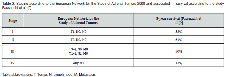
Adrenalectomy was performed by left subcostal laparotomy, confirming low grade adrenal cortex carcinoma (pT3NxM0, stage III, 2% Ki67). She was referred to the Oncology consultation, presenting pulmonary and hepatic metastasis, started mitotane for palliation with a slow and progressive increase in the dose that was suspended due to the progression of the disease. Later, the patient resumed mitotane associated with carboplatin and etoposide, presenting infectious complications on the tenth day of chemotherapy. She was referred for Palliative Care consultation. In this patient, CAC progressed quickly to stage IV, despite complete resection, using mitotane with chemotherapy for palliation without success. However, there are cases described for preventing recurrence and improving survival with the use of mitotane and adjuvant chemotherapy.

Keywords: Adrenocortical Carcinoma; Mitotane; Cushing’s syndrome; Virilization

Abbreviations: CAC: Adrenocortical Carcinoma; CT: Computed Tomography; MRI: Magnetic Resonance Imaging

Introduction

Adrenocortical carcinoma (CAC) represents only 0.02% of all neoplasms, with an approximate incidence of 1 to 2 cases/million people/year, although it can develop at any age, it has two peaks, before 5 years of age and in the age group between 40 to 50 years old. It is predominant in females (1.5-2.5:1), there are studies that suggest a proliferative effect of estrogen on adrenal neoplastic cells [1].

Most cases are sporadic, with a tumor progression model similar to colorectal cancer, it is common to find loss of heterozygosity in locus 17p13 where the tumor suppressor gene TP53 (mutated in one third of sporadic CACs) and others unidentified suppressor genes. Mutations of the CTNNB1 gene that cause activation of β-catenins and at the 11p15 locus that increase IFG-2 expression also play an important role in the development of adrenal carcinoma [2]. A minority of cases are hereditary, associated with syndromes, such as Li-Fraumeni, Beckwith-Wiedemann and MEN-1 [3].

Approximately 60% of the tumors have hypersecretion of steroids, causing symptoms of Cushing’s syndrome, weight gain, weakness and violet streaks and/or symptoms of excess androgens: virilization, acne, hirsutism and menstrual irregularities. In a small group of men it presents as feminization, loss of libido, testicular atrophy, gynecomastia. Hyperaldosteronism is rare with hypokalemia and hypertension [1].The rest do not secrete enough hormones and are manifested by abdominal/low back pain due to the rapidly progressive mass effect, constitutional symptoms or may be incidentalomas [1].

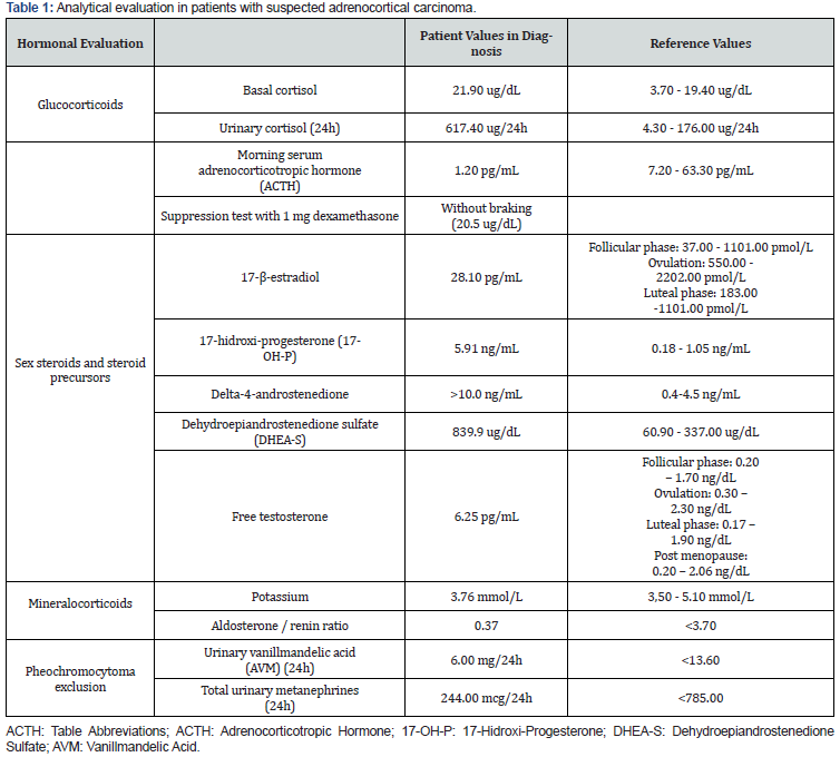
The diagnostic gait should include the assessment of tumor secretory activity (Table 1) [4], which is also essential for monitoring recurrences and adrenal insufficiency after adrenalectomy, and computed tomography (CT) with magnetic resonance imaging (MRI), to assess local invasion and/or FDGPET, to distinguish adenoma and carcinoma [5]. Regarding fine needle aspiration biopsy its indication is controversial, it does not distinguish benign from malignant lesions, however it differentiates primary from secondary adrenal tumors, using markers such as α-inhibin, melan A and SF-1[6] nevertheless its use is not recommended by ENSAT due to the high risk of complications, such as the spread of disease. Metastasis of adrenal carcinoma occurs to the liver, lungs, lymph nodes and bone, with thoracoabdominal CT and bone scintigraphy necessary for staging (Table 2) [7].

Clinical Report

The authors describe the case of a 41-year-old woman, regularly followed in an Endocrinology consultation, since 2013 for left lobectomy and isthmectomy for adenomatoid goiter and obesity. In September 2019 she presented a fifteen-kilogram weight gain, alopecia, hirsutism, violet streaks, secondary amenorrhea with about 6 months of evolution (Figure 1).

Result

The hormonal evaluation carried out in this context revealed hypercortisolism with emphasis on reduced adrenocorticotropic hormone, elevated urinary cortisol, without suppression in the braking tests with 1mg dexamethasone and Liddle’s test. 17-hydroxyprogesterone, delta-4-androstenedione, free hydroxydiandrostenedione and testosterone-free hydroxyprone were increased (Table 1). In this context, in October 2019, a thoracoabdominopelvic CT scan revealed an atypical formation in the left suprarenal gland measuring 13.5x8.5x8.0cm, with calcifications and heterogeneous contrast uptake, causing deviation of the homolateral kidney, being suggestive of cortical adrenal carcinoma, confirmed in MR and without secondary lesions [8].

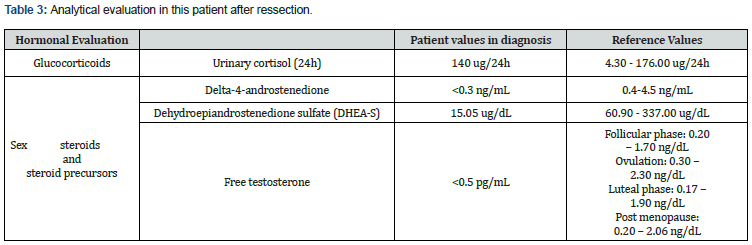
In January 2020, the patient was submitted to an adrenalectomy by left subcostal laparotomy. Histologically, low grade adrenal cortex carcinoma (pT3NxM0, stage III, 2% Ki67) was observed. The patient kept the follow up in General Surgery and Endocrinology consultation. On a control CT scan done in February 2020, the patient had a hypodense left renal mass measuring 5.5x4.2x5.4cm, hepatic parenchymal nodular images hypodense in the left lobe segment IV with 1.8 and 1.4 cm, a nodule with 1.2 cm, adjacent to the right lobar hepatic capsule and several pulmonary parenchymal nodules at both bases, with dimensions between 0.7 and 1.4cm (Figure 2). Findings suggested tumor recurrence/remnant associated with liver and lung metastasis, however, analytically without evidence of secretory activity [9].

Subsequent imaging tests were suggestive of unfavorable progression, especially at the expense of the increased number of secondary deposits in the lung parenchyma. The patient was referred to the Medical Oncology consultation, starting mitotane for palliation with a slow and progressive dose increase. Suspended due to exuberant tumor recurrence at the adrenal site, with invasion of adjacent organs and structures and homolateral renal deposits and perirenal fat documented on CT performed due to complaints of abdominal pain, in August 2020 (Figure 3) (Table 3).


The patient resumed mitotane associated with carboplatin and etoposide in September 2020, on the tenth day of chemotherapy she was admitted to the Intermediate Care unit for febrile pancytopenia, septic shock with a starting point in phlegm on the right forearm and bacteremia caused by Enterobacter and Staphylococcus aureus resistant to methicillin. After the second line treatment ceased, she was referred to the Palliative Care consultation. Having progressively worsened, she died in January 2021.

Discussion

In this article is reported the clinical case of a patient with lowgrade adrenal cortex carcinoma treated with adrenalectomy, the only potentially curative option. The laparotomic approach utilized is recommended by the literature [10]. For potentially resectable tumors, stage I to III, surgery generally requires en-bloc resection of the adjacent organs, [11] however in this patient despite the tumor dimensions, about 14cm, at the time of adrenalectomy, there was no invasion of the adjacent structures and the resection was complete, with free margins. Paradoxically, after one month, the presence of liver and lung metastases was verified. After seven months an extensive recurrence in the surgical site with invasion of the homolateral kidney and associated compressive symptoms, both findings are justified by the high relapse power of this tumor with probable micro metastases even in early stages [12]. The early detection of recurrence, either analytically or imagiologically, is other impacting factor in the prognosis of this neoplasia10 and may allow an intervention with a curative intent, when the lesion is unique and resectable or palliative, for multiple or unresectable injuries. The adjuvant use of mitotane is recommended by the European Society of Endocrinology for patients at high risk of histological recurrence (Ki67 greater than 10% and high mitotic rate, more than 20 mitoses per 50 high-power fields regardless of tumor size), intraoperative rupture of the tumor capsule, invasion of the capsule or vascular and for primary treatment of unresectable lesions and recurrence [13]. Its indication in low to intermediate risk patients with resected disease is being evaluated by ADIUVO, a randomized clinical trial [14]. In extensive disease, rapidly progressive and high-grade disease it is often associated with cisplatin, doxorubicin and etoposide regimens [13].

Radiotherapy is indicated for palliation of bone metastases or locally advanced disease [15]. The presentation of CAC associated with Cushing’s syndrome is associated with a poor prognosis due to frequent infectious and metabolic complications [9]. The evolution of this case, even though it was low-grade, followed the typical course of adrenal carcinomas with rapid progression, early metastasis and poor survival. In the future, molecular markers may help to define the risk of recurrence and individualize therapy, [16,17] together with new treatments, like immunotherapy, may improve the outcome of CAC15. More clinical trials, such as ADIUVO, are needed to define clear indications for the use of adjuvant treatments and their impacts on the evolution of the disease. However, progress is slow and studies are scarce due to the high mortality and rarity of this neoplasia.

Conclusion

Progress is slow and studies are diminutive due to the high mortality and rarity of this neoplasia. Clinical trials can only be useful if there is cooperation between referral centers so that the sample is significant and the orientation of patients is transversal. Ideally, there should be at least one reference center in each country coordinated with the European Network for the Study of Adrenal Tumors.

To Know more about Gynecology and Womens Health

To Know more about our Juniper Publishers

Click here:
 https://juniperpublishers.com/index.php  

Therapeutic and Prognostic Factors of Upper Gastrointestinal Bleeding in the Intensive Care Unit in a Sub-Saharan African Country- Juniper Publishers

  Anesthesia & Intensive Care Medicine- Juniper Publishers Abstract Background: The aim of our study was to determine the therapeutic a...