Cardiology & Cardiovascular Therapy - Juniper Publishers

The field of Cardio-oncology is rapidly growing with
significant advances in research leading to better understanding of the
underlying pathogenesis with implications in the diagnosis and
management of cancer-related cardiomyopathy.
Parallel to advancement in cardio-oncology is an increased awareness of
the incidence of congestive heart failure and cardiomyopathy associated
with malignancy. While specific cardiotoxic profiles exist for certain
chemotherapeutic agents, there is increasing evidence of unexpected
cardiotoxic side effects of some therapeutic modalities, combination
chemo- and radiotherapy with large analyses identifying a strong
association between malignancy and Takotsubo cardiomyopathy. Takotsubo
Cardiomyopathy, also known as “broken-heart” syndrome or stress
cardiomyopathy, is characterized by transient and reversible, regional
or global, myocardial dysfunction without inciting ischemic perfusion
defect from obstructive coronary artery disease. While direct causative
pathophysiologic mechanisms continue to be investigated, much of the
postulated pathways center on the high emotional and physical burdens of
cancer and the related emotional stress associated with the diagnosis
of cancer as well as the corporal effects of anti-neoplastic therapies,
radiation, and oncologic surgery. In this manuscript we review the most
current data in this rapidly emerging field highlighting the
epidemiology, the postulated pathogenetic mechanisms as well as the
current guidelines by major societies addressing malignancy -associated
heart failure and cardiomyopathy, a rather complex disease entity with
high morbidity and mortality.
Keywords: Heat Failure; Cardiomyopathy; Takotsubo Cardiomyopathy; Stress Cardiomyopathy; Cancer; Cardiotoxicity
Among the greatest stories in modern medicine,
research and advancements in diagnostics and therapeutics is the field
of oncology, with continuously evolving management and diagnostic
modalities, and establishment of regional and national centers of
excellence in clinical investigation and care for patients with cancer,
the second leading cause of death in the United States [1].
Highlighting the remarkable figures of increasing
survival, reductions in mortality and annual incidence of cancer is a
recent report with 2020 malignancy-associated morbidity and mortality
projections by the American Cancer Society demonstrating the
slowing of malignancy associated death and the notable gains in
prevention and definitive treatment in cancer [2].
While malignancy survival, mortality, incidence, and
prevalence figures have prominently improved since the 1970s,
cardiovascular disease mortality risk, both in active cancer patients
and cancer survivors, is prominently increased [3]. With the
identification of malignancy associated cardiovascular morbidity and
mortality, and known cardiotoxic profiles of cancer related therapies
including chemotherapy, targeted antineoplastic agents, and
radiotherapy, the field of cardio-oncology has become one of the most
rapidly developing subspecialties in cardiology, with
increasing numbers of fellowship training, clinical, and research
programs, and dedicated scientific conferences, symposia, and
publications [4-7].
Given the direct cardiomyocyte effects associated with
chemotherapy, immunotherapy, other targeted antineoplastic
agents, and radiotherapy, broad interest and attention has been
given to cardiomyopathies and congestive heart failure (CHF)
among the diverse cardiovascular pathologies identified in
malignancy [8].
A particular cardiomyopathy and etiology of CHF of importance
to cardiologists and oncologists is Takotsubo cardiomyopathy
(TCM). TCM, also known as “broken-heart” syndrome or stress
cardiomyopathy, is a syndrome of acute, transient, reversible,
regional myocardial dysfunction with resulting ventricular
hypokinesis, dyskinesis, or akinesis in the absence of underlying
obstructive epicardial coronary artery disease, typically preceded
by a physical or emotional stressor [9,10]. Large analyses of
international TCM databases and registries have identified a
strong association between malignancy and TCM [11]. Similar
to investigations of pathophysiologic mechanisms of TCM in the
absence of malignancy, postulations on TCM pathways in cancer
patients center on identifiable emotional and physical triggers,
noticeably abundant in malignancy, and include the emotional and
mental stress of diagnosis and living chronically with cancer, and
the physical stress associated with the symptoms of the disease
itself and effects of therapies [12].
In this review, we present a comprehensive summary of the
modern understanding of heart failure and cardiomyopathy
through the lens of malignancy, analysis of epidemiology,
cardiotoxic profiles of chemotherapy, radiotherapy,
immunotherapy, and other targeted antineoplastic therapies,
cardiovascular outcomes and diagnostic management strategies
in the setting of direct therapy associated cardiomyopathy and
in TCM, and consider current and future perspectives on areas of
investigation in cardio-oncology.
Historical trends and clinical observations have highlighted
an increasingly apparent relationship between malignancy
and cardiovascular disease. Among high income countries,
cancer death rates exceed cardiovascular death rates. In the US,
cardiovascular disease and malignancy are first and second leading
causes of death, respectively. Therefore, from an epidemiological
standpoint, identification of potential interactions between
cancer and cardiovascular disease is of great importance [1,13].
National measures of cardiovascular disease indicate 12.1%
prevalence of adults with diagnosed heart disease, corresponding
to approximately 30 million people with an annual mortality rate
of approximately 200 people per 100,00 population [14]. Within
cardiovascular pathologies, CHF prevalence has been estimated to
be 2.2% among US adults above the age of 20, with 1-year and
5-year mortality rates estimated at 22% and 42.3%, respectively
[15].
National measures of cancer-related incidence and survival
point to overall improvements in morbidity and mortality, but
highlight improving outcomes are not shared equally across
malignancies or genders. Male and female mortality rates have
similarly decreased since 1990, with 5-year survival rates as
of 2015 at 67% [2]. However, since 2007, the incidence of new
cancer diagnoses in males has declined to a rate of approximately
500 per 100,00 population, while the incidence rate among
females has remained stable at approximately 400 per 100,000
population.
In parallel to the expanding research on the pathophysiology
of cardiovascular disease in the setting of malignancy are data
and epidemiologic profiles of cardiovascular complications in
patients with cancer. Cardiovascular risk and burden of disease
among cancer patients, both in terms of preexisting comorbidities
(diabetes mellitus, smoking, etc.) and risk associated with specific
malignancies and treatment modalities, have been increasingly
identified as prevalent clinical vulnerabilities in malignancy
[16,17].
Among coexisting cardiovascular diseases in the US
population of patients with lung, colorectal, breast, and prostate
cancer, the prevalence of CHF exceeded the combined prevalence
of cerebrovascular disease and myocardial infarction (MI) [17].
While HF prevalence was observed to be highest among Chinese
cancer patients with hematologic malignancies, the hazard ratio
of all-cause mortality was significantly increased for patients with
CHF [16]. HF carries a lower 5-year survival rate than gender
specific malignancies, such as prostate cancer in men and breast
cancer in women.
Along with being at an increased risk for developing cancer,
patients with CHF later diagnosed with malignancy have been
shown to have higher all-cause mortality demonstrating an
additive effect [18,19]. Epidemiology of HF and cardiomyopathy
associated with chemotherapy and targeted antineoplastic
therapies has been well documented in literature and has prompted
major national and international cardiology and oncology
societies to develop dedicated guidelines for the evaluation
and management of cardiomyopathy and CHF attributed to
malignancy and chemotherapy, or incorporate recommendations
on chemotherapy induced CHF and cardiomyopathy into larger
CHF guidelines [20-22]. Historically, anthracyclines and human
epidermal growth factor receptor 2 (Her2) inhibitors have been
commonly cited for their potent cardiotoxicity, with adjuvant use
of these agents increasing the overall risk of cardiotoxicity [23-
25].
Moreover, in patients presenting with mild to end stage CHF,
or in those who are clinically asymptomatic but with clinical
evidence of cardiomyopathy, doxorubicin, other anthracyclines,
and non-anthracycline chemotherapeutic and anti-neoplastic
agents were implicated as the etiology of up to 3% of initially
unexplained cardiomyopathies [26,27].
Cardiotoxic Chemotherapy, Immunotherapy, Antineoplastic
Targeted Therapy, and Radiotherapy
Cardiomyopathy
While anthracyclines and Her2 inhibitors have the
most cardiotoxic profiles and are frequently implicated
in the development of cardiomyopathy and CHF, multiple
chemotherapies, immunotherapies, and targeted antineoplastic
therapies have also been highlighted by major cardiology societies
and in national and international CHF guidelines [22,28].
Since the release of the American and European CHF guidelines
in 2013 and 2016, respectively, numerous guideline updates and
position papers have pointed to additional information on the
pathophysiology and importance of monitoring of ventricular
dysfunction in previously and newly implicated cardiotoxic
chemotherapeutic drugs [20,22,28-30]. While the management of
malignancy continues to transition and incorporate increasingly
novel targeted antineoplastic therapies, the extensive use of
chemotherapy over the last 50 years has resulted in a large body
of evidence specifically on cardiomyopathy and CHF, the most
worrisome of cardiovascular complications of chemotherapy.
Anthracyclines
Since first observations of myocardial dysfunction were
made in the 1970s, anthracyclines, particularly doxorubicin, have
been shown to possess a potent dose-dependent cardiomyocyte
toxicity profile resulting in left ventricular (LV) dysfunction and
CHF [20,23,31]. The transition from asymptomatic ventricular
dysfunction to overt symptomatic CHF is highly variable with
often long latencies between anthracycline exposure and the
clinical manifestations of cardiomyopathy, with similarly diverse
cardiac structural and functional findings including LV wall
thinning, chamber dilation, increased LV wall stress, reduced
ejection fraction (EF), and diastolic dysfunction [23,32,33].
Current understanding of mechanisms of anthracycline
induced cardiomyocyte injury center on the generation of toxic
reactive oxygen species (ROS), inhibition of Topoisomerase 2ß
(Top2ß) and the resulting breaks in DNA [23,34]. Doxorubicin
increases oxidative stress in the cytoplasm and mitochondria
via direct catalysis and formation of irreversible complexes with
cardiolipin in the inner mitochondrial membrane susceptible
to peroxidation, promoting cardiomyocyte death via caspase
mediated apoptosis and disrupting mitochondrial ATP metabolism,
further contributing to cardiomyocyte death through cytoplasmic
and mitochondrial swelling and sarcomere lesions [34]. Top2ß, a
DNA replication enzyme that is highly expressed in both highly
proliferating cancer cells and active quiescent non-proliferating
cells like cardiomyocytes, is inhibited by anthracyclines, resulting
is double-stranded DNA breaks and activation of p53 mediated
apoptosis pathways [23,35].
Anthracycline’s dose dependent cardiotoxic profiles show
similar dose dependent LV dysfunction rates, best displayed by
wide incidence ranges of 3-5% and 18-48% for low and high dose
doxorubicin, respectively [20].
Predisposing risk factors for anthacycline-induced
cardiomyopathy include female gender, African-American
heritage, age > 65 years, kidney disease, and concurrent radiation
therapy at or near the heart as seen in malignancies of the chest
and mediastinum such as lymphoma, lung, and breast cancer
[20,23].
While there is high variability in onset of cardiomyopathy after
anthracycline exposure, large retrospective analysis conducted
by Cardinale et al. on the timing of anthracycline cardiotoxic
manifestations, both symptomatic and asymptomatic, show 98%
of cases of cardiomyopathy develop within a year of anthracycline
exposure, with a median of 3.5 months [23,36]. The temporal
relationship between anthracycline exposure and manifestations
of cardiotoxicity has led to different approaches to classification.
Early effects are classified as cardiomyopathic changes that
develop within the first year of treatment, subcategorized
as either acute (after single dose or course) or early-onset
chronic progressive (within first year), with late-onset chronic
progressive effects developing after the first year of treatment
[20,36,37]. However, the timing and degree of physiologic and
structural LV abnormalities are largely viewed as a continuous
progressive cardiotoxicity with decline in LVEF where patients
who are initially asymptomatic but with clinical manifestations
and evidence of cardiomyopathy eventually develop symptoms
[20,38].
Human Epidermal Growth Factor Receptor 2 (Her2)
Inhibitors
Her2 inhibitors, namely trastuzumab and more recently
developed agents including pertuzumab, lapatinib, and
adotrastuzumab emtansine, target Her2, a cell surface tyrosine
kinase receptor that is overexpressed in up to a quarter of breast
cancers, with Her2 positivity associated with a more aggressive
malignancy course, reduced survival, and increased risk of
recurrence [39]. While most commonly connected to breast
cancer, Her2 overexpression has also been seen and studied
in gastric, gastroesophageal, and bony metastases of prostate
malignancies [40-42].
In the absence of concurrent anthracycline use, trastuzumab
carries a considerably low risk of cardiotoxicity, with rates of
asymptomatic systolic dysfunction and overt CHF occurring
at 3.2 and 0.5%, respectively [39]. When included in adjuvant
anthracycline-based regimens, the rates of both asymptomatic
reductions in ejection fraction and symptomatic CHF increase to
4.0 and 18.6%, respectively [39,43]. Each Her2 targeted therapy
inhibits Her2 mediated pathways in different ways, but share a
common cardiomyopathic pathophysiology of disrupting the
cardiomyocyte homeostatic functions of Her2 signaling pathway,
disturbing cardiomyocyte responses to hemodynamic stress,
interfering with sarcomeric organization and hypertrophy, and
triggering accumulation of cardiotoxic reactive oxygen species
[39,44,45].
In contrast to anthracyclines that are directly cardiotoxic
causing cardiomyopathy thats is generally progressive and
irreversible, Her2 inhibitors disrupt the cardioprotective
functions of the Her2 pathway, with the overwhelming cases
of newly developed cardiomyopathies and overt CHF being
reversible [39,46]. Beyond antecedent exposure to anthracyclines,
risk factors associated with trastuzumab related cardiotoxicity
include age, cardiovascular comorbidities like hypertension and
diabetes mellitus, and African American ethnicity [39,47,48].
Additional Therapies
Beyond the frequently implicated anthracyclines and Her2
inhibitors, numerous additional conventional chemotherapies,
immunotherapies, and targeted therapies have been shown
to induce myocardial dysfunction resulting in reversible and
irreversible cardiomyocyte changes [20].
Vascular Endothelial Growth Factor (VEGF) signaling
pathway inhibitors, both direct VEGF inhibitors like bevacizumab,
and downstream signaling pathway tyrosine kinase inhibitors
(TKIs) have exhibited varying rates of LV dysfunction and CHF
[43,49,50]. VEGF pathway inhibitors have been approved and are
under investigation in multiple malignancies, particularly solid
tumors, including renal, lung, gastroesophageal, breast, cervical,
ovarian, and gastrointestinal stromal cancers [50]. The incidence
of LV dysfunction with bevacizumab is approximated at 1.6-4.0%,
with large variability in incidence and severity of HF (higher
New York Heart Association [NYHA] classification) influenced
by dose, underlying malignancy, and previous or concurrent
use of other cardiotoxic chemotherapy [20,51]. Different
cardiomyopathic mechanisms have been suggested, including
thinning of ventricular walls, depressed contractility, and loss of
cardioprotective function mediated by VEGF [52]. Among VEGF
pathway TKIs, the relative risk of developing both all grade and
high grade (NYHA III-IV) CHF, was similar between specific
TKIs like axitinib and non-specific TKIs like sunitinib, sorafenib,
vandetanib, and pazopanib, with a collective all grade CHF risk
of 2.69 [20,53]. Prospective echocardiographic studies and large
meta-analyses on sunitinib approximate a CHF incidence of 4.1%
with relative risk of 1.8 compared to placebo, driven by a 9.7%
incidence of LV dysfunction [49,54].
As VEGF pathway inhibitors are typically used in metastatic
disease on patients with limited life expectancies that often
undergo interruption and discontinuation of therapies, the
degree of reversibility, approximated at 60-80%, and overall
prognosis associated with VEGF pathway inhibitor mediated
cardiomyopathy and CHF is difficult to assess [20,55].
Other conventional chemotherapies have been associated
with cardiotoxicity, LV dysfunction, and CHF with wide ranging
incidences and dose-dependent relationships [20]. While
infrequent, the cardiomyopathy associated with alkylating agents
such as cyclophosphamide, ifosfamide, and cisplatin is typically
irreversible [20,56]. The risk CHF secondary to cyclophosphamide
is dose-dependent, with LV systolic dysfunction occurring
shortly after initial administration and irreversibility setting
in at doses greater than 1.55 g/m²/day [56,57]. The incidence
of cardiomyopathy in cyclophosphamide rangers from 7-28%,
showing a similar dose dependent relationship to ifosfamide,
with pathophysiologic mechanisms based on gross pathology and
autopsy suggesting therapy induced hemorrhagic cardiomyocyte
necrosis, interstitial edema, fibrin deposition, subendocardial
hemorrhage, and epicardial petechial lesions [20,49,56]. Beyond
bolus and total dose, additional predisposing risk factors for the
development of LV dysfunction include older age and concurrent
or previous use of other chemotherapeutic agents and mediastinal
radiation [57]. Cisplatin has been associated with a later onset of
LV dysfunction, with the appearance of symptomatic CHF more
common in patients with preexisting myocardial disease that is
exacerbated by the high volumes of intravenous fluids administered
to mitigate cisplatin related oto- and nephrotoxicity [20,56].
Furthermore, cisplatin mediated myocardial ischemia strongly
contributes to the development of ischemic cardiomyopathy and
CHF as opposed to direct cardiomyocyte toxicity [20].
Antimetabolites, such as 5-fluorouracil and capecitabine,
and anti-microtubule agents, such as docetaxel and paclitaxel,
have low cardiomyocyte toxicity profiles with rare instances of
cardiomyopathy and CHF [58]. Cardiomyopathies associated
with antimetabolites and anti-microtubules are exceedingly
rare and manifest in patients exposed to more commonly
implicated therapies like anthracyclines, trastuzumab, and
cyclophosphamide, rendering assessment of the individualized
cardiomyocyte toxicity profiles of these agents difficult [20].
Studies have hypothesized different mechanisms of antimetabolite
mediated direct cardiomyocyte toxicity, including increased
reactive oxygen species and toxic metabolite formation, Krebs
cycle disruption, myocardial dysfunction and resulting apoptosis
and necrosis from caspase-activation [58,59]. While the literature
on the cardiomyocyte toxicity profiles of antimetabolites and antimicrotubules
continues to evolve, the prominent multifactorial
cardiac effects associated with these chemotherapeutic
agents, including arrhythmogenesis, high grade conduction
abnormalities, endothelial dysfunction, coronary vasospasm,
platelet aggregation and thrombotic risk, contribute to the
development of cardiomyopathy and CHF [58,60,61].
Two additional classes of cancer therapies with emerging
evidence of cardiovascular toxicity include the BCR-ABL tyrosine
kinase and proteasome inhibitors [20,62,63]. Tyrosine kinase
enzymes have roles in myocardial, vascular, and metabolic
physiology. Analysis of BCR-ABL tyrosine kinase inhibitors,
including imatinib and newer formulations like nilotinib, dasatinib,
bosutinib, and poratinib, have demonstrated an increased risk
of adverse cardiovascular events, primarily progression of
atherosclerosis and related complications, especially in those
with pre-existing disease [20,62,64]. While early studies on
imatinib raised concern for cardiomyopathic toxicity, subsequent
research on imatinib and the other aforementioned BCR-ABL
tyrosine kinase inhibitors have not demonstrated therapy
related myocardial dysfunction, but implicate these agents in
the development of other non-atherosclerotic cardiovascular
complications, including pulmonary arterial hypertension and
QT interval prolongation, [62,65,66]. Given the high protein
turnover and proteasome activity in cardiomyocytes, proposed
mechanisms of proteasome inhibitor associated cardiotoxicity,
specifically carlfizomib and bortezomib, have centered on caspase
mediated apoptosis and activation of the unfolded protein
response, a signaling pathway accelerated by the accumulation
of misfolded and unfolded proteins typically degraded by the
proteasome [20,63,67]. Among multiple myeloma patients
exposed to proteasome therapy, the incidence of cardiomyopathy
and CHF among those treated with carfilzomib was considerably
higher than those managed with bortezomib, likely secondary
to robust proteasome inhibition and cardiomyocyte injury given
the more potent irreversible interaction of carfilzomib at the
chymotrypsin-like site of the 20S proteasome [20,63,68].
Radiotherapy Cardiotoxicity and Cardiomyopathy
Incidence and prevalence of radiation induced toxicity is
difficult to establish due to several factors, most notably temporal
delay between radiation therapy and onset of symptoms, previous
or concurrent exposure to cardiotoxic therapies, and overall failure
to properly attribute cardiac pathophysiologic complications to
previous radiation treatment [20].
While diverse pathophysiologic mechanisms are proposed
to drive the cardiotoxicity of radiotherapy, the constellation of
these mechanisms appears to be a synergistic cardiotoxicity
from the cumulative long term exposure to both chemotherapy
and radiotherapy. Previous studies in patients exposed to both,
particularly anthracyclines in malignancies like breast cancer
and lymphoma, have significantly higher rates of cardiomyopathy
than in patients treated with chemotherapy alone [20,32,69].
Radiotherapy cardiotoxicity is primarily driven by endothelial
damage and resultant inflammation causing accelerated
atherosclerosis, coronary artery disease, and myocardial ischemia
[69,70]. Furthermore, a marked cytokine mediated inflammatory
process that increases collagen deposition with a diffuse pattern of
fibrosis involving the pericardium, myocardium, and conduction
system has been noted [69,71].
Beyond the direct myocardial effects of previous or
concurrent chemo-, immuno-, and antineoplastic therapy, the
cardiomyopathy resulting from radiation is a combination of
fibrotic complications, including diastolic dysfunction secondary
to LV stiffening, and chronic pericardial effusions and constrictive
pericarditis [69,72]. Diastolic dysfunction arises from myocardial,
perivascular, and pericellular fibrosis caused by micro-ischemia
and alterations of ventricular distensibility and compliance
[69,73]. Pericardial radiation injury causes neovascularization
that furthers micro-ischemia, fibrosis, and inflammation, leading
to disrupted pericardial venous and lymphatic drainage and a
chronic exudative pericardial effusion [69]. While some cancer
patients may develop rapidly accumulating or large volume
malignant effusions presenting with cardiac tamponade, this
is more typical of metastatic involvement of the pericardium or
direct local invasion of mediastinal and thoracic malignancies
[74,75]. Chronic inflammation and fibrosis of the pericardium
results in constrictive pericarditis and restrictive cardiomyopathy,
disrupting ventricular hemodynamics through normalization of
right ventricle (RV) and LV pressures, and RV encroachment on LV
diastole with impaired filling and output, and loss of inspiratory
negative thoracic pressure assisting in biventricular filling [72].
Pre-, Peri-, and Post-treatment Approach to Cardiotoxic
Chemotherapy
Given the wide cardiotoxic profiles of chemotherapeutic,
immunotherapeutic, and targeted antineoplastic agents and risk
of developing of asymptomatic cardiomyopathy and symptomatic
CHF, especially in malignancies where multiple therapies are
used simultaneously or in succession, a thorough and careful
cardiovascular workup and assessment of cardiovascular risk
factors and predisposing comorbidities is recommended in the
clinical practice guidelines of the major American and European
oncology organizations [21,29]. The 2016 American Society of
Clinical Oncology (ACSO) guidelines [29] and the 2012 European
Society for Medical Oncology (ESMO) guidelines [21] give specific
recommendations regarding cardiotoxic risk stratification,
specifically with anthracycline and Her2 inhibitor use, and general
approaches and strategies for prevention and surveillance before,
during, and after treatment.
In addition to screening for cardiovascular risk factors and
predisposing comorbidities like smoking, obesity, hyperlipidemia,
hypertension, and underlying coronary artery disease, all patients
should undergo an echocardiogram to assess for pre-existing
structural abnormalities and LV systolic or diastolic dysfunction.
Regardless of comorbidities and preexisting coronary artery or
structural heart disease, the ACSO (Table 1) and ESMO (Table 2)
classify cancer patients as high risk for cardiotoxicity depending
on the cumulative dosage of chemotherapy and radiotherapy,
specifically anthracyclines and trastuzumab. Lower dosages of
other therapeutic agents combined with radiotherapy result in
the same high-risk categorization.
Takotsubo Cardiomyopathy and Malignancy
Recent data from international registries and large metaanalyses
have identified a strong association between Takotsubo
cardiomyopathy (TCM), malignancy and particularly poor
outcomes [11,76].
Epidemiologically, patients with TCM are more likely than
age and gender matched peers to have a cancer diagnosis and
with increased probability of having malignancy diagnosed in
subsequent follow up [77]. Data from the InterTAK Registry, a
multi-center collaboration across 9 countries, demonstrated a
16.6% prevalence of malignancy in patients with TCM [11].
Taken alongside other reviews, observational studies, metaanalyses,
the prevalence of cancer in TCM has been approximated
to be 1.3-25.5% [11,76-78]. Similar to investigations of
pathophysiologic mechanisms of TCM in the absence of
malignancy, postulations on TCM pathways in cancer patients
center on identifiable emotional and physical triggers, noticeably
abundant in malignancy, and include the emotional and mental
stress of diagnosis and living chronically with cancer, and the
physical stress associated with the symptoms of the disease itself
and effects of therapies [12].
In the InterTAK Registry study from 2019, TCM in malignancy
was more likely to have been caused by new pharmacological
or procedural interventions than an emotional trigger and was
associated with a higher in-hospital mortality and poorer longterm
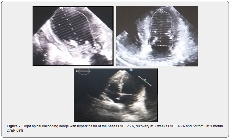
survival [11] (Figures 1 & 2).
Given the primary and secondary rankings of cardiovascular
disease and malignancy among annual national and international
mortality, and the incidence of development of heart failure in
patients with cancer, and vice versa, there has been an increasing
focus on investigating the connections between cardiomyopathies
and malignancy. Beyond known and postulated potential
cardiotoxicities associated with specific chemotherapeutic,
immunotherapeutic, and targeted antineoplastic agents,
properties of particular hematologic and neuroendocrine
malignancies increase the risk of cardiomyopathy, and highlight a
bidirectional nature to cancer and heart failure [43].
Carcinoid heart disease, a rare manifestation of neuroendocrine
tumors, is primarily characterized by endomyocardial fibrotic
plaques and fibrotic remodeling of the endocardium [43,79].
Fibroblast growth and fibrogenesis typically involves the
valves, and given the efficient inactivation of neuroendocrine
mediators in the pulmonary vasculature and liver, the resulting
valvular stenosis or regurgitation is greatly influenced by the
anatomic location of the tumor, with bronchial carcinoids
resulting in rare mitral, aortic, and left ventricular dysfunction,
or the presence of a right-to-left intracardiac shunt [43,79,80].
Tricuspid, pulmonic, and right ventricular carcinoid heart
disease arises from gastrointestinal neuroendocrine tumors
with metastases to liver that have significantly disrupted hepatic
clearance of serotonin, tachykinin, and kallikrein, or rare primary
ovarian neuroendocrine tumors that avoid the portal venous
system [79,81]. Myocardial metastasis of neuroendocrine
cancerous cells is rare, and while direct tumor deposition and
proliferation in the myocardium can contribute to systolic and
diastolic dysfunction, right, left, or biventricular heart failure in
the presence of neuroendocrine malignancies more typically
arises from valvular involvement and resulting disruptions in
normal physiology [79,82].
In hematologic malignancies and plasma cell dyscrasias
like chronic lymphocytic leukemia, macroglobulinemia, nonlymphoplasmacytic
lymphoma, monoclonal gammopathy
of unknown significance, smolder myeloma, and multiple
myeloma, cardiac deposition of light chain immunoglobins and
AL amyloidosis in the myocardium and valves contributes to
the development of restrictive cardiomyopathy and heart failure
from cardiac amyloidosis [43,83]. In cardiac amyloidosis, amyloid
fibril infiltration causes increased wall thickness with restrictive
physiology, diastolic dysfunction, smaller end-diastolic volumes,
and despite preserved ejection fractions and systolic function,
overall lower stroke volumes and cardiac output [84].
More recent early experimental work on cardiotoxic
oncometabolites has elucidated additional pathophysiologic
pathways of cardiomyopathy in cancer [43]. In acute myeloid
leukemia, alterations in genetic and global expression of
isocitrate dehydrogenase 1 and 2 has been shown in retrospective
observational studies to be associated with a higher prevalence
of coronary artery disease, and to mechanistically exacerbate
doxorubicin mediated cardiotoxicity [85].
The discipline of cardio-oncology continues to emerge with
the expansion of dedicated subspecialty fellowship programs
and cancer and cardiovascular institution based cardio-oncology
centers and services. As cardiologists and oncologists learn more
and more about these maladies and the evolving understanding
of connections between them, the clinical care of patients
with asymptomatic cardiomyopathy, symptomatic congestive
heart failure, and malignancy will be continually refined. It
is unquestionably an exciting and privileged time for cardiooncology,
and through continued collaboration, the longitudinal
and personalized cardiac and cancer care of these high-risk
patients will mature and improve.
Click here: https://juniperpublishers.com/index.php