Showing posts with label orthopedics. Show all posts

Thursday, May 9, 2024

Implants in Orthopedy - Juniper Publishers

 Orthopedic & Orthoplastic Surgery - Juniper Publishers


Abstract

Orthopedics is a branch of surgery that deals with disorders and conditions that involve the musculoskeletal system. Orthopedic surgeons use surgical and non-surgical agents to treat musculoskeletal trauma, spinal diseases, sports injuries, degenerative diseases, infections, tumors, and congenital disorders.

Keywords: Orthopedics; Bone; Ligament; 3D; CT

Introduction

The quality of human life can be dramatically improved with the use of biomaterials [1]. Rapidly advancing technologies are allowing new and improved biomaterials to be developed with unprecedented performance behaviour.

Biomedical Engineering

There has been significant improvement in technologies to reconstruct musculoskeletal defects as a result of trauma or disease [2]. During the last few decades, there has been widespread use of bone-banked, processed skeletal allografts to reconstruct large deficits of bone and cartilage with outcomes at intermediate follow-up providing 85% satisfactory results. However, there still is a significant incidence of nonunions and graft failures, which usually require additional surgical intervention and result in additional morbidity. Additionally, the cost and availability of graft materials and some immunological issues still have not been completely resolved. Although great strides have been made to improve materials and surgical techniques, the failure rate in these younger patients still approaches 10% in long-term follow-ups. The ultimate goal of any treatment that addresses musculoskeletal tissue loss is the restoration of the morphology and function of the lost tissue. The recent emergence of a new discipline, defined as tissue engineering, combines aspects of cell biology, engineering, materials science, and surgery with the outcome goal to regenerate functional skeletal tissues as opposed to replacing them. Repair and regeneration of skeletal tissues are fundamentally different processes. In many situations, scar, which is the result of rapid repair, can function satisfactorily, such as in the early phases of bone restoration. By contrast, regeneration is a relatively slow process that ultimately results in a duplication of the tissue that has been lost. Regeneration is rarely seen in adults but is evident in very young children. Such regeneration appears to recapitulate some of the key steps that occur in embryonic development. Our approach to musculoskeletal tissue regeneration is to use principles of tissue engineering that are based upon the premise that there are important constituents that distinguish the fetal environment from that in adults and by mimicking aspects of these fetal microenvironments, we can engineer the restoration of adult tissue. The basic component of any tissue engineering strategy is the use, either in combination or separately, of cells, biomatrices or scaffolds/delivery vehicles, and signaling molecules that provide the biological cues for the progression of cellular differentiation and its site-specific functional modulation. Significant issues remain for each component that must be addressed to develop successful and realistic tissue engineering treatment strategies. Central to our strategies is the need for cells. Significant issues that remain include the source of these cells, the number and density, and, most important, their age, phenotypic character, and developmental potency. We have put forth the hypothesis that mesenchymal stem or progenitor cells possess the appropriate developmental potential, are responsive to local cueing, and are capable of ultimately differentiating into the appropriate required phenotype. By contrast, adult differentiated cells are generally less responsive to mechanical and biological cues and may not be available in the appropriate quantities to achieve the desired tissue density.

Tooth

Tooth is a biological organ originating from ectomesenchymal cells composed of enamel, dentin, and viable pulp tissue which is altogether called as tooth organ [3]. These tissues usually arise from the interaction of oral epithelium and mesenchyme of cranial neural crest.

Bone

As a highly specialized and dynamic tissue, bone is characterized by its mineralized matrix, rigidity and hardness with certain degree of elasticity [4]. Bone provides support and protection to internal organs and also aids in locomotion.

Ligament

Ligaments are specialized connective tissues whose biomechanical properties allow them to adapt to and carry out the complex functions required of the body [5]. While ligaments were once thought to be inert, it is now recognized that they are in fact responsive to many local and systemic factors which influence their performance within the organism. Injury to a ligament results in a drastic change in its structure and physiology and may resolve by the formation of scar tissue, which is biologically and biomechanically inferior to the ligament it replaces. T1 weighted images are particularly useful at demonstrating the normal anatomy [6]. Ligaments will appear black against the adjacent fat which will be white. In case of injury, T2 weighted images will show edema in the soft tissues and if fat suppression is used then this can easily be differentiated from fatty structures. Therefore, T2 weighted images with fat suppression, or perhaps more sensitively, Fast STIR images should be employed. Because the anatomy of the lateral complex is variable, the choice of imaging planes is difficult. True axial images are particularly useful for looking at both the anterior and posterior tibiofibular ligaments. The anterior talofibular ligament will also be seen on most axial images although arguably an oblique axial running along the plane of this ligament may be more precise. Much more difficult is the calcaneofibular ligament. This is unfortunate as it is the most structurally important and therefore where we would like to image most accurately. The calcaneofibular ligament runs in oblique plane from the calcaneus running anteriorly and superiorly to the fibula. The angle varies with individuals and the shape of the hind foot. It is very difficult to judge the inclination of the best imaging plane to produce a true axial of this ligament. It is common that axial images will show the ligament on multiple slices, and it is difficult to follow its integrity even with MIP reconstructions. Alternative strategies are to place the foot in an equinus position, which elevates the calcaneus, making the calcaneofibular ligament a more horizontal structure. In this position a true axial is more likely to show the calcaneofibular ligament in its full length, but this may be a difficult position for the patient to achieve and hold, particularly if the ankle is painful. Therefore, it may be easier to examine the foot in a neutral position and incline the imaging plane with the anterior margin more cranial. The difficulty is how to assess the degree of angulation that would be required for an individual. Careful palpation of the ankle and judgement of the imaging plane by the examining technician or radiographer may assist. True 3D volume imaging of this region has an advantage that reconstructions can be made in different planes. However, 3D volume is most effectively achieved using gradient echo imaging and the contrast between the ligament and the adjacent structure is not as effective as it is on spin echo imaging. Therefore, 3D volume images are more difficult to interpret.

Reconstruction

A mechanically stable and bioactive substance would dramatically change the practice of reconstructive fields, such as orthopedic, plastic and oromaxillofacial surgery [7]. Percutaneous procedures with injectable, bioactive and resorbable cements could replace invasive treatments of acute fractures, chronic nonunions, and critical-sized bone defects. Management of soft tissue defects that also require mechanical strength, such as rotator cuff patches, anterior cruciate ligament (ACL) reconstruction, and cartilage or meniscal repair could likewise be performed with minimally invasive procedures and incur little functional loss during recovery.

For this reason, there has been considerable research in nanotechnology, which considers the biomaterial properties such as chemistry, charge, wettability, and surface roughness. These determine the extracellular protein interactions and mediate cell interactions at the tissue/matrix interface, which are critical for biocompatibility and longevity of the implant. In vitro research of surface morphology has suggested the importance of nanometer roughness. Up to four times the calcium-mineral deposition occurs when osteoblasts were cultured for 28 days in the presence of ceramics with grain sizes below 100 nm, compared with conventional alumina surfaces. Even greater osteoblast performance has been reported in grain sizes below 60 nm. This has been correlated to osteoblast interactions with vitronectin, which shares a linear dimension of approximately 60 nm. Multiple techniques are now being explored, such as e-beam lithography, polymer demising, chemical etching, cast-mold techniques and spin casting to fine-tune surface characteristic for optimal biologic interactions. In addition, three-dimensional (3D) printers can construct 3D organic-inorganic composite matrices with a defined internal architecture. These have also demonstrated osteoblast ingrowth and proliferation in vivo.

3D

Obviously, the use of the computer and associated software has benefited the orthopedic surgeons in other aspects, such as preoperative planning, preoperative 3D imaging, intraoperative computer navigation in total joint and spine surgery, besides trauma surgery, more recently, virtual intraoperative impingement and stability testing in ACL reconstruction in the field of sports medicine [8].

CT

Computed tomography, or CT, greatly facilitates 3D viewing of the internal morphology of soft tissue and skeletal structures [8].

Clinical Pathways

The adoption of clinical pathways in patient care has grown from the necessity of providing consistently high quality of care for an increasing demand for clinical services [9]. Clinical pathways are structured multidisciplinary care plans that detail the essential steps in the care of patients with specific clinical problems. Clinical pathways provide hospitals with a consistent template for patient care by creating a predetermined standardized approach to care that should be adhered to by each member of the healthcare team. Clinical pathways are especially suited to the high volume and elective nature of much of orthopedic surgery. In our specialty quality and efficiency must be optimized. To help achieve this clinical pathways are used as standard protocols. Each process, in a clinical pathway, is followed in order to ensure that the desired end results are achieved. The pathway also ensures that each patient is receiving optimum levels of care pre, intra-, and postoperatively. Clinical pathways are evidence based using the common international experience but must be adapted to the culture of any given hospital. Clinical pathways are effective because they standardize care, help develop measures for prevention of patient discomfort and harm and provide ongoing performance measures that promote effective and useful change in practice.

Adoption of clinical pathways can be met with skepticism and resistance from any member of the multidisciplinary team involved in patient care. Because clinical pathways standardize care they reduce reliance on individual decision making or traditional approaches to care. Every effort must be made in adopting new clinical pathways to educate and inform the multidisciplinary team of the evidentiary basis on which the principles of the new pathway are based. Physician, nursing, and administrative champions must work together to develop and institute new pathways. The process should be communicated in a completely transparent manner. The thought processes involved should be clearly documented, and every member of the patient care team must be trained and oriented to the new process. Following implementation documentation of important clinical indicators should be monitored and regular reports of outcome must be communicated back to the hospital staff. The pace of implementation must be geared to the tolerance of the staff at each individual hospital. Often implementation should be conservative with realistic expectations. As success is garnered more progressive modifications to the pathway based on real outcomes can be pursued. It is critical that the clinician champions involved in this process be sensitive and realistic as well as willing to devote their time and energy to the process.

Education

At a time of groundbreaking medical advances in the diagnosis and treatment of arthritis and musculoskeletal diseases, patient education has become an essential component in providing comprehensive care and in achieving positive clinical outcomes [10]. These advances, coupled with novel education delivery systems such as the Internet, have created consumer demand for information from patients, their families, and the general public.

Conclusion

Traumatological implants are used for the surgical treatment of fractures, deformities, and tumor diseases of the bones. In addition to products intended for the fixation of long bone fractures, trauma implants for the shoulder, hand, pelvis and hip are also produced. It should certainly be noted that innovations in orthopedics and traumatology serve to complement and enhance existing implants thereby improving the final outcome for patients. The basis for making an implant is a doctor’s request for making such an implant and a CT scan in order to precisely shape the bone that needs to be replaced. Such implants are created in close collaboration with the surgeon-operator with whom each individual feature is analyzed and coordinated. After the doctor agrees on the final design, the implant is produced by additive manufacturing technology, popularly called 3D print technology, which represents a revolution in the production of medical implants because of its speed, accuracy, and economy.


To Know more about Juniper Online Journal of Orthopedic & Orthoplastic Surgery

To Know more about our Juniper Publishers

Thursday, September 28, 2023

Palmer and Pellegrini-Stieda Disease, Two Late Complications of Medial Collateral Ligament Rupture of the Knee - Juniper Publishers

Orthopedics & Rheumatology - Juniper                                             Publishers

Summary

Introduction: Palmer and Pellegrini-Stieda disease are rare complications of traumatic rupture of the medial collateral ligament of the knee with a different behavior.

Objective: To assess the pathophysiology, clinical, radiological, arthroscopic picture, and treatment of two late complications of traumatic injury to the internal collateral ligament of the knee.

Method: The search and analysis of the information was carried out in the period from January 1 to May 31, 2023, in the PubMed, Hinari, SciELO and Medline databases using the EndNote search manager and reference manager, using Keywords: rupture, internal collateral ligament, disease, Palmer, Pellegrini-Stieda. A total of 38 articles were obtained, of which 18 from the last five years were selected for discussion.

Development: Palmer and Pellegrini-Stieda disease are late complications resulting from traumatic rupture of the internal collateral ligament but with a different behaviour, the first with intra-articular manifestations with repercussions on the functionality of the joint simulating a rupture of the internal meniscus and the second with extra-articular manifestations with ossification of the thickness of the internal collateral ligament and notable impairment of knee function.

Conclusion: Internal collateral ligament rupture, undiagnosed or insufficiently treated, can result in Palmer’s or Pellegrini-Stieda’s disease as a late complication, the treatment of which is cumbersome, hence the importance of its correct diagnosis and treatment.

Keywords: Rupture; Internal collateral ligament; Disease; Palmer; Pellegrini-Stieda

Introduction

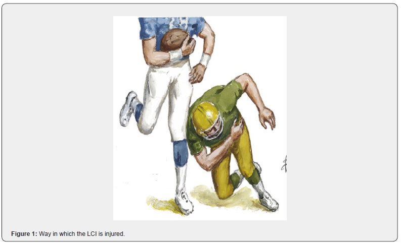
Internal collateral ligament (LCI) injury is a common trauma in young people, especially because of both recreational and high-performance sports in contact sports such as soccer in its different variants (Figure 1) as well as activities work, it is considered exceptional in children and older adults [1,2]. According to Busto-Villarreal [3] quoting Márquez [4] in a study of 500 people with knee ligament injuries, 29% corresponded to LCI ruptures and between 13-18% combined injuries of the LCI and ACL due to what the LCI is involved between 42-47% of all ligament injuries of the knee.

The diagnosis of these lesions is basically clinical and is achieved with exhaustive questioning and a physical examination consisting of the valgus stress test or Böhler maneuver [5,6]. This maneuver can be done radiologically by subjecting the knee to stress by performing an Rx in anteroposterior (AP) view [7,8]. Other complementary tests that can be performed for the diagnosis of these lesions are Ultrasound and Nuclear Magnetic Resonance (NMR), the latter limited by not existing in all hospitals and its high cost. The treatment of traumatic injuries of the LCI is basically conservative with immobilization with an inguinopedic cast for 3-4 weeks and then starting an energetic rehabilitation program [3]. Some authors undertake ligament repair in patients with high demand on their knee using primary repair techniques and even plasties [3,7].

If the lesion is not diagnosed or an incomplete treatment is carried out, two late complications could occur: Palmer’s disease (PD) and Pellegrini-Stieda disease (PD-S), so the objective of this article is to assess these two late complications. consequence of a traumatic injury of the LCI of the knee to better understand its pathophysiology, clinical, radiological, arthroscopic picture as well as the best treatment to carry out for its solution.

Method

The search and analysis of the information was carried out in a period of five months (from January 1 to May 31, 2023) and the following words were used: Rupture, internal collateral ligament, disease, Palmer, Pellegrini-Stieda. Based on the information obtained, a bibliographic review of a total of 38 articles published in the PubMed [https://pubmed.ncbi.nlm.nih.gov/], Hinari [https://www.who .int/hinari/es/], SciELO [https://scielo.org/ es/] and Medline [https://medlineplus.gov/spanish/] through the EndNote search manager and reference manager, of which They selected 18, from the last five years, to carry out the discussion. The words selected for the search were taken from MeSH (Medical Subject Headings).

Development

Palmer’s disease

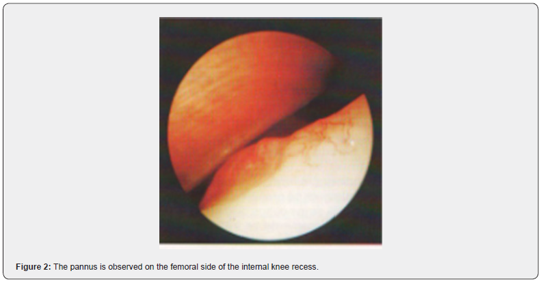
Described by the Swedish doctor Ivar Palmer in 1936, cited by Eriksson [9], it is a rare and unknown pathology that originates from traumatic rupture of the LCI in the thickness of the ligament towards the femoral insertion and whose subsequent healing with consequent retraction of the ligament originates in the internal recess of the knee towards the internal femoral condyle in arthroscopic visualization a mamelonating lesion with bulge, synovial proliferation and pannus formation (Figure 2) that also causes pain on the internal side of the knee on the joint spacing, limitation to the extension of the last few degrees and references to episodes of blockages simulating a meniscal lesion [10,11].

It generally occurs in young male patients because of practicing sports, whether recreational or high performance, between one and two years after the occurrence of a traumatic event in the knee, hence the importance of exhaustive questioning of the patient [10]. Patients complain of pain on the inside of the knee and of suspected blockages, occasionally they may present joint effusion. The physical examination reveals a limitation to the extension of the knee compared to the contralateral one. It simulates a meniscus lesion with positive Böhler, Mc Murray and Appley maneuvers [5,10]. But there is a maneuver discovered and practiced by Palmer, also known as the Wobble Test’s, brought to the present day by Carnes and Malanga [5,6] and cited by Morales et al. [10].

Palmer’s maneuver or Wobble Test’s

It consists of placing the patient in a supine position and the hip flexed between 30-45°, the performer of the maneuver standing next to the patient holds the knee with both hands and holds the leg between the thorax and the examiner’s arm, place the balls of the thumbs on the medial and lateral joint lines to gain space and sensation, then alternate valgus and varus repeatedly starting with the knee at 30° and slowly extending it. If the procedure is painful or there is apprehension, the maneuver is positive.

Mistakes that can be made.

Move the knee from front to back.

Move very fast.

Applying too much valgus and varus to the knee.

Don’t make it repetitive.

Do not make it comparative with the contralateral knee.

Meaning of the maneuver

If the ligament is lax, it is a premonitory of hyperlaxity or ligament rupture (acute phase), if there is no laxity and it is limited in relation to the contralateral knee, it is synonymous with anterior rupture, healing, and retraction of the ligament (chronic phase).

Pathophysiology

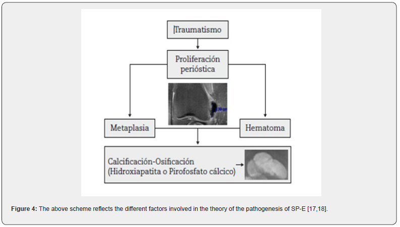
The superficial internal collateral ligament has abundant irrigation (Figure 4). When it is injured, the healing of this ligament complies with the classic model that consists of hemorrhage, inflammation, repair and remodeling. The rupture must occur in the thickness of the ligament, since if it occurs in After insertion, bleeding can be accompanied by osteogenic cells and create conditions for ossification [12-14]. Although the phenomenon occurs in the thickness of the LCI, translation is intra-articular, forming a small promontory in the internal recess of the internal face of the femoral condyle. internal surrounded by pannus and that is the cause of the supposed “blockages”.

Diagnosis

It is achieved with an exhaustive questioning, detailed physical examination where, in addition to practicing the classic knee exploration maneuvers, we must practice the Palmer or Wobble Test’s maneuver, many times the finding is accidental when performing a routine arthroscopy due to the suspicion of an injury of the internal meniscus.

Treatment

The treatment is arthroscopic and consists of the ablation of the pannus and the promontory that causes the blockages with an energetic post-surgical rehabilitation.

Pellegrini-Stieda disease

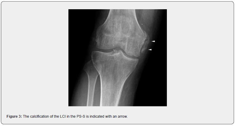
Also known as Pellegrini-Stieda Syndrome, described in 1905 by the former and 1908 by the latter [15]. It consists of calcification of the LCI (Figure 3) and occurs because of the rupture of this ligament as a result of a trauma in the femoral insertion that does not was misdiagnosed or undertreated. Mostly young patients, men who are active at work or in sports, who report a history of knee trauma and complain of pain on the inner face and present limitation mainly to knee flexion [15]. The physical examination revealed pain on the inner side of the knee with greater sensitivity in the path of the LCI and pain with some limitation to flexion. Böhler and Palmer maneuver are positive.

Diagnosis

The diagnosis is basically made by simple X-ray of the AP and lateral knee, observing the calcification of the LCI. In 2006 Mendes et al. [16] cited by Forriol [17] published a classification of the radiological characteristics of LCI calcifications characterized by:

Grade I: Peak-shaped with inferior orientation and union to the femur.

Grade II: Drop-shaped with inferior orientation and parallel to the femur.

Grade III Elongated with superior orientation.

•Grade IV: With upper and lower orientation attached to the femur.

Ultrasound and MRI are also used, the latter being more expensive and not within the reach of many. pathophysiology.

Treatment

Once the diagnosis has been established, treatment is basically conservative, aimed at eliminating calcification, using rest, NSAIDs and physiotherapy in its different variants [17,18]. Other authors recommend needle aspiration under echosonographic vision and infiltration on steroids. Surgical treatment consisting of excision of calcifications is also recommended, with the drawback that they can be reproduced [18].

Conclusion

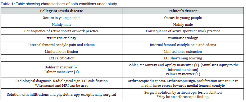
Finally, and as a conclusion, we condense in the following synoptic table the most significant characteristics of both conditions under study (Table 1).


Monday, July 10, 2023

Surgical Treatment of Thumb UCL Injuries with Suture Augmentation - Juniper Publishers

 Orthopedics and Rheumatology - Juniper Publishers


Opinion

It is common for elite athletes to sustain thumb ulnar collateral ligament (UCL) injuries while playing their respective sport. Typically, the mechanism of injury is a radially directed force to the thumb, usually from a fall on the abducted thumb through means such as a ski fall or from sliding into a baseball base. The main goal for this patient population is to return them to sport the quickest and safest way possible.

In addition to the athlete population, many individuals rupture their UCL through every day activities, which can be acute or chronic. With the thumb providing up to 40% of hand function, stability and prevention of post-traumatic arthritis are of utmost importance to the hand surgeon. Injury to the structures surrounding the MCP joint results in a significant impairment of the hand and has been shown to lead to a loss of 22% of hand function. Due to the commonality of the injury and the importance of the MCP joint in the function of the thumb, the treatment of UCL ruptures is significant in the practice of orthopedics. This is done by many means, including direct repair and tendon and free tendon graft. Here we will highlight our preferred method which is surgical UCL repair with suture augmentation.

The current technique we prefer, and use is the Arthrex Suture Tape Augmentation originally described by Giacomo & Shin et al. [1], which has been shown to give increased stability into the thumb after surgery with limited postoperative immobilization. This technique involves creating drill holes for anchors into the proximal phalanx and metacarpal head at the origin and insertion of the ulnar collateral ligament, then loading Fiber Wire suture and Suture Tape onto a 3.5 mm Swivel Lock Anchor and inserting the loaded anchor into the proximal phalanx. A stitch is then thrown into the UCL at its distal free torn end with suture from the anchor at the proximal phalanx. The suture augmentation “Internal Brace” is then completed when the second anchor is inserted in the metacarpal head with suture tape coming from the distal anchor while the thumb is held in 30 degrees of flexion for appropriate tensioning. The only variation from the original technique is use of a 3.5 mm Swivel Lock Anchor in the proximal phalanx rather than a 2.5 mm Push Lock Anchor as we feel the Swivel Lock is easier for insertion and has been shown to have greater pull-out strength. Postoperatively, the patient is seen in hand therapy on post op day #4 and placed in a hand based thermoplastic thumb spica orthosis. The patient begins motion on post op day 10 with the guidance of a hand therapist. The patient begins using the hand without the splint at 4 weeks postoperatively and returns to sport at 6 weeks postoperatively without restriction.

In previous research it has been shown that there is a wide range of time frames for return to sports utilizing the suture augmentation technique for repair of the thumb UCL. Carlson recommended a 6-8 week return to sport for basketball athletes [2]. Werner et al. [3] showed a mean 7 week return to play for collegiate football players. For athletes in season, Sochacki et al. [4] showed 34.8 days in the National Football League and Jack et al. [5] showed a mean of 56.2 days for Major League Baseball Players.

In the past five years, we have performed 55 UCL repairs using the Suture Tape Augmentation. We have had no patients return with re-ruptured UCLs and returned all patients to activity without any complications or laxity in the thumb. Four patients were high level alpine skiers, who returned to racing 4-6 weeks from surgery without complication. We believe that the increased strength and stability gained from suture augmentation allows athletes to return to their sport quicker and safer than conservative treatment or traditional surgical repair relying only on the integrity of the suture to ligament repair. The thumb MCP joint functions to provide a stable base for flexion and extension motion as well as a post for opposition and pinch. Because of this, stability of the MCP joint is essential for hand function. The UCL provides critical stability to the thumb MCP joint during pinch and grip and is therefore important to the function.

Summary

In conclusion, the stability of the MCP joint is important to both the general population and the elite athletes. In our experience, surgical fixation with suture augmentation has led to increased stability postoperatively with excellent results. It is therefore our recommendation to perform this procedure to release athletes back to their sport and the general population back to activities of daily living quicker and safer.

To Know more about Orthopedics and Rheumatology Open Access Journal  

Click here:  https://juniperpublishers.com/oroaj/index.php

To Know more about our Juniper Publishers

Click here: https://juniperpublishers.com/index.php 

Thursday, September 10, 2020

Preventive Program for Optimal Performance in Elite Wheelchair Basketball Players - Juniper Publishers

 Orthopedics and Rheumatology - Juniper Publishers


Abstract

Health intervention in adapted sport has been a critical issue in order to decrease musculoskeletal injuries. In this regard, shoulder injuries are a common problem among wheelchair basketball players (WB). Research in adapted sport injuries is increasing all over the years; however, it is necessary to implement protocols to treat and prevent injuries in wheelchair sports. Shoulder home-based preventive program has demonstrated to be useful for maintaining shoulder health conditions of WB players throughout their sport training regimes when preparing for an international competition. This allows it to be useful complementary tool in combination with teams´ technical strategy.

Keywords: Adapted sport; Paralympic sport; Wheelchair injuries; Shoulder biomechanics; prevention; Exercise

Abbreviations: SHEP: Shoulder Home-based Exercise Program; WB: Wheel chair Basketball

Introduction

In order to improve the performance of elite wheelchair basketball players, experts in sport and health science have suggested exercise programs to prevent and treat injuries. Studies suggest that exercise program is a useful tool as an element to prevent and to treat shoulder pain as a result of injuries [1,2]. Being biomechanical aspects, related to mechanics of the sport, the impairment type and level a standing point to develop a strategy. Practical guidelines and recommendations are published to the prevention and management of exercise in manual wheelchairs users; however, only a few evidence of general recommendations for wheelchair basketball athletes. In this regard, some researches [3,4] show how exercise program could influence in SP in wheelchair population. On other hand, previous researches are developed in elite WB population including prevention of shoulder injury in collegiate WB payers [5,6].

Interventional Study

An interventional study show that a shoulder home-based exercises program (SHEP) [7] appears to be useful tool for maintaining shoulder conditions of WB players throughout their sport training regimes when preparing for elite WB competitions, such as the Paralympic Games. In a group of WB players who received a 10 week SHEP, the changes in SP and ROM were not significantly different from the changes observed in the CG that did not receive the intervention. For both groups, no increase in neither SP nor shoulder injuries was observed, so the functionality and the health of the shoulder were preserved. However, in females WB players there were a significantly reduction of shoulder pain after 10 weeks of the SHEP intervention and an increase of the shoulder functionability.

Discussion

Preventive programs for optimal performance establish the incidence and severity of the problem, the etiology and mechanism of injury, implement the preventive measure and determine the effectiveness of the intervention. A preventive program for wheelchair basketball appears as a convenient tool to maintain the shoulder health condition during the training process working in line with teams´ technical strategy, where players realize a high level of training [7], high impact loading and repeated actions that can develop injuries. In this regard, WB players require regular evaluations, also, multidisciplinary team need to be coordinated to implement and promote an adequate intervention, being necessary the synergy between clubs and institutions. Futher perspectives needed to develop technological innovations that include a protocol with aspects related to the musculoskeletal condition in wheelchair athletes. Also, is needed to determine the effectiveness of the SHEP in a larger sample and include prevention programs in the junior players’ preparation.

To Know more about Orthopedics and Rheumatology


To Know more about our Juniper Publishers
Tuesday, March 10, 2020

Recalling Incident of Own Accident for Suggesting Better Insights-Juniper Publishers

Orthopedics & Rheumatology-Juniper Publishers


Opinion

Quick access to emergency services for any reason is necessary that might be fatal for patients. Clients especially with fractures are quickly guided to services so that they can have an emergency treatment and other diagnostics for a better safety from that injury and secures infectious organisms to attack or enter the site. In many areas of Pakistan there are lack of facilities that can immediately tackle an accident or a fracture. In big and busy cities of Pakistan the increase in road traffic accidents (RTAs) has been so common and it’s estimated that many of the deaths and disabilities occur just because of accidents around the globe and Pakistan specifically.

I recall a serious RTA that affected me during the beginning of year 2017. As a usual practice of more than fifteen days I was crossing the road similarly and was hit by a motor bike where two of the young people were recklessly riding. The hit broke my left lower leg seriously and, in few seconds, I was on ground. I checked my senses and felt like nothing else has happened and I am conscious too, but my leg is now only supported by the nerve supply and the blood supply has somewhat leaked and dropping on the ground and the remaining some blood has become a huge blood clot like a size of tennis ball.

My younger brother who was studying to be a nurse (had witnessed such a serious thing with his eyes for the first time) was in panic and started to shout and cry. We both can’t recall how we moved, what else happened, who was around and what we were thinking to do. Just we can recall a huge feeling of adrenaline rush in our body and palpitations. Fortunately, the incident happened in front of a tertiary hospital in Karachi (which had good protocols and high-quality health services/standards). Somehow with a help I was managed to get to their emergency services.

As a young male, who was working to earn for a family I really felt sad that how miserable incident happened with us and recalling the event itself creates a storm in our daily life. But what we realized is I was fortunate to be so near to the hospital and because of that I was saved from complications. No doubt I was transferred in an improper way, but I was timely reached and my brother who was studying to be a nurse though panicked and got disturbed with this event was there all the time to make my transport and stay so easy and satisfying.

Unfortunately, not every case has that similar kind of situations. Other patients had very bad experiences of accidents and orthopaedic hits or fractures. They had complications and other issues due to improper emergency care and exposure to infectious agents. This is what we have heard from the victims and the clients along with us in the orthopaedic care. In my opinion, it is really necessary to have quick ambulances to pick accident or fracture victims and take them to the emergency care. Secondly, public should be trained so they must know basic things like how to immobile victim of fracture in beginning, how to transfer and where to seek help in emergency. Continuing with this, small emergency points for specific dealings regarding orthopaedic fractures must be developed and maintained in order to achieve a better care.

In addition, hospitals or emergency settings should have well versed resuscitation and trauma staff to handle the case and have an aseptic environment to secure client from microorganisms. The more we work on quick service, safe service, awareness of public and quality standards of trauma emergency care for fractures we can ensure a better outcome in victim’s health.

To know more about Journal of Orthopedics & Rheumatology

To know more about Juniper Publishers

Monday, February 10, 2020

Recent Development of Laser Photo-Chemotherapy (LPC) for Bone Tumors-JuniperPublishers

Journal of Orthopedics and Rheumatology-Juniper Publishers

Introduction

Photodynamic therapies (PDT) have become increasingly popular in the adjuvant treatment of different tumor entities [1]. Chemotherapeutic agents, such as cisplatin may be used in combination with laser-induced thermal therapy (LITT) in an improvement to PDT, known as laser photo chemotherapy (LPC) [1,2]. Based on recent reports on the application of laser photo chemotherapy (LPC) on malignant bone cells under chemotherapeutic conditions with cisplatin or zolendronic acid, the authors feel compelled to describe in this mini-review some relevant aspects of such combined therapy as a potential therapeutic strategy for osteosarcoma [3].
Chemotherapy is regularly used for treating Ewing sarcoma and osteosarcoma, but it isn't often used for other bone cancers, like chordomas and chondrosarcomas, because they aren't very sensitive to chemo [4]. However, it can be useful for some special types of chondrosarcoma, like the dedifferentiated and mesenchymal lineag [3,4]. Anti-cancer agents are sometimes used for bone cancer that has spread through the bloodstream to the lungs and/or other organs [5]. The drugs mainly used for this condition include: Doxorubicin (Adriamycin®), Cisplatin, Carboplatin, Etoposide (VP-16), Ifosfamide (Ifex®), Cyclophosphamide (Cytoxan®), Methotrexate and Vincristine (Oncovin®) [3,5]. In this regard, a number of investigators have shown that some of the above anti-cancer agents are likely candidates for light and or heat activation in cancer cells, as laser photo chemotherapy (LPC) has been consistently used for treatment of retinoblastoma since 1996 [3,6-8].
The propensity for survivors of heritable retinoblastoma to develop second primary osteosarcomas at substantially greater frequency than either the general population or survivors of nonheritable retinoblastoma is well known [9,10]. There is some molecular genetic evidence that the development of these two disparate tumor types involves specific somatic loss of constitutional heterozygosity for the region of human chromosome 13 that includes the RB1 locus [11]. In regards to chemotherapy a number of investigators have shown that anthracyclines and cis-platinum are likely candidates for light or heat activation in cancer cells [1,2,12]. In this sense, Heyman et al. [3] have recently reported a significant decrease of cell bioviability and histomorphological alterations suggestive of higher apoptical activity in osteosarcoma cell lines (Saos-2) treated by cisplatin and zolendronic acid followed by diode laser irradiation, when compared with non-irradiated cells. Therefore, LPC outcomes for retinoblastoma may suggest that a conceptual approach towards osteosarcoma treatment may be possible based on recent clinical studies on combined therapy [12,13-18].
Photo chemotherapy with lasers is an alternative therapy which consists of using a monochromatic light delivered via external irradiation or via interstitial fiber optics to enhance the "killing" threshold in tumors containing light and/or heat-sensitive anticancer agents [12]. The development of photoactivatable pro-drugs of platinum-based antitumor agents is aimed at increasing the selectivity and thereby lowering toxicity of this important class of antitumor drugs [19-21]. Hence, laser photo chemotherapy explores three distinct mechanisms of antitumor action: direct anti-cancer effect
    i. Additionally: thermal
    ii. Light sensitizer
    iii. Effects [1,2,22].
These drugs may be injected intravenously at concentrations lower than normal chemotherapeutic levels, or at higher intratumor doses reducing systemic toxicity while enhancing local tumoricidal effects by laser photoactivation in situ [2,23,24]. Anthracyclines have also been identified that have greater photosensitization potential than daunomyucin [25]. With all the supporting evidence of translational and clinical protocols laser photo chemotherapy has established itself as an alternative treatment for retinoblastoma [18,26,27]. Most of these studies were conducted in children where there has been a few standardized clinical protocols, in particular for unilateral retinoblastoma [27,28]. One ofthese studies by Ventura et al. [29] was sophisticated enough to direct intra-arterial chemotherapy for combined laser photo activation in an advanced unilateral case in an 8-year old girl with no other options for treatment [29].
In sum, bone tumors are rare neoplasm that causes significant morbidity and mortality that despite important medical advances in the past 20 years produced few significant changes in function or survival for patients affected with these diseases. Based on the successful establishment as an alternative treatment for retinoblastoma LPC may become an alternative option for this devastating disease.


To know more about JuniperPublishers

Friday, September 21, 2018

Bone Fractures and Analgesia: Figures from the Orthopedic Emergency-Juniper Publishers

Juniper Publishers-Open Access Journal of Orthopedics and Rheumatology


Introduction: Accidental injuries are part of human life and present in a variety of styles ranging from soft tissue damage to complexed bone fractures. We observed the emergency presentations of the orthopedic patients with different body part injuries. The nature of injury and associated damage was assessed by clinical and radiological tools. Analgesia and fracture management was done according to nature of trauma using general and local anesthesia as needed.

Methodology: Both genders presenting with any sort of trauma at any age were included and non-traumatic non orthopedic emergencies were excluded. Centre for study was liaquat university of medical and health sciences orthopedic section of the emergency department. X-rays were obtained for ruling out fractures and soft tissue injuries. Name, age, gender and other biodata were collected from patients or attendants as found convenient. Sample size was taken 99 and sampling technique was non probability, purposive sampling.

Data Analysis: information received from subjects was analyzed in terms of frequency, percentage, mean age was also calculated.

Results: A figure of 42(42.42%) was found with bony fracture while 57(57.5%) had no bone injury. 16(16.16%) were female 04(4.04%) out of them had fractures while 12(12.12%) were having soft tissue injuries, 83(83.83%) were male 38(38.38%) had fractures while 45(45.45%) had their bones saved. NSAIDs were mostly used analgesics with tramadol as alternative other techniques were not in common practice here.

Conclusion: Male were the predominant subjects to trauma and the same gender was more prone to have bone fractures as compared to female with lower limb at more risk and NSAIDs still remain the first choice as analgesia.


Artificial Intelligence System for Value Added Tax Collection via Self Organizing Map (SOM)- Juniper Publishers

  Forensic Sciences & Criminal Investigation - Juniper Publishers Abstract Findings:  Based on our experiments, our approach is an effec...