Showing posts with label Acute coronary syndrome. Show all posts

Friday, February 18, 2022

Ldl-c Levels In Secondary Prevention: The Lower The Better and The Sooner The Better - Juniper Publishers

 Cardiology & Cardiovascular Therapy - Juniper Publishers

Abstract

Ischemic heart disease continues to be the leading cause of death worldwide. Low-density lipoproteins cholesterol (LDL-c) is the main responsible factor in its pathophysiology. Despite the great farmacological advance in lipid-lowering treatments (estatins, ezetimibe, PCSK9 inhibitors and those that remain to come bempedoic acid and inclisiran) most of our patients in secondary prevention present LDL-c levels over the recommended by the Clinical Practice Guidelines. For this reason, we developed a specific program in an attempt to achieve the goal of lowering LDL-c the lower and the sooner the better.

Keywords: Low-density lipoproteins cholesterol; LDL-c; PCSK9 inhibitors; Acute coronary syndrome; Secondary prevention

Abbreviations: ACS, acute coronary syndrome; CPGs, Clinical Practice Guidelines; LDL-c, low-density lipoproteins cholesterol; PCSK9i, inhibitors of proprotein convertase subtilisin/kexin type 9

Introduction

The WHO reminds us that ischemic heart disease continues to be the leading cause of death worldwide. Over the last few years, a relevant scientific impulse has been developed aimed at investigating its pathophysiology, managing to identify different causes that, to a greater or lesser extent, are responsible for it. However, up to date, its main causal factor continues to be low-density lipoproteins cholesterol (LDL-c) [1-6].

For this reason, LDL-c reduction therapies are postulated as the best tool available for the secondary prevention of patients who have suffered an acute coronary syndrome (ACS). Since the initiation of statin therapy with lovastatin in 1987, great scientific evidence has been built on cholesterol-atheroma plaque causality in parallel with the therapeutic arsenal used, with the incorporation of ezetimibe [4,7] and inhibitors of proprotein convertase subtilisin/kexin type 9 (PCSK9i) [5,8], capable of reducing LDL-c levels to unusual values with previous therapies. In a few months we will have two new molecules: benpedoic acid [9], for patients intolerant to statins or as a complement in those patients bordering on the fulfillment of objectives and Inclisirán, a new drug based on RNA interference technology with promising results to change the routine of lipid-lowering treatment, with a biannual dosage [10].

Keeping in mind this impressive advance in cardiovascular prevention, it should be considered that clinical practice has already internalized that, when it comes to a reduction in LDL-c, “the lower the better”. Consequently, it should have resulted in a higher success rate in achieving the lipid targets recommended by the Clinical Practice Guidelines (CPGs) based on the patient's risk score [11]. However, the latest multicenter secondary prevention studies such as EUROASPIRE [12] and DA VINCI [13] show poor lipid control in our patients despite the disponibility of drugs capable of achieving this goal. This is due to an inappropriate use of available treatments with an insufficient number of combination treatments (statins with ezetimibe) and a low use of PCSK9i.

In addition to the paradigm of "the lower the better", it is necessary to reduce LDL-c levels "the sooner the better". Obviously, this requires a closer follow-up of patients, with more frequent analytical controls and a greater dedication to meeting the lipid threshold, than is usually used in a conventional cardiology consultation. In this sense, in the Cardiology Service of the Puerta del Mar University Hospital, we have developed a specific program called “Post-ACS Virtual Consultation”. It is aimed not only at achieving the LDL-c levels recommended by the CPGs [11] in the majority of patients in post-ACS secondary prevention, but also to accomplish it “the sooner the better”. This idea emerged as an improvement action after analyzing our results from a previous series (2018). In this series, we achieved the goal of LDL-c <70 mg/dL (still in force in 2018-2019) in 52% of our post-ACS patients, after 1 year of follow-up [14]. Although these results improved those of the Euroaspire V study, they were insufficient, especially if we take into account the time elapsed until its achievement (almost 1 year) and that the new 2019 CPGs lowered the lipid target for LDL-c from 70 to 55 mg/dL. Therefore, a more energetic and faster lipid control strategy was required than the one we had been using.

In 2020, we developed the “Post-ACS Virtual Consultation” and despite the COVID pandemic, we included more than 300 patients in this program. It basically consists of monthly monitoring and analytical control of patients who have suffered an ACS. These patients are discharged from hospital with high intensity statins, together with a request for a lipid profile within 1 month post ACS and are also included in a specific database. Analysis and telephone consultation are carried out every month. In these monthly telephone consultations, the treatment is escalated (increase in statins and/or ezetimibe and/or PCSK9i) until the lipid goal of LDL-c <55 mg/dL is achieved. At this time, the “time until reaching the lipid goal” is computed, which is the main variable of this program.

We are currently finalizing the follow-up of the last patients included, but, in a preliminary way, we can anticipate that the vast majority of them achieved an LDL-c <55 mg/dL and that the average time for this was around 3 months. Although this results are still preliminary, we believe that this more committed approach in the control of LDL-c will allow the achievement of the recommended objectives by the European CPG [11] in a greater percentage of patients and, especially, in a much shorter time than usual.

Conclusion

Given the known causal relationship between LDL-c and cardiovascular risk, a more conscientious strategy to reduce LDLc the lower and the sooner the better should help reduce the incidence of new ischemic hospitalizations after ACS.

To Know more about Cardiology & Cardiovascular Therapy

Monday, July 19, 2021

Ldl-c Levels In Secondary Prevention: The Lower The Better and The Sooner The Better - Juniper Publishers

Cardiology & Cardiovascular Therapy - Juniper Publishers


Abstract

Ischemic heart disease continues to be the leading cause of death worldwide. Low-density lipoproteins cholesterol (LDL-c) is the main responsible factor in its pathophysiology. Despite the great farmacological advance in lipid-lowering treatments (estatins, ezetimibe, PCSK9 inhibitors and those that remain to come bempedoic acid and inclisiran) most of our patients in secondary prevention present LDL-c levels over the recommended by the Clinical Practice Guidelines. For this reason, we developed a specific program in an attempt to achieve the goal of lowering LDL-c the lower and the sooner the better.

Keywords: Low-density lipoproteins cholesterol; LDL-c; PCSK9 inhibitors; Acute coronary syndrome; Secondary prevention

Abbreviations: ACS, acute coronary syndrome; CPGs, Clinical Practice Guidelines; LDL-c, low-density lipoproteins cholesterol; PCSK9i, inhibitors of proprotein convertase subtilisin/kexin type 9

Introduction

The WHO reminds us that ischemic heart disease continues to be the leading cause of death worldwide. Over the last few years, a relevant scientific impulse has been developed aimed at investigating its pathophysiology, managing to identify different causes that, to a greater or lesser extent, are responsible for it. However, up to date, its main causal factor continues to be low-density lipoproteins cholesterol (LDL-c) [1-6].

For this reason, LDL-c reduction therapies are postulated as the best tool available for the secondary prevention of patients who have suffered an acute coronary syndrome (ACS). Since the initiation of statin therapy with lovastatin in 1987, great scientific evidence has been built on cholesterol-atheroma plaque causality in parallel with the therapeutic arsenal used, with the incorporation of ezetimibe [4,7] and inhibitors of proprotein convertase subtilisin/kexin type 9 (PCSK9i) [5,8], capable of reducing LDL-c levels to unusual values with previous therapies. In a few months we will have two new molecules: benpedoic acid [9], for patients intolerant to statins or as a complement in those patients bordering on the fulfillment of objectives and Inclisirán, a new drug based on RNA interference technology with promising results to change the routine of lipid-lowering treatment, with a biannual dosage [10].

Keeping in mind this impressive advance in cardiovascular prevention, it should be considered that clinical practice has already internalized that, when it comes to a reduction in LDL-c, “the lower the better”. Consequently, it should have resulted in a higher success rate in achieving the lipid targets recommended by the Clinical Practice Guidelines (CPGs) based on the patient's risk score [11]. However, the latest multicenter secondary prevention studies such as EUROASPIRE [12] and DA VINCI [13] show poor lipid control in our patients despite the disponibility of drugs capable of achieving this goal. This is due to an inappropriate use of available treatments with an insufficient number of combination treatments (statins with ezetimibe) and a low use of PCSK9i.

In addition to the paradigm of "the lower the better", it is necessary to reduce LDL-c levels "the sooner the better". Obviously, this requires a closer follow-up of patients, with more frequent analytical controls and a greater dedication to meeting the lipid threshold, than is usually used in a conventional cardiology consultation. In this sense, in the Cardiology Service of the Puerta del Mar University Hospital, we have developed a specific program called “Post-ACS Virtual Consultation”. It is aimed not only at achieving the LDL-c levels recommended by the CPGs [11] in the majority of patients in post-ACS secondary prevention, but also to accomplish it “the sooner the better”. This idea emerged as an improvement action after analyzing our results from a previous series (2018). In this series, we achieved the goal of LDL-c <70 mg/dL (still in force in 2018-2019) in 52% of our post-ACS patients, after 1 year of follow-up [14]. Although these results improved those of the Euroaspire V study, they were insufficient, especially if we take into account the time elapsed until its achievement (almost 1 year) and that the new 2019 CPGs lowered the lipid target for LDL-c from 70 to 55 mg/dL. Therefore, a more energetic and faster lipid control strategy was required than the one we had been using.

In 2020, we developed the “Post-ACS Virtual Consultation” and despite the COVID pandemic, we included more than 300 patients in this program. It basically consists of monthly monitoring and analytical control of patients who have suffered an ACS. These patients are discharged from hospital with high intensity statins, together with a request for a lipid profile within 1 month post ACS and are also included in a specific database. Analysis and telephone consultation are carried out every month. In these monthly telephone consultations, the treatment is escalated (increase in statins and/or ezetimibe and/or PCSK9i) until the lipid goal of LDL-c <55 mg/dL is achieved. At this time, the “time until reaching the lipid goal” is computed, which is the main variable of this program.

We are currently finalizing the follow-up of the last patients included, but, in a preliminary way, we can anticipate that the vast majority of them achieved an LDL-c <55 mg/dL and that the average time for this was around 3 months. Although this results are still preliminary, we believe that this more committed approach in the control of LDL-c will allow the achievement of the recommended objectives by the European CPG [11] in a greater percentage of patients and, especially, in a much shorter time than usual.

Conclusion

Given the known causal relationship between LDL-c and cardiovascular risk, a more conscientious strategy to reduce LDLc the lower and the sooner the better should help reduce the incidence of new ischemic hospitalizations after ACS.

To Know more about Cardiology & Cardiovascular Therapy


To Know more about our Juniper Publishers

Click here: https://juniperpublishers.com/index.php  

Thursday, September 17, 2020

Acute Coronary Syndrome Revealing Non-AA Amyloidosis: Case-Report and Review of Literature - Juniper Publishers

 Cardiology & Cardiovascular Therapy - Juniper Publishers  


Abstract

Amyloidosis forms a set of diseases characterized histologically by extracellular deposition of insoluble proteins with fibrillar conformation in the tissues. In diffuse forms, heart, kidney and digestive tract are preferentially affected, more than the skin and the peripheral nervous system. Three types of amyloidosis predominate: AL that contains immunoglobulin light-chains, AA or serum amyloid A protein, and transthyretin amyloidosis (ATTR). We report the case of a 65-year-old male hospitalized for Acute Coronary Syndrome and in whom clinical investigations will find non-AA type amyloidosis. Amyloidosis AL is the commonest type, and is associated with cardiac involvement of variable extent in almost 70% of the cases. The diagnosis can be challenging since echocardiographic and cardiac MR findings are nonspecific. Coronary involvement may be due to the presence of infiltration of amyloid deposits at the vascular level. However, only a biopsy of a coronary vascular fragment can confirm the diagnosis with the contribution of histology but this can only be done in post mortem. Cardiac amyloidosis is a rare condition with a poor prognosis, the diagnosis of which should be made early. Coronary artery disease should be systematically sought by cardiac catheterization, in order to improve management.

Keywords: Amyloidisis; Acute coronary syndrome

Introduction

Amyloidosis forms a set of diseases characterized histologically by extracellular deposition of insoluble proteins with fibrillar conformation in the tissues. In diffuse forms, heart, kidney and digestive tract are preferentially affected, more than the skin and the peripheral nervous system. Three types of amyloidosis predominate [1]: AL that contains immunoglobulin light-chains, AA or serum amyloid A protein, and transthyretin amyloidosis (ATTR). Cardiac involvement is mainly manifested by hypertrophic cardiomyopathy or heart failure with preserved ejection fraction (HEFPeF) more rarely by coronary artery disease. We report the case of a 65-year-old male hospitalized in the Cardiology Department of Ibn Rochd Universitary Hospital of Casablanca, Morocco, for Acute Coronary Syndrome and in whom clinical investigations will find non AA type amyloidosis.

Case Report

A 65-year-old male patient, chronic smoker at 40 packs/year, was admitted to our cardiology department for chest pain, a typical angina suggesting an acute coronary syndrome complicated by heart failure.

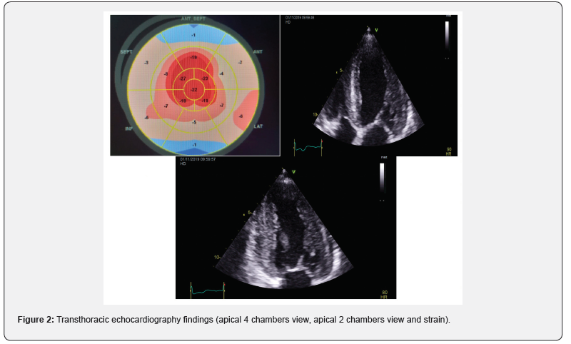
Upon admission, the patient presented typical anginal chest pain with class III NYHA dyspnea, at his clinical examination, his blood pressure was at 130 / 80 mmHg, heart rate at 80 bpm. He presented a global cardiac decompensation, with right heart failure signs that were predominant: retro malleolar edema, ascites of great abundance, turgor of the jugulary veins and hepatic jugular reflux and crackles in both pulmonary bases. The interrogation found a notion of low abundance rectorragia and right hypochondrium pain. The EKG recorded a regular sinusal rhythm at 75, with extreme low voltage in the limb leads (all ,0.5 mV) with a pseudoinfarction pattern in the anterior leads (QS waves from V1 to V3) (Figure 1). Transthoracic ultrasound, noted a symmetrical concentric hypertrophy with a septal wall thickness at 15mm and a posterior wall thickness at 15mm and a discreet inferolateral and anterolateral hypokinesia with a left ventricular ejection fraction (LVEF) at 55%. A longitudinal systolic dysfunction (S’LVseptal = 4cm / S, S’LVlateral = 6cm / S) with alteration of the GLS (global longitudinal strain (-11.5) at the expense of the bases with conservation of the apical kinetics (appearance in roundel of apical spearing). A sparkling and granite appearance of the interventricular septum, without an intra Left ventricular (LV) gradient. Type II diastolic dysfunction; with grade I diastolic dysfunction, impaired relaxation (E / A = 0.7) with an increase in filling pressures (E / E’= 17). The left atrium was dilated to 37ml / m2. The right ventriculi was dilated and hypertrophied (right ventriculi lateral wall = 10mm), with a preserved longitudinal function and without pulmonary hypertension (systolic pulmonary artery pressure SPAP at 30mmHg) and a dilation of the Inferior vena cava to 22 mm compliant (Figure 2).

In the assessment, we noted positive ultrasensitive troponins at 3857ng/L, Pro BNP at 270 ng/L. The ultrasound aspect allowed us to evoke the diagnosis of a cardiac amyloidosis nevertheless in front of the anginal chest pain, the troponins positive we carried out a coronarography by radial way that objectified a long subacute occlusion of the proximal left anterior descending artery, recovered through the right network with a good downstream bed and a tight long stenosis of the proximal right coronary artery. With indication for medical treatment. In front of the digestive manifestations, and the rectorragia, an esogastroduodenal fibroscopy was indicated with biopsies objectifying a fundic and antral erythematous gastritis and a congestive bulbitis. The anatomopathological study showed a chronic pangastritis with presence of intestinal metaplasia without malignancy signs and red congo positive interstitial amyloid deposits, found also in the vascular walls, with phenotyping showing non-AA amyloidosis. The patient was put on treatment of his ischemic disease with double anti platelet aggregation, Aspirin 75mg per day, Clopidogrel 75mg per day, angiotensin-converting enzyme inhibitors 5mg per day, Beta-Blocker 2,5mg per day, Statins 10mg per day and Furosemide 40mg per day with good clinical progress, improvement of dyspnea and regression of chest pain, nevertheless he kept ascites in relation to his intestinal amyloidosis.

Discussion

The amyloid diseases are protein misfolding diseases in which the pathogenic protein misfolds and aggregates as insoluble amyloid fibrils, most commonly in the extracellular space. Three types of amyloidosis predominate [1]:

• AL amyloidosis, characterized by k or λ monotypic deposits and associated with lymphoproliferation and especially with monoclonal gammopathy.
• AA amyloidosis, occurring in a chronic inflammatory context.
• Transthyretin amyloidosis (ATTR), which is either senile or hereditary.
• Cardiac amyloidosis is a rare disease, which is characterized by infiltration of insoluble proteins in cardiac, muscular and / or vascular tissues. Coronary amyloidosis is still a rarer entity, the diagnosis is confirmed post-mortem [2].

Amyloidosis AL is the commonest type, and is associated with cardiac involvement of variable extent in almost 70% of the cases. Cardiac amyloidosis has a poor prognosis, requiring early diagnosis [2,3]. The disease typically presents as a restrictive cardiomyopathy secondary to interstitial amyloid deposits; Hypertrophic cardiomyopathy, Heart failure with preserved ejection fraction (HEFPeF) however it can uncommonly present ischemic symptoms [4].

Extracardiac clinical signs should be sought on clinical examination: carpal tunnel syndrome, macroglossia, periorbital bruising, neuropathy. Electrocardiogram EKG can show micro voltage, conduction disorder or pseudo-Q wave. Transthoracic ultrasound objective:
• Atrial septal thickening considered a characteristic feature of cardiac amyloidosis,
• Characteristic granular/sparkling appearance of the left ventricular (LV) myocardium: not specific and need to differentiate from other infiltrative diseases,
• Increased Left Ventriculi wall thickness which results from amyloid infiltration of interstitial space and may relate to amyloid burden [5].

The biological assessment includes protein electrophoresis, immunofixation, determination of free immunoglobulin light chains, Bence Jones proteinuria, NT-pro BNP and troponin. The genetic analysis of TTR is performed when there is an argument for transthyretin amyloidosis. MRI (late enhancement of gadolinium) and scintigraphy (cardiac fixation of the tracer) anomalies are also very suggestive of cardiac amyloid involvement but the diagnosis of amyloidosis is purely anatomopathological [6,7]. The least invasive biopsies are performed as a first intention. If they are negative, a cardiac biopsy may be necessary to confirm the diagnosis [7]. A retrospective study involving histopathological review of myocardial tissue from 98 patients with AL amyloidosis showed that 66% patients had intramural amyloid deposits. However, only 25% of these patients had any symptoms suggestive of ischemia [8,9]. The extent of vascular involvement in cardiac amyloidosis is variable and depends on the type of amyloidosis [4].

The diagnosis can be challenging since echocardiographic and cardiac MR findings are nonspecific [4,10]. Likewise, the mild elevation left ventricular filling pressures, which may be noted in cardiac catheterization, are nonspecific. Coronary angiography is typically normal as the infiltration is predominantly microvascular [4]. Consequently, the diagnosis of this form of cardiac amyloid is often delayed. The cardiac localization of AA amyloidosis is rare and exceptionally symptomatic, whereas that of non-AA in particular AL is very frequent as it can reach 60% of cases [3,11]. The prognosis is serious with symptomatic cardiac involvement, the average survival is only a few months [12].

Authors suggest that amyloid deposits can sometimes be confined to the coronary arteries [9]. On the other hand, several autopsy studies have been reported, among which, the study by Smith et al. which examined 47 hearts of patients with cardiac amyloidosis (21 primary and 26 senile) the vascular component a was found in 19 patients with primary amyloidosis and 1 patient with senile amyloidosis. Indeed, coronary amyloidosis was present in most of the patients (66%) with primary cardiac amyloidosis. Because only 3 patients had an isolated vascular attack. This study suggests that vascular involvement and ischemia are more important than previously thought [13]. Cardiac involvement of amyloid origin is therefore due to its protein infiltration into the cardiomyocytes, thereby achieving rigidity and thickening of the myocardium, most often causing restrictive cardiomyopathy evolving in three stages [8,12]. Coronary syndrome is also part of one of the entities of cardiac amyloidosis by infiltration of amyloid deposits in the various coronary vessels [7,9,12].

These various hypotheses evoking vascular damage of an ischemic nature in cardiac amyloidosis corroborate with our diagnosis on the possibility of coronary attack in our patient suffering from cardiac amyloidosis. Coronary involvement may be due to the presence of infiltration of amyloid deposits at the vascular level. However, only a biopsy of a coronary vascular fragment can confirm the diagnosis with the contribution of histology but this can only be done in post mortem [14].

Conclusion

Cardiac amyloidosis is a rare condition with a poor prognosis, the diagnosis of which should be made early. Coronary artery disease should be systematically sought by cardiac catheterization, in order to improve management.

To Know more about Cardiology & Cardiovascular Therapy  


To Know more about our Juniper Publishers

Therapeutic and Prognostic Factors of Upper Gastrointestinal Bleeding in the Intensive Care Unit in a Sub-Saharan African Country- Juniper Publishers

  Anesthesia & Intensive Care Medicine- Juniper Publishers Abstract Background: The aim of our study was to determine the therapeutic a...