Showing posts with label Juniper publishers group. Show all posts

Tuesday, February 10, 2026

What Happens After You Submit a Paper to Juniper Publishers? 2025 Workflow Explained

Submitting a manuscript is only the first step in the academic publishing journey. In 2025, Juniper Publishers follows a structured editorial workflow designed to balance transparency, ethical review, and timely publication.

This guide explains what authors can expect after submission, outlining each stage of the Juniper Publishers workflow from initial screening to final publication.


Initial Submission and Editorial Screening

After submission, manuscripts undergo an initial editorial assessment before being sent for peer review.

What is checked at this stage:

  • Alignment with the journal’s scope
  • Compliance with formatting and submission guidelines
  • Originality and proper citation
  • Authorship validity and ethical declarations
  • Confirmation that the manuscript is not under review elsewhere

Manuscripts that do not meet these basic requirements may be returned or declined at this stage. Submissions that pass screening proceed to peer review.


Peer Review Process

Manuscripts approved for review are evaluated by qualified reviewers with relevant subject expertise.

Key features of the review process:

  • Typically conducted under a double-blind model
  • Reviewers assess scientific soundness, originality, clarity, and ethical compliance
  • Recommendations may include acceptance, revision, or rejection

Review timelines can vary depending on reviewer availability and manuscript complexity.


Editorial Decision and Author Notification

Once reviewer feedback is received, the handling editor evaluates the reports and makes an editorial decision.

Possible outcomes include:

  • Acceptance
  • Minor revisions
  • Major revisions
  • Rejection

Authors are notified of the decision along with reviewer comments. If revisions are requested, authors are expected to respond point-by-point and submit a revised manuscript for further evaluation.


Revision and Re-Evaluation (If Required)

When revisions are submitted, the editor reviews the changes to ensure reviewer concerns have been addressed.

  • Minor revisions may be assessed editorially
  • Major revisions may be sent back to reviewers for re-evaluation

Only manuscripts that meet scientific, ethical, and editorial standards proceed to acceptance.


Article Processing Charges (APC) and Publication

As an open-access publisher, Juniper Publishers applies Article Processing Charges (APCs) to accepted manuscripts.

Important points for authors:

  • APCs vary by journal and author region
  • Payment is requested only after acceptance
  • Publication proceeds after APC processing

Once finalized, the article is prepared for online publication and made freely accessible worldwide.


Production and Online Publication

Accepted manuscripts enter the production stage, which includes:

  • Typesetting and formatting
  • Quality checks for figures, tables, and references
  • Author review of galley proofs

After final approval, the article is published online and assigned a DOI, enabling citation and long-term accessibility.


Typical Timelines (What Authors Should Know)

Publication timelines vary depending on several factors, including:

  • Quality and completeness of the initial submission
  • Reviewer availability
  • Number of revision cycles
  • Administrative processing

While Juniper Publishers emphasizes efficient workflows, actual turnaround times may differ between journals and manuscripts.


Ethical and Procedural Responsibilities

Authors are expected to meet ethical and procedural requirements throughout the process.

Key responsibilities include:

  • Submitting original work only
  • Providing accurate citations and disclosures
  • Including ethics approval and consent statements where applicable
  • Understanding that withdrawal may be restricted after peer review or DOI assignment

General policies related to review, publication, and fees are outlined in the publisher’s official resources:
👉 https://juniperpublishers.com/juniperpublishers-frequently-asked-questions.php


What This Workflow Means for Authors in 2025

Authors submitting to Juniper Publishers should be prepared to:

  • Follow submission guidelines carefully
  • Respond constructively to reviewer feedback
  • Plan for APCs after acceptance
  • Monitor manuscript status during review and production

Understanding each stage helps reduce uncertainty and supports smoother publication planning.


Conclusion

In 2025, the Juniper Publishers submission workflow is designed to provide a structured, transparent, and ethically guided publishing experience. From initial editorial screening through peer review, revision, and final publication, each stage supports quality control and global dissemination.

By understanding this process in advance, authors can navigate submission with greater confidence and align their expectations with the realities of open-access academic publishing.

Juniper Publishers Submission Guide 2025: How to Prepare a Manuscript That Gets Accepted

 Juniper Publishers Submission Guide 2025: How to Prepare a Manuscript That Gets Accepted

Preparing a manuscript for academic publication requires more than strong research findings. Authors must also meet journal expectations related to ethics, structure, formatting, and transparency. In 2025, Juniper Publishers continues to emphasize originality, ethical compliance, and clear presentation across its multidisciplinary journal portfolio.

This guide outlines the key submission requirements and best practices authors should follow to improve the likelihood of smooth editorial handling and successful peer review.


Core Ethics and Submission Conditions

Ethical compliance is a foundational requirement for manuscript consideration.

Authors must ensure that:

  • The manuscript is original work and has not been published elsewhere
  • The same manuscript is not under simultaneous review at another journal (in any language)
  • All sources, data, and ideas from prior work are properly cited and credited
  • Any conflicts of interest are fully disclosed
  • Studies involving human participants include ethics committee approval and informed consent where applicable

Meeting these conditions helps protect research integrity and author credibility.


Manuscript Types Accepted in 2025

Juniper Publishers journals accept a variety of manuscript formats. Selecting the correct type before writing helps ensure proper structure and review alignment.

Common manuscript categories include:

  • Research Articles
  • Review Articles
  • Case Reports
  • Short Communications or Mini-Reviews
  • Opinions or Commentaries
  • Letters to the Editor
  • Editorials or journal-specific formats

Authors should choose the manuscript type that best matches the scope and intent of their work before submission. Submission instructions are available through the official portal:
👉 https://juniperpublishers.com/submit-manuscript.php


What Your Submission Package Should Include

A complete and well-prepared submission package helps minimize delays during initial screening.

Required components typically include:

  • Main manuscript file (properly formatted and complete)
  • Cover letter, including:
    • Full author and co-author details
    • Corresponding author contact information
    • Disclosure statements (conflicts of interest, ethics approval if applicable)
  • Copyright transfer form, submitted after acceptance

Providing complete documentation signals professionalism and readiness for review.


How to Structure and Format Your Manuscript

While Juniper Publishers does not mandate a single universal template for all journals, clarity and consistency are essential.

Recommended formatting best practices:

  • Use clear, concise academic English
  • Follow a structure appropriate to your manuscript type

For original research articles, a common structure includes:
Title Page → Abstract & Keywords → Introduction → Materials & Methods → Results → Discussion → Conclusion → Acknowledgments → References → Tables/Figures

Additional formatting guidance:

  • Label all tables and figures clearly
  • Include ethics and consent statements where required
  • Ensure references are complete and accurately formatted
  • Avoid plagiarism and ensure originality throughout

Consistency and readability are prioritized even when formatting rules vary by journal.


Submission Process Overview

The submission workflow is designed to be straightforward and efficient.

Typical steps include:

  • Manuscript submission via the online portal (or journal-specific email, where applicable)
  • Initial editorial screening for scope and compliance
  • Peer review if the manuscript passes screening
  • Copyright transfer request after acceptance

Authors are kept informed throughout the process.


Practical Pre-Submission Checklist

Step

What to Verify

Originality

Manuscript is unpublished and not under review elsewhere

Manuscript File

Prepared in Word (DOC/DOCX) with all required sections

Cover Letter

Includes author details, rationale, disclosures

Ethical Compliance

IRB/ethics approval and consent statements if applicable

Citations & Permissions

All sources cited; permissions obtained where needed

Declarations

Ready to submit copyright transfer post-acceptance

Format & Clarity

Clear language, accurate references, labeled tables/figures

Completing this checklist before submission can significantly reduce editorial delays.


Conclusion

Successfully publishing with Juniper Publishers in 2025 requires careful preparation, ethical rigor, and close attention to submission requirements. Authors who submit original, well-structured manuscripts supported by accurate citations and transparent ethical disclosures are more likely to progress smoothly through editorial screening and peer review.

By ensuring that manuscripts, cover letters, figures, tables, and permissions are complete at submission, researchers demonstrate professionalism and readiness for publication. Following the practices outlined in this guide helps authors present their work effectively and improves the likelihood of acceptance across Juniper’s scientific journals.


Friday, March 13, 2020

What is Freshness?-JuniperPublishers

Journal of Nutrition & Food Science-Juniper Publishers

Summary

All nutritional advisors recommend that an ample portion of fresh food be eaten regularly. Many can define it, but very few understand what it achieves. Underlying principles, that are yet to be scientifically acknowledged, are the subject of this paper.

Biochemistry

We can be proud of the accomplishments of biochemistry, particularly during the past century. We now know the structure of most of the substances that occur in living things, and a great deal about how they interact metabolically. However, these substances and their interactions are manifold and very complex; so far we know less about how order and grace are created and maintained in the structures and functions of living organisms. A major part of the reason for this deficiency is that we know almost nothing about how organisms arrange their substances into tissues, so fashioning, developing and maintaining their forms.
Genes get the credit for this, and certainly do explain how one generation of organisms reproduces another, and how ageing proteins within each organism are replaced with faithful replicas, day by day. But none of that gets us nearer understanding the organisation and maintenance of each organism's form in the first place - let alone metamorphosis, in species that exhibit it. This is particularly puzzling when we realise how large a fraction of the genome is shared between creatures with hugely differing bodily forms and habits. Even within a single organism, we cannot account genetically for the differences between the structures of different cell lines and organs, when the genome is basically identical in all the cells of that organism. We can propose genetic switches, but all these too would be the same in every cell. Chemical gradients and electromagnetic fields are plausible reasons for simple intercellular differences, but we struggle in vain to see how they can ever be complex enough - let alone discontinuous enough - to account for the specialised tissues, organs and systems visible to histologists, anatomists and taxonomists. As the wisest biochemists freely admit, form is beyond the scope of biochemistry.

Agronomy

Farming and horticulture have also developed massively over the past hundred years, devising novel crops and methods of animal husbandry; ever intensifying both inputs and yields. Agronomists and physicians lead closely parallel lives.
Besides their dependence on modern agronomy, however, farmers and gardeners still have enormous regard for the way things grow. For most of them, this is set in the wider context of a deep love and reverence for nature writ large - in a word, for life itself. They observe how neighbouring plants take account of each other; how each draws moisture and minerals from the soil, and gradually unfolds through simple to more complex forms, arriving at length at the mature crop they harvest.
Most growers will testify to the delightful flavour of a fruit or vegetable, picked fresh and eaten there and then. It explodes in the mouth. Milk straight from the cow or goat is sweet and rounded in flavour, qualities dulled by pasteurisation and storage.
But this is the stuff of cookery books and gastronomy, little of which betrays any debt to chemistry. Unlike physicians, chefs-de-cuisine has nothing professional in common with agronomists.
Biochemists have occasionally puzzled about this. Albert Szent-Gyorgyi is probably the most famous example. Having isolated vitamin C, he noted how much less potent it was weight-for-weight than in the tissue from which he isolated it. He stressed to his students, how differently chemistry proceeded in the context of a live tissue than between purified reagents in vitro. One of them went on to manufacturer nutritional supplements by growth rather than chemical synthesis, and to demonstrate their greater effectiveness as nutrients compared with their purified equivalents. Happily, this habit is spreading.
The allusion from this is that in agronomy, too, there is a gulf between the formal chemistry we understand, and the growth processes in nature which we do not yet comprehend.

Growth

We can, however, observe the difference imparted by the growth, say, of a lettuce or a cabbage. From germination to maturity, the seed unfolds through a succession of forms, fed by soil and air, and energised by sunlight. Each layer of leaves is displaced outwards to make place for fresh axial shoots. The leaves green progressively as they enlarge and ripen. But a cabbage takes much longer to ripen than a lettuce, and in the process acquires a more complex structure, more intense colour and far stronger flavour. So we can observe at least two properties that growth contributes to a plant. One is vitality, a measure of the dynamism of its growth in any moment. The other is structure, an accumulative property achieved over time through repeated cycles of this dynamic growth. Yet, if analysed chemically, the plant can still be reduced to small dead piles of purified ingredients, and water. Which tastes better, and why?
Here I submit speculations of my own, though they have some basis in Kirlian photography and circular chromatography of fresh living specimens.
Vitality is far more intense in plants grown in clean, healthy soil without recourse to chemical fertiliser or biocides. It explains the more intense flavour people report, and justifies the distinction of organic from chemical agriculture. More intense vitality, in turn, creates more robust and detailed structure in the tissue of the plant, enhances its immunity to attack and therefore its structural integrity.
Cooking plants releases their vitality in a matter of hours; hence the rather dull flavour of a baked potato, after overnight storage. Out of the oven, a similar potato tastes more gratifying because the vitality is radiating away from its tissue, to be intercepted by the taste organs of the consumer. (Cooking also bursts indigestible cellulose, breaking cell walls and making cytoplasm available to taste and digest. This offers an alternative explanation of the flavour just after cooking, but does not account for the loss of flavour in storage).
Vitality, in this sense, is an important nutrient. It can be eaten, and enhances the vitality, structure and immunity of the consumer - just as of the food it came from. This not only deters invasive micro-organisms but diminishes the risks that the structure of the body will decay or stray - which lead to ageing and cancer.
Freshness is synonymous with vitality. Raw food may possess vitality, but may have lost it gradually in storage or rapidly in cooking, refining or attack by fungi. Tubers, seeds, nuts, corms and bulbs, formed in nature to be dormant between seasons, are live and therefore fresh despite storage. Their vitality may be released by cooking, chewing or sprouting into fresh shoots.
I have not, in this brief essay, dealt with foods from animal sources. The same principles apply in a far more complex way.

Conclusion

Freshness is a necessary but insufficient property of the diet. It is vitality that coveys the benefit. This is ensured in any plant food item by harvesting without physical damage or fungal decay, then eating fresh or very shortly after cooking. Crops that will grow in the next season retain their vitality despite prolonged winter storage. Consuming vitality every day is vital (sic) to general immunity, and the key to preventing or diminishing all illness - in particular slowly progressive "consumptions" such as tuberculosis, leprosy ageing and cancer.

To know more about Juniper Publishers

Thursday, March 12, 2020

Metal Weapons of “Warrior’ Burials” Found in the Middle Bronze Age II Southern Levant – Economical and Social Aspects-JuniperPublishers

Journal of Archaeology & Anthropology-Juniper Publishers

Abstract

Copper-based weapons including battleaxes, daggers, and spearheads from the Middle Bronze Age II (c. 1900-1600 B.C.E.) have been unearthed mainly in burials found in the southern Levant. Archaeological and metallurgical analyses of these metal weapons done recently, make it clear that in the beginning of the period, in the decorated weapons were made of tin bronze with and without arsenic while during the second part of this period we can see a decrease in the number of weapons found in graves as well as changes in the metallurgical composition into the usage of tin bronze, arsenic copper and copper with tin and arsenic. To explain these results, there is a need to look further on the possible social economical context of the “Warrior’ burials” phenomenon using metal weapons made of copper, arsenic, and tin, which are unavailable metals in the southern Levant at this period.
Keywords: Metal Weapons; Warrior Burials; Middle Bronze Age; Tin Bronze, Arsenic copper; Southern Levant; Kit; Tin; Archaeological; Funerary contexts; Society; Copper sources

Case Report and Results

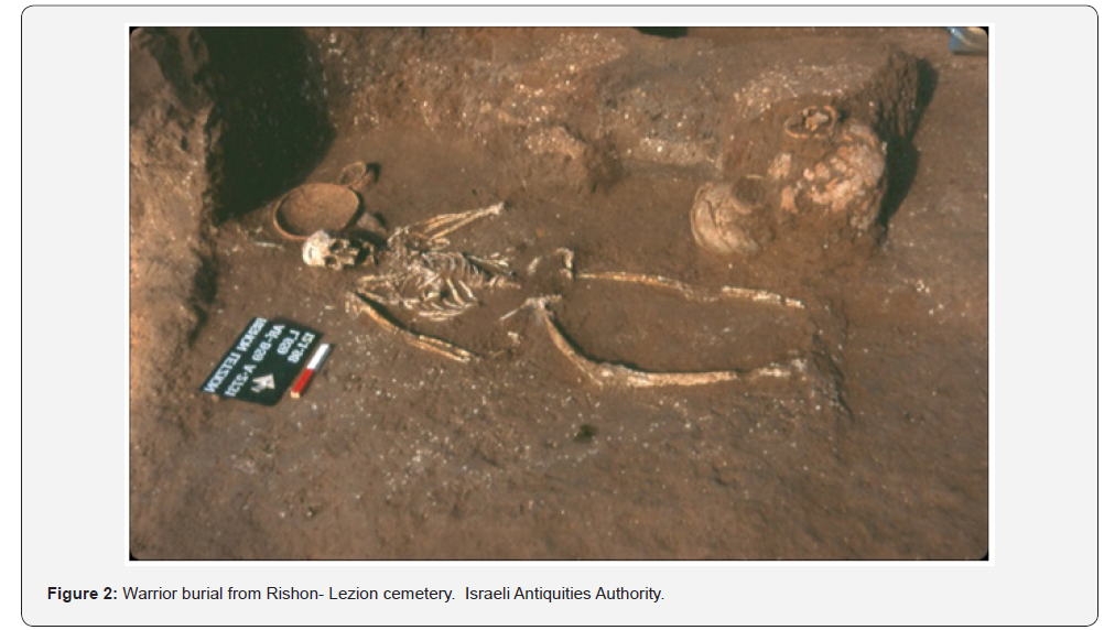
More than 1000 copper-based weapons associated with the Middle Bronze Age II (MB II; ca. 1950–1550 BCE) culture have been recovered, primarily in burials, throughout the Levant (Figure 1); [1-7]. These, funerary contexts have generally been referred to as “warrior burials”, and contained individuals buried with a presumed “kit”, comprising weapons, such as daggers, axes and spearheads found on the deceased’s waist and/or next to their head (Figure 2). The “warrior burials” are dated mainly to the first half of the MBII period (MB IIA; 1950–1750 BCE) and decline in occurrence in the Middle Bronze IIB (MB IIB; 1750–1550 BCE) [6-8].


Recently, it has been shown [6,7] that less than 25% of all the MB IIA burials can be defined as “warrior burials”, and they should rather be considered to reflect high-ranking members of the contemporary society, i.e., an elite social class . The weapons in the MB IIA “warrior’ burials” were well-made, elaborate and composed of copper alloyed with up to 14% tin, both with and without low arsenic concentration [5-7].
The use of tin bronzes is considered the most important technological innovation of the Middle Bronze Age II (MBII). Tin Bronze objects are known from earlier periods, but in small quantities while the use of arsenic copper was more common [5,9]. During the Middle Bronze Age II, tin bronze was widely used to create metal objects in general and weapons [5,6,7,10]. While for production of arsenic copper one metal source containing copper with arsenic was needed, the production of tin bronze required two metal sources, one of tin and one of copper, which were in far distance from the southern Levant [11,12]. Nevertheless, no tin sources were found in the Levant and there is no evidence that local copper sources were exploited at this time, in contrast to the former periods [13-15]. In addition, almost no ingots and complete workshops from this period were found in the Levant [1,2,14-16]. This raise a series of significant questions concerning the factors that led to the widespread use of tin bronzes at this period, the sources of copper and tin, and the trading systems that brought the raw materials and the finished products to the Levant.
In addition, to date, the transition from the use of arsenical copper to tin bronze was perceived as a linear development; It was assumed that at the beginning of the Middle Bronze Age (MBIIA), most weapons, in continuation of the former period, were made of arsenical copper, while in the later part of the period (MBIIB), most of the weapons were made of tin bronze [5]. Through a detailed analysis of the available metallurgical data we have shown that the situation was in fact quite the opposite. It was demonstrated that the transition was highly complex; Tin bronze appeared quite abruptly in the MBIIA, with only few antecedents, and decreased during the MBIIB [6,7].

To know more about Juniper Publishers

Wednesday, March 11, 2020

Simultaneous Estimation of Aliskiren and Amlodipine in Combined Tablet Formulation by Simultaneous Equation and First Derivative Spectroscopic Methods-JuniperPublishers

Journal of Organic & Medicinal Chemistry-Juniper Publishers

Abstract

Simple, sensitive and accurate UV-spectroscopic methods were developed and validated for simultaneous estimation of aliskiren and amlodipine in tablet formulation using simultaneous equation and first derivative spectroscopic methods. Developed methods include direct estimation of amlodipine at 365nm without any interference, since aliskiren has zero absorbance at this wavelength. Estimation of aliskiren through simultaneous equation was performed at 279 nm, while 236.8 nm were selected as zero crossing point for estimation of aliskiren by first order derivative spectroscopic method. Linearity was found to be satisfactory over the concentration range of 25-300μg/ml and 5-100μg/ml for aliskiren and amlodipine respectively. The mean percentage label claim of aliskiren and amlodipine using simultaneous equation was 99.84 and 99.85 % respectively, while for first derivative spectroscopic method it was found to be 100.36 and 99.85% respectively. The developed methods are economical and reproducible for routine analysis of aliskiren and amlodipine in tablet formulation.
Keywords: Aliskiren; Amlodipine; First order derivative method; Simultaneous equation Method; Validation

Introduction

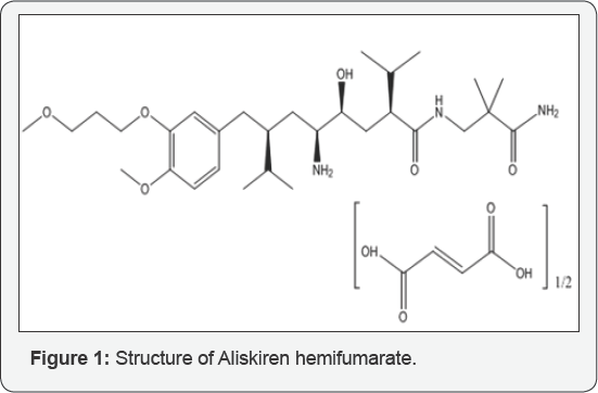
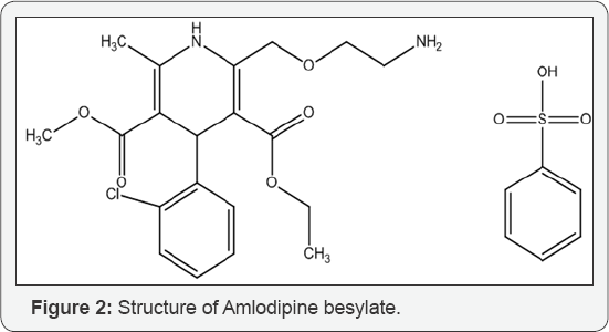
Chemically aliskiren (ALS) is (2S, 4S, 5S, 7S)-5-Amino-N-(3- amino-2, 2-dimethyl-3-oxopropyl)-4-hydroxy-7-[[4-methoxy- 3-(3-methoxypropoxy)phenyl]methyl]-8-methyl-2-propan-2- ylnonanamide [1]. It is a is a white to slightly yellowish crystalline powder. Aliskiren is the first in a class of drugs called direct renin inhibitors. It is used for essential (primary) hypertension [2]. It is highly soluble in water, ethanol and DMSO [3,4]. Amlodipine (AML) is chemically 3-Ethyl-5-methyl (±)-2-[(2-aminoethoxy) methyl]-4-(2- chlorophenyl)-1, 4-dihydro-6-methyl-3,5- pyridinedicarboxylate. Amlodipine besylate is white to off white powder, crystalline and has long-acting 1, 4-dihydropyridine calcium channel blocker [5,6]. It acts primarily on vascular smooth muscle cells by stabilizing voltage-gated L-type calcium channels in their inactive conformation. By inhibiting the influx of calcium in smooth muscle cells, amlodipine prevents calcium-dependent myocyte contraction and vasoconstriction. Amlodipine is used to treat hypertension and chronic stable angina [7,8]. Several analytical methods have been reported for estimation of ALS and its combination with other drugs which includes spectrophotometry and HPLC [9-13]. Similarly, various spectrophotometric and HPLC methods have been reported for estimation of AML and its combination with other drugs [14-18]. In the present work, a successful attempt has been made to estimate both these drugs simultaneously using dual wavelength UV spectrophotometric method. Structures of both the drugs ALS and AML are given in (Figures 1 & 2).

Materials and Methods

Instrumentation

A double beam UV spectrophotometer (UV-1800, Shimadzu, Japan) with UV probe software version (2.31) and 10mm quartz cells was used. All weights

Reagents and Chemicals

Pure drug, Aliskiren hemifumarate and amlodipine besylate was procured from Swapnroop Drugs and Pharmaceuticals, Aurangabad, Maharashtra, India. Marketed formulation was procured from local Pharmacy. All the chemicals and reagents used were of A.R. grade.

Method Development

Preparation of Standard Stock Solution: The standard stock solutions of Aliskiren and Amlodipine were prepared by dissolving 110.5mg of aliskiren hemifumarate (110.5mg of aliskiren hemifumarate is equivalent to 100mg of aliskiren) and by dissolving pure drug of amlodipine besylate equivalent to 100mg of amlodipine in separate 100 mL volumetric flask containing sufficient quantity of distilled water, the solutions were sonicated for 5 min then volume was made up to the mark with distilled water to get a concentration of 1000μg/mL of each solution. The standard stock solutions were further diluted to obtain desired concentrations.
Preparation of Sample Solution: Twenty tablets were weighed and powdered. The quantity of the powder equivalent to 150mg of ALS was transferred to 100 ml volumetric flask. The content was mixed with sufficient quantity of distilled water and sonicated for 20min to dissolve the drug. The solution was then filtered through a Whatman filter paper no. 41 and made up to the mark with distilled water An aliquot of solution (1.0ml) was transferred to a 10 ml volumetric flask and the volume was adjusted up to the mark with distilled water to obtain required concentration of ALS (150μg/ml) and AML (10μg/ml).

Simultaneous Equation Method

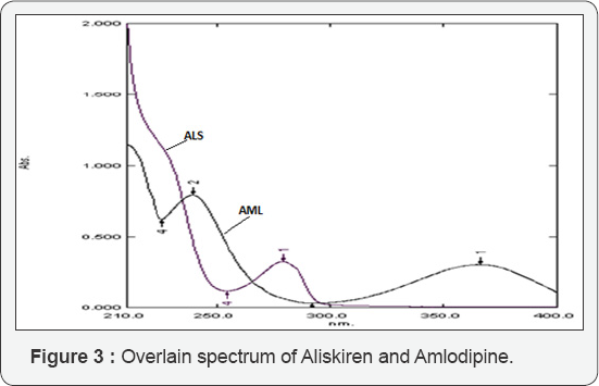
For simultaneous estimation of ALS and AML using simultaneous equation method (SE method) the solutions of ALS (50μg/ml) and AML (20μg/ml) were prepared from the standard stock solutions of ALS and AML and scanned over the range of 200 nm to 400 nm. An overlain spectrum was studied for development of suitable method for analysis. The overlain spectrum of ALS and AML is shown in (Figure 3). From the overlay spectra, 279 nm wave length was selected for the estimation of ALS using simultaneous equation method. Estimation of AML was done as a single component at 365nm. The absorptivity values were calculated and were applied in framed simultaneous equation 1, which is presented as,]
Where, A is absorbance of sample solution at 279 nm, CX and CY are concentrations of ALS and AML, respectively in μg/ml.

First Order Derivative Method

Estimation of AML was performed similarly as in simultaneous equation method. For estimation of ALS first order derivative method (DR method) was applied. The zero order spectrum was then derivatised to obtain first order derivative spectrum (Figure 4). From this spectrum of ALS and AML zero crossing point of 236.8 nm was selected using 2 nm as wavelength interval (Δλ = 2) and scaling factor taken as 1 for estimation of ALS.

Analysis of ALS & AML in Tablet Formulation

The absorbance of final sample solution was measured against distilled water as blank at 279 nm for SE method and at 236.8nm for DR method while the estimation of AML was done directly at 365nm. The analysis procedure was repeated five times for marketed formulation.

Method Validation

Linearity and Range: Aliquots of standard solution of ALS (0.25, 0.5, 0.75, 1.0, 1.25, 1.5, 2.0 and 3.0 ml) and AML (0.05, 0.1, 0.2, 0.3, 0.4, 0.5, 0.6 and 1.0 mL) were transferred in a series of 10 ml volumetric flasks. The volume was adjusted up to the mark with distilled water and mixed. Absorbance values were recorded at 279 nm for SE method and at 236.8 nm for DR method against distilled water as blank for determination of ALS. Absorbance values of AML were recorded at 365 nm for all dilutions. The calibration curves were plotted between the concentration of component and absorbance values of ALS for SE method and between concentration and dA/dX for DR method. Calibration curve for AML was plotted between the concentration of component and absorbance value of AML.
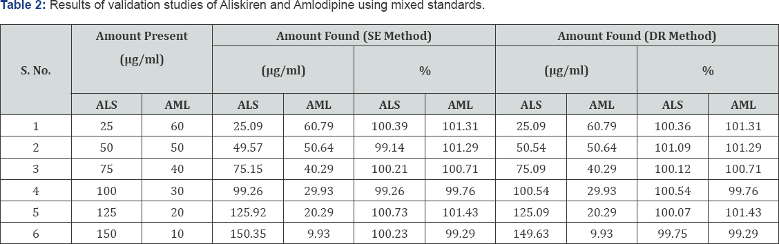
Standardization of the Method by Analysis of Mixed Standard Solutions: To check the validity of the selected methods, mixed standard solutions of ALS and AML were prepared. The solutions were subjected to determine absorbance values at respective wavelengths and concentration of the components were calculated.
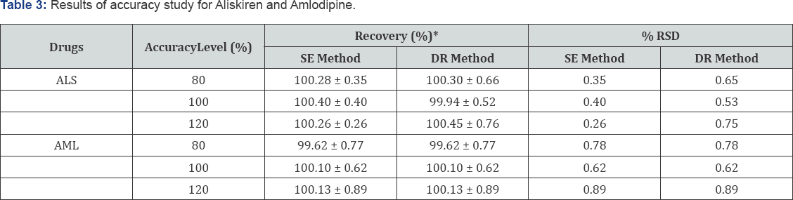
Accuracy: The accuracy of the method was determined for both the methods by calculating recoveries of ALS and AML by the standard addition method. Known amount of standard solution of ALS and AML were added at 80%, 100% and 120% levels to pre-quantified tablet sample solutions of ALS and AML. The results are reported in terms of % Recovery

Results and Discussion

Method Development and Validation

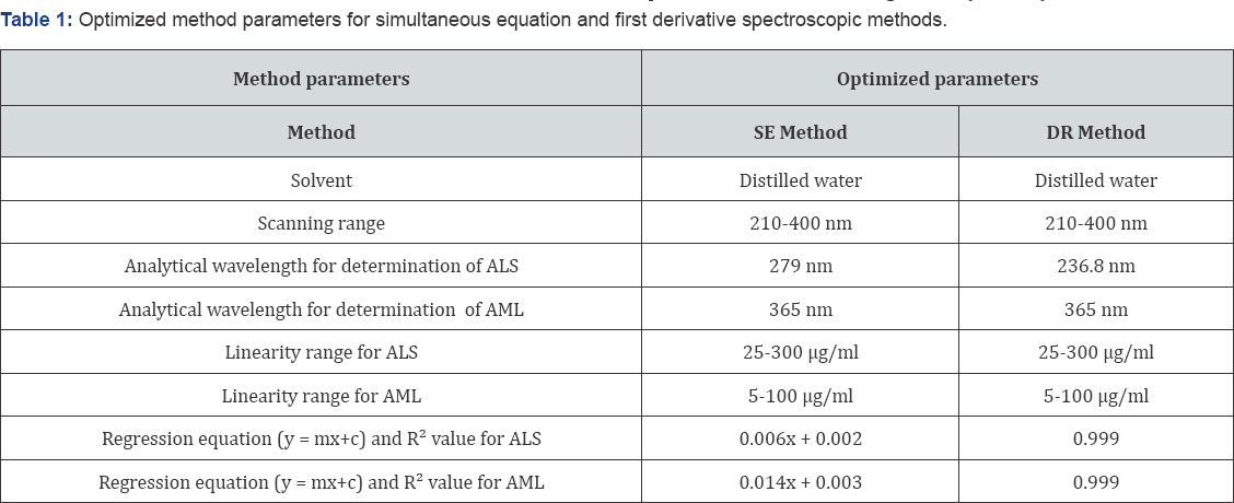
Two simple, sensitive and accurate UV-spectroscopic methods were developed and validated for simultaneous estimation of aliskiren and amlodipine in tablet formulation using SE and DR spectroscopic methods. From the overlain spectra of the drugs it was observed that SE and DR spectroscopic methods were suitable methods for simultaneous determination of ALS and AML. Distilled water was taken as solvent system, as both the drugs were soluble in this solvent and reduce the cost of the method. In SE method and DR method, wavelengths 279 nm and 236.8 nm respectively were selected for determination of ALS, whereas AML was estimated directly at 365 nm as ALS has zero absorbance at this wavelength. Optimized method parameters for simultaneous equation and first order derivative spectroscopic methods are shown in Table 1.

Linearity

The calibration curves of ALS and AML were linear in the range of 25-300μg/ml and 5-100μg/ml respectively. Regression equation and R2 values are given in (Table 1).

Standardization of the method by analysis of mixed standard solutions

The concentration of ALS and AML recovered from mixed standard solutions for both methods was within range and are given in Table 2.

Accuracy

The percentage recoveries of drugs from sample were determined by standard addition of pure drugs at three known concentrations and recoveries were obtained at each level.The percent recoveries for ALS were found to be in the range of 100.26- 100.40% for SE method and 99.94-100.45 % for DR method. Percent recoveries for AML were found to be in the range of 99.62-100.13% for both methods. The results of accuracy studies are shown in Table 3.

Application of the Method in Assay of Tablets

The proposed UV method was applied for the determination of ALS and AML in their combined pharmaceutical formulation and the results are shown in Table 4.

*Mean ± SD (n=3), SD (Standard deviation), %RSD (Percent relative standard deviation).


*Mean ± SD (n=5), SD (Standard deviation), %RSD (Percent relative standard deviation).

Conclusion

The proposed simultaneous equation method and first order derivative gives accurate and precise results for determination of aliskiren and amlodipine in marketed formulation (tablet) without prior separation and is easily applied for routine analysis. Method validation has been demonstrated by variety of tests like linearity, accuracy and validation through mixed standard. The proposed method can be successfully applied for determination of these drugs in commercial tablet formulation.

Acknowledgement

The authors express their sincere gratitude to Swapnroop Drugs and Pharmaceuticals, Aurangabad, Maharashtra, India for providing the pure drug samples of Aliskiren Hemifumarate and Amlodipine Besylate and are also thankful to colleagues and authorities of Department of Pharmacy, SRMSCET, Bareilly, U.P. who helped us in this work.

To know more about Juniper Publishers

Therapeutic and Prognostic Factors of Upper Gastrointestinal Bleeding in the Intensive Care Unit in a Sub-Saharan African Country- Juniper Publishers

  Anesthesia & Intensive Care Medicine- Juniper Publishers Abstract Background: The aim of our study was to determine the therapeutic a...