Showing posts with label Neurology. Show all posts

Thursday, August 6, 2026

Effective converting ratio when switching between Ona-botulinumtoxin-A and Abo-botulinumtoxin-A in cervical dystonia. A patient assessment cross over study- Juniper Publishers


Neurology & Neurosurgery- Juniper Publishers

Introduction

In the 1950s it was hypothesized that Botulinum Neurotoxins (BoNT-A’s) could be used to reduce activity in muscle disorders [1]. Botulinum neurotoxin is made of a gram-positive anaerobic bacterial exotoxin-

clostridium botulinum [2]. When injected in the skeletal muscle, the botulinum toxin inhibits vesicular neurotransmitter (acetylcholine) release at the neuromuscular junction at the presynaptic membrane and a local chemo-denervation appears [1]. Thereby the toxin can reduce muscular contraction, which is the desire to reduce overactive muscle activity [3-5]. The medical therapies for dystonia are effective, and studies have shown that botulinum toxin is the most effective treatment for cervical dystonia [6]. The evidence from several Class I and Class II studies has led to a level A recommendation for use of BoNT-A for treating focal dystonia such as cervical dystonia, hemifacial spasm and blepharospasm [7]. Three main BoNT-A products are available on the market today: Ona-botulinumtoxin-A (Botox®), Abo-botulinumtoxin-A (Dysport®) and Inco-botulinumtoxin (Xeomin®). Due to different production methods, the biological nature of the three BoNT-A’s are different (e.g. the protein load, formulation, pH, dose and immunogenicity). Moreover, the BoNT-A’s are not interchangeable on an equivalent-dose basis [1, 7].

Study background

The goal of this study was to find the optimal conversion ratio for patients with cervical dystonia when switching treatment between Abo-botulinumtoxin-A and Ona-botulinumtoxin-Awhile obtaining a stable satisfying symptom improvement based on a subjective patient assessment. There is heterogeneity in treatment strategies and on how doses and the effect of BoNT-As are reported until now. This study represents a retrospective (follow-up), cross-over study (each patient acts as his/her own control). Effect of treatment was based on individual patient assessment. Individual based conversion rates were calculated using the optimal treatment dose for Abo-botulinumtoxin-A and Ona-botulinumtoxin-A. Due to the length of the present study with a follow-up time from 1991 to end 2014, it was possible to find stable toxin doses where each patient felt the best effect (min. 90%, VAS-score).

Methods

Study design

The clinical data was sampled from the patients’ medical records. Dystonic muscles and injection sites were identified by using electromyography (EMG). The neurologist determined the toxin doses based on each patient´s disease condition and the dystonic muscles activity (EMG). Each treatment (dosing) pr. clinical visit was individualized. At each consultation the neurologist reported the data (doses of toxin and the patient assessed effect and side effects) in the internal IT-system (Danish electronical patient journal) and also used standard sketches of the face/neck-regions to note which muscles have been injected

Botulinum toxin treatment

Ona-botulinumtoxin-A was diluted with saline to a concentration of 40U/ml. Abo-botulinumtoxin-A was dilutedwith saline to a concentration of 200 U/ml. For cervical dystonia the injection sites were between 2-8 sites. The injection doses in the specific muscles pr. patient and the total botulinum toxin doses pr. consultation were registered.

Effect of the procedure

The patients received treatment with BoNT-A´s every 3rd month at the neurological movement disorder clinic by an experienced neurologist. The subjective assessment of maximal treatment effect on dystonia symptoms between each treatment visits, were evaluated at each visit by the patient on a horizontal 100-mm Visual Analogue Scale (VAS) ; where the left end point indicated 0% treatment effect (no change in dystonia symptoms and no effect of the botulinum toxin) and the right end point indicated a total 100% treatment effect (indicate no dystonia symptoms and a maximal effect of botulinum toxin) between treatments)[8-11].

Data collection
Open Access Journal of Neurology & Neurosurgery

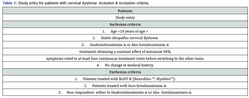
In this study, data was only obtained from patients with a stable idiopathic cervical dystonia, and no change in relevant medical treatment over the data recording period. (Patient study entry, Table 1). The mean individual dose of Onabotulinumtoxin- A was obtained from 4 continuous consultations from each patient with a symptom relief of >90%. The treatment was then changed to Abo-botulinumtoxin-A after filtration to maximal effect and the mean individual dose of this toxin was obtained in the same manner as Ona-botulinumtoxin-A. The conversion ratio of Ona- & Abo-botulinumtoxin-A could then be calculated for each individual by dividing the mean individual Ona-botulinumtoxin-A dose with the mean Abobotulinumtoxin- A dose. The final average conversion ratio (Ona- BoNT-A: Abo-BoNT-A) in the study was found by using all the individual conversion ratio data from the 48 patients (Figure 1).

Statistical analysis

For our statistical analysis of our data we used the software package SPSS Statistics. All averages are in mean± standard deviation.

Results

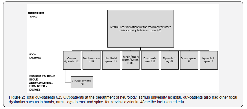
625 out-patients in total were registered at the movement disorder clinic and received treatments with botulinum toxins. Of these, 48 patients with cervical dystonia patients met the inclusion criteria (Figure 2).

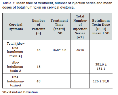
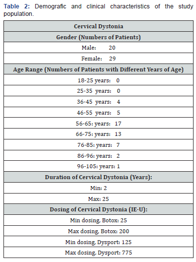
Cervical treatment was determined by the level of dystonia and ranged between 25- 200 units (Ona-botulinumtoxin-A) and 125- 775 units (Abo-botulinumtoxin-A) pr. clinical visit. More females had dystonia and most of the patients’ ages were between middle 50s to middle 70s (Table 2). All data wascollected in a period of 25 years from the movement disorder clinic. The VAS-outcome for each patient was minimum 90%. This retrospective data formed a cross-over design as each patient acted as his/her own control. Therefore, patients with confounders such as smoking, obesity etc. and comorbidities such as heart and lung diseases were not excluded, due to the cross over design. The average treatment time for patients with cervical dystonia receiving Ona-botulinumtoxin-A and Abobotulinumtoxin- A was 15,8±4,6 years. This average treatment time is the total period before and after switching botulinum toxin-A. 2546 injection series (total) were analyzed. Doses were 126 ± 38,8 IE-U for Ona-botulinumtoxin-A and 381,6 ± 151,1 IE-U for Abo-botulinumtoxin-A (Table 3). The mean conversion ratio for cervical dystonia was 1: 2.56. (Table 4).
Open Access Journal of Neurology & Neurosurgery

Discussion

We are reporting 55 patients who converted treatment between Abo- and Ona-botulinumtoxin-A. All data wascollected in a period of 25 years from the movement disorder clinic. Patients have been stabilized with the Ona- or Abobotulinumtoxin- A treatment for at least a year before crossing over to treatment with the other toxin. Due to the long followup and moderate amount of data, we were able to find the average conversion ratio based on each patient´s subjective assessment. Former reports show no statistically significant difference on duration of the effect or adverse effects between Ona-botulinumtoxin-A and Abo-botulinumtoxin-A [12]. Data in these topics were excluded and not evaluated here. Treatment effect of Ona-botulinumtoxin-A and Abo-botulinumtoxin-A was in this study assessed by the treated patients. Due to the routine feedback from each of the treated patients over several years it was possible to achieve regular measurement of treatment effect from a subjective patient perspective without extra time for objective observations and evaluation in between toxin treatment sessions. The conversion ratio between the toxins (Ona-BoNT´s and Abo-BonT´s) using a patient-based assessment has in the present study shown to be equal to the study where they were using multicenter double-blind randomized trials [13]. Due to risk of different treatment techniques, toxin diffusion and especially individual variation in differentiating on relevant pain, a 90% margin was chosen as cut off for registration of desired effect.

Botulinum toxin effect & adverse effect

One unit of Ona-botulinumtoxin-A is not bioequivalent to one unit of Abo-botulinumtoxin-A. The ratio- Ona-botulinumtoxin-A: Abo-botulinumtoxin-A varies between 1:3 and 1:6 [14]. An earlier double-blinded study showed that the conversion ratio of Ona-botulinumtoxin-A and Abo-botulinumtoxin-A was 1:3 [14]. Abo-botulinumtoxin-A has been shown to have a doserelated significant longer duration of the effect rather than Onabotulinumtoxin- A, with a ratio 1:4 (Ona-botulinumtoxin-A: Abobotulinumtoxin- A, p-value= 0,02) [15]. Similar to former reports 12, this report have shown that the effect on dystonia, stabilizes after a period of treatments when changing between different toxin doses. In the present study the toxin doses were stabilized when the patients felt an effect of at least 90% measured on a VAS-score (0-100%) minimum at 4 continuous consultations.

Experimental results have shown that Abo-botulinumtoxin-A diffuses faster than Ona-botulinumtoxin-A which was explained by Abo-botulinumtoxin-A´s lighter molecular weight; Abobotulinumtoxin- A 3-400 kDa vs. Ona-botulinumtoxin-A 900kDa [16], and therefore the side effects in Abo-botulinumtoxin-A compared to Ona-botulinumtoxin-A was assumed due to a higher diffusion rate [17-19]. Other studies stated that botulinum toxin products dissociate under physiological conditions, and therefore the time of diffusion is not related to the toxin size [20]. In a study using Abo- and Ona-botulinumtoxin-A in the same patient with palmar hyperhidrosis, the patient was treated with Ona-botulinumtoxin-A in one palm, and Abo-botulinumtoxin-A in the other palm at the same session for 8 months. The patientswere their own control- like in the present study. The conclusion of that study was that the efficacy and safety of treating with Ona-botulinumtoxin-A and Abo-botulinumtoxin-A was by using the toxins in the conversion ratio of 1: 2.5 [21] which is similar to our conclusion. It has also been shown that conversion ratios of 1:3 or lower could be appropriate for the treatment of spasticity, cervical dystonia, hemifacial spasm, and blepharospasm 22. In the present study we found the conversion ratio of cervical dystonia to be 1:2, 56 which is similar to the former findings [21, 22].

The repeated administration and documentation of the usage of toxin doses (every 3rd month) at the movement disorder clinic reduced the information and selection bias. Due to the long follow-up period with a positive outcome and rare expected side effects, it was found to be effective to use a conversion ratio of 1: 2,56 (Ona-BoNT-A : Abo-BoNT-A) for botulinum toxin-A. The present study did not exclude any patients if they had confounders or comorbidities in behalf of that each patient was his/her own control in this cross-over design. The study design intended to prevent informational bias when evaluating the effect shortly after the toxin injections. To prevent selection and treatment variation bias the use of data was from patients who have been stabilized first with either Ona- or Abo-botulinumtoxin-A treatment for at least a year before crossing over to treatment with the other BoNT-A.

Conclusion

Considering the positive treatment effect, and the long followup period, we concluded that it is safe and clinically useful to use a conversion ratio at 1: 2,56 based on a patient assessment when changing treatment in between Ona- and Abo-botulinumtoxin-A for cervical dystonia. We consider the conversion ratios in our study applicable in other settings as well thus the effect of the botulinum toxin treatments was evaluated on a VAS-score (each patient is his/her own control) in this long follow-up study on dystonic out-patients.

Thursday, April 8, 2021

Diffuse Large B-Cell Lymphoma of the Sphenoid Sinus Initially Presenting with Binocular Diplopia - Juniper Publishers

Neurology & Neurosurgery - Juniper Publishers 

Mini Review

A 59 year old man was referred by his neurologist with symptoms of twelve days of binocular, horizontal diplopia and decreased visual acuity. His medical history was notable for hypertension and hyperlipidemia, adequately controlled with medication. He was a former 1 pack per day smoker but had stopped smoking 20 years previously. He was recently hospitalized for work-up of his symptoms in a community affiliate hospital where a magnetic resonance angiogram of the head was performed, which was unremarkable. Visual acuity was 20/40 in the right eye and 20/30 in the left eye. In addition, he had a trace afferent pupillary defect of the right eye and 12 Prism Diopter (PD) esotropia and 10 PD hypertropia in primary gaze which increased to 20 PD esotropia and 20 PD hypertropia in right gaze. The remainder of the eye exam was normal. He was diagnosed with sixth nerve palsy versus skew deviation, advised to patch the eye for symptomatic relief, and return in 8 weeks.

The patient experienced decreasing vision of the right eye and developed right-sided headache and periorbital pain, prompting a follow-up visit two weeks later. His visual acuity had decreased to light perception, and his pupillary and motility exams remained unchanged. Dilated fundus exam was unremarkable. The differential diagnosis included ophthalmic artery occlusion, optic neuritis or giant cell arteritis, and he was admitted to the same community affiliate hospital and received high dose intravenous steroids. Computed tomography imaging revealed near complete opacification of the right sphenoid sinus and enhancement and thickening of the right optic nerve. On follow-up visit, his visual acuity in the right eye had improved to 20/400, but he still had an afferent pupillary defect and there was no improvement in ocular motility. He was then referred to a tertiary care hospital for dedicated Magnetic Resonance Imaging (MRI) of the brain and orbits and further evaluation and management. The MRI scan revealed a 3.6 x 2.2 x 2.8 cm enhancing sphenoid sinus mass, with extension through the right orbital apex and annulus of Zinn with 270 degrees of encasement and superior displacement of the right optic nerve (Figure 1). Subsequent sphenoid sinotomy and biopsy demonstrated diffuse large B-cell lymphoma with high proliferation index (Figure 2). The patient was referred to neuro-oncology for chemotherapy.

Diffuse large B cell lymphoma is the most common subtype of non-Hodgkin’s lymphoma but rarely arises from the paranasal sinuses [1,2]. The sphenoid sinus as site of origin is especially rare, with only 19 reports in English literature; incidence is slightly more common in Asian patients [1]. Yoshihara et al. reviewed 15 reports and found 13/15 (86.7%) presented initially with ptosis or diplopia caused by CN III, IV, or VI palsy [1]. This is the fourth report in the English literature to describe sphenoid lymphoma with orbital apex syndrome and optic neuropathy. Imaging findings that can be demonstrated with sphenoid lymphoma include permeative bone destruction, hemorrhage, or fibrous dysplasia [2]. As seen in our patient, lack of classical radiologic findings can lead to delay in diagnosis. Vision loss, diplopia affecting multiple cranial nerves, and an indolent course should alert clinicians to suspecting this entity. Prompt consultation with otolaryngology and medical oncology can afford the patient the greatest chance at preventing additional morbidity and mortality.

Material Science


Material Science

Tuesday, May 26, 2020

Biomarkers of the Innate and Adaptive Immune System Responsible for the Pathogenesis of Multiple Sclerosis - A Narrative Review - Juniper Publishers

Neurology & Neurosurgery - Juniper Publishers  


Abstract

Introduction: Multiple Sclerosis (MS) is a complex neurodegenerative disease whose pathophysiology involves demyelinating changes, axon injury, oligodendrocytic death, neuron apoptosis, and inflammation and demyelination of both white and gray brain matter.
Discussion: The role of T cells/B cells has been fairly well documented in MS patients. Analysis of inflammatory infiltrates reveals that CD8+ T cells are found in active lesions while B cells are mainly concentrated in the meninges. Secretory B cell products play an important role in the apoptosis of neurons and the death of oligodendrocytes in MS patients. However, MS cannot be characterized by immune responses; there are genomic components as shown by gene knockout studies. Although there is no cure for MS, it has recently been discovered that increased doses of melatonin may have therapeutic effects.
Conclusion: While the research of MS is ongoing, it is clear that the role of innate and adaptive immunity should not only be the central factor in further understanding the progression of the disease but also the focus of future therapeutic targets. In this review, we focus primarily on immune responses characterized by lesion analysis and inflammatory response in MS patients.

Keywords: Multiple sclerosis; Innate immunity; Adaptive immunity; Biomarker; T cell; B cell; Interleukin

Abbrevations: MS: Multiple Sclerosis; CNS: Central Nervous System; GWAS: Genome-Wide Association Studies


Introduction

Multiple Sclerosis (MS) involves an immune-mediated process in which an abnormal response of the immune system is directed against the body’s Central Nervous System (CNS). Demyelination and inflammation are the primary factors of MS, affecting the formation of plaque in the CNS. This results in MS’s clinical manifestations, which are vision impairment, fatigue, weakness, depression, numbness, bowel changes, loss of muscle coordination and bladder dysfunction. Depending on the affected areas of the CNS, most MS patients will experience phases of deteriorations, flare-ups or exacerbations before the emergence of a new symptom [1,2]. The cause or etiology of MS is not fully understood. However, there is a likely correlation between various factors, influenced by genetic and environmental dynamics. Despite this, research confirms that MS is an autoimmune disease. These studies are corroborated by Genome-Wide Association Studies (GWAS), which identify over 100 MS risk loci, numerous of which intersect in many gene activations of other autoimmune diseases [3,4]. Diseases that cause dysregulation in the immune system are at the highest risk of causing contraction of MS. The progression of MS is found to be correlated with various biomarkers including immune cells and members of the IL1 cytokine family [5]. In this review we attempt to identify the role of innate and adaptive immunity and provide a comprehensive role of biomarkers in the pathogenesis of MS. In addition, we hope to highlight possible strategies/therapeutic targets in order to slow the progression of the disease and improve the quality of life of MS patients.

Discussion

The role of innate immunity in multiple sclerosis

Multiple sclerosis is generally accepted as an autoimmune disease that causes neurodegeneration in the brain; however, some recent studies have suggested that neurodegeneration in progressive MS may be independent of inflammation. In a study exploring the validity of these claims, researchers investigated the correlation between neurodegeneration, inflammation, and disease development in MS stages. They studied 67 MS autopsies from various phases of the disease, comparing it to 28 controls free of neurological disease and brain lesions. Analysis of inflammatory infiltrates in relation to lesion activity revealed that T cells are actually the source of the most prominent inflammation in the active lesions. Moreover, T-cell infiltration in the meninges was highly noticeable in comparison to the cortical parenchyma, which had little to no T cell infiltrates. It is also interesting to note that CD8 and CD4 positive T cell infiltration supported previous studies, which found that CD8 positive T cells infiltrate the most in MS lesions. Similarly, B-cells and HLA-Dpositive microglia cells and macrophages revealed a comparable pattern of inflammation; B-cells were mainly seen in meninges, with only a few isolated in the parenchyma in concentrations ten times lower than T cells. The most intense inflammation was found in patients with relapsing/acute disease, followed by patients in which the disease was progressing. T- and B-cell occurrence was similar in those patients, but plasma cell ratio was different-they were seen more prominently in patients suffering from a progressive form of the disease [6].

Link between the adaptive and innate immunity

Factors that trigger inflammation are crucial for the pathogenesis of MS. Given this, single nucleotide polymorphisms play an important role as they can amplify the expression of inflammatory cytokines and mediators. Multiple studies have indicated that MS patients have high pro-inflammatory and inflammatory cytokine serum levels. One of the most potent and crucial components of innate immunity is the IL-1 family [7]. Vigne and colleagues found that IL-36α, IL-36β, and IL-36γ, members of the IL-1 family are crucial in the production of proinflammatory cytokines as most CD4 T lymphocytes and murine bone marrow-derived dendritic cells constantly expressed IL- 36R. This was substantiated by high levels of IL-6, IL-12, IL-1β, IL-23 and TNF-α seen in bone marrow-derived dendritic cells. These pro-inflammatory cytokines were affected by IL-36 more than any other IL-1 cytokine. IL-36 was also found to stimulate the production of IFN-γ, IL-17 and IL-4 by CD4 T cells. This clearly demonstrates the key role that IL-36R ligands play, not only in the relationship between innate and adaptive immunity, but also in the stimulation of T-helper responses [8].

Importance of serum level of interleukin 36 in relapsing-remitting MS

In a study recruiting 49 relapsing remitting MS patients and 41 healthy individuals, researchers evaluated the implications of Interleukin (IL)-36 in the pathogenesis of MS. The results of their study indicated that there is a significantly higher concentration of IL-36 serum levels in patients with MS [9]. In a separate study examining the expression of IL-36 colonic epithelial cells in patients suffering from inflammatory bowel syndrome, it was revealed that ulcerative colitis patients had higher expression of IL-36α and IL-36γ in comparison with IL-36β. Monocytes plasma cells and T cells were the main sources of this increased expression of IL-36α and IL-36γ. IL- 36α is found to be largely responsible for the formation of acute phase proteins and expression of CXC chemokine, along with the stimulation of MyD88 adaptor proteins TRAK1, IRAK1 and TRAF6 [in conjunction with IL-36γ. This stimulation of adaptor proteins triggers activation of AP-1, NF-κB and phosphorylation of MAPKs. Furthermore, it was found that siRNAs and MAPK inhibitors for c-Jun, AP-1 and NF-κB considerably downregulated IL-36-induced expression of the XCX chemokine [10].

Melatonin effects in peripheral t-helper lymphocytes in RR-MS

Melatonin is known to be a modifier of T helper (Th) 1, Th17 and Treg, the presence of which, along with increased Th22 cells in peripheral blood, are associated with MS progression. In fact, the gene encoding the Th22 receptor il22ra2 actually makes patients more susceptible to MS disease [11]. In a 2017 study, researchers investigated the effects of in vitro administration of melatonin on T-helper (Th) 1, Th9, Th17, T22, and Treg responses. Results of the study showed that melatonin effectively decreased: CNS infiltration of T cells expression of adhesion molecules and the following chemokines: IL2, IL12, IFN-gamma, and TNF. In-vitro melatonin in phytohemagglutinin-stimulated peripheral blood mononuclear cells showed a reduced Th1 response. In addition to Th1, Th9 and Th22 responses were also found to be reduced by melatonin treatment [12]. The study further suggested that blocking IL9, IFN gamma, TNF, and IL17A could be potential forms of treatment since all these cytokines increase T-cell infiltration of the CNS, causing oligodendrocytic and neuronal death [12]. Thus, melatonin may be able to improve the quality of life of MS patients through regulation of Th cells and chemokines.

Naïve, memory, and effector T cells in progressive MS

In order to better understand T-cell activation, researchers studied CD26-dipeptidyl peptidase IV and CD49d. CD26 is associated with T helper (Th) 17 cells and the activation of T cells while CD49d is seen when T cells enter the central nervous system. In one study to help determine the role of CD49 in the progression of MS, researchers used Natalizumab, a monoclonal antibody that attaches to the CD49d receptor. At the end of the 15-month treatment period with natalizumab, there was a statistically significant reduction of inflammatory mediators along with less damage to tissues CD28+ and CD4+ terminally differentiated effector memory T cells were increased in primary progressive MS patients [13].


Researchers analyzed the difference in percentages and absolute numbers of T cells before and after natalizumab treatment, along with their expression of CD26 and CD49d. They found that the absolute number of circulating CD4+ and CD8+ CD28+ EM and TEMRA T cells increased post-treatment, which is consistent with the idea that natalizumab prevents these T cell subsets from entering the CNS. Additionally, treatment of natalizumab decreased the percentage of cells expressing CD26 in all cell subsets of CD4+ and CD8+ T cells besides CD26+ CD28- TEMRA T cells, which already show the lowest expression of CD26 in untreated patients and controls. Lastly, they found a decrease in the frequency of T cells expressing CD49d in all cell subsets of CD4+ and CD8+ [13]. These findings encourage future studies to look into additional benefits that natalizumab may provide MS patients.

B lymphocytes role in MS

Lisak et al. [14] investigated the role of secretary B cells on the apoptosis of neurons and oligodendrocytes. Secretory products of B cells were taken from both patients with RRMS and control patients and then were treated onto oligodendrocytes. Investigators found that in the secretory product secreted from 13 MS patients, 58% oligodendrocytes died from exposure compared to only 4% in oligodendrocytes treated with secretary product in control patients. Human neurons, when treated with secretory products from RRMS patients, showed a similar trend with over half experiencing neuronal death compared to only ten percent of neurons when treated with control samples. As a result, it is evident that although the mechanism of B cell interaction is not fully understood, it plays a major role in cell death especially in RRMS patients and needs to be further investigated [14].

Hydroxylase effects on immunity

25-hydroxycholesterol (25-OHC) not only initiates a signaling cascade that suppresses the production of IgA but also has antiviral properties. While 25 OHC needs to be further investigated, 24S-Hydroxycholesterol 24S-OH-chol has been fairly well documented. Leoni and colleagues had shown that 24S-OH-chol is synthesized in the brain and its distribution through plasma may serve as a biomarker for the progression of MS. In a study conducted on 118 patients who suffered from MS it was demonstrated that older patients, presumably those who had suffered from MS for a longer duration, had reduced 24S-OH-chol plasma levels compared to age matched controls. As a result, the progression of MS may be correlated with a loss of neuronal cells that synthesize oxysterols [15].

The role of oxysterols

Chalmin and colleagues investigated both how oxysterols modulate/contribute to T lymphocyte morphology and play a role in MS autoimmunity through the use of MS Ch25h -/- mice models. Ch25h is a gene that is highly involved in both cholesterol and lipid metabolism. They reported that 16 days post immunization, only 15 % of Ch25h-/- mice developed MS and 43 % had remained symptom free. The Ch25h knockout had no influence on immune system activation in the periphery as CD 4+ T cells and wild type T cells with immunization of myelin oligodendrocyte glycoproteins revealed the same number of leukocytes, IFN-g, IL-17A and IgG proliferation. Moreover, the study demonstrated that Ch25h may actually intensify inflammatory signals and impair trafficking of CD 44+ and CD4 + T cells [16]. While here we have highlighted the importance of the CH25H gene, there are over 100 genetic markers that may increase the likelihood of MS in a given population.

Conclusion

investigating the role of T cells/B cells and cytokines. These essentially have served as biomarkers for the progression of MS and much of the clinical focus has been on finding ways to regulate them. Currently two of the most effective treatments available to clinicians are increased use of melatonin and administration of the monoclonal antibody Natalizumab, both of which work to regulate inflammation and cytokine activity. Current clinical trials have also noted the importance of immune system regulation in slowing down the progress of MS and as a result the majority of phase 3 clinical trials running today have a focus on immune regulation. While over 100 genetic markers have been discovered for MS, gene therapy has yet to become a viable form of treatment, but perhaps may be an alternative in the future. 

To Know more about Neurology & Neurosurgery

To Know more about our Juniper Publishers
Click here: https://juniperpublishers.com/index.php

Anthropology, Participatory Research and Development Cooperation: A Necessary Dialogue- Juniper Publishers

  Archaeology & Anthropology- Juniper Publishers Abstract Le domande sul “perché e per chi” if it generates conoscenza [1] sono da tempo...