Showing posts with label Allergy and Immunology. Show all posts

Wednesday, January 22, 2020

Stem Cells to Improve the Wound Healing-Juniper Publishers

Journal of Head Neck & Spine Surgery

The cellular therapy using Mesenchymal Stem Cells, specially its two subtypes - Bone-marrow Mesenchymal Stem Cells and Adipose Derived Stem Cells - can benefit by virtue of the possibility of differentiating in specialized cells that secrete and suppress growing factors and cytokines necessary in the lesion niche. When attracted by the pro-inflammatory sinalization of the lesion, they act using the paracrine signaling, decreasing the inflammation, increasing the angiogenesis and the cell migration and proliferation. The development in the researches regarding the association of the application of MSCs, with reconstructive surgery practices, leads to effective future results that can bring more benefits to the clinic practice of this field. This paper has the objective of briefly reviewing the literature about the usage of MSCs and its subtypes, the ADSCs, to improve the skin cicatrization.
Keywords: Mesenchymal stem cells; Adipose derived stem cells; Bone-Marrow Mesenchymal Stem cells; Skin flaps; Skin grafts; Wound contraction; Paracrine signaling; Pro-inflammatory Interleukins; Anti-inflammatory Interleukins.
Abbreviations: MSC: Mesenchymal Stem Cells; HLA-DR: Human Leukocyte Antigen; ADSC - Adipose derived stem cells; BM-MSC: Bone-Marrow Mesenchymal Stem Cells; SDF1: Stromal Cell-Derived Factor; PDGF: Platelet-Derived Growth Factor; IL: interleukin; TNFα: Tumor Necrosis Factor-α; IFNγ: Interferon-γ; VEGF: Vascular Endothelial Growth Factor; BFGF: Basic Fibroblast Growth Factor; EGF: Epidermal Growth Factor; KGF: Keratinocyte Growth Factor; HGF: Hepatocyte Growth Factor; PDGF: Platelet-Derived Growth Factor; TGF-β: Transforming Growth Factor-β; FGF: Fibroblast Growth Factor; α-SMA: Alpha-smooth muscle actin's; IRI: Ischemia-Reperfusion Injury

Introduction

Mesenchymal stem cells (MSCs) were first described by Friedenstein et al. [1,2]. MSCs are heterogeneous cell populations, called multipotent adult cells or somatic stem cells [3,4], capable of differentiation in most cell types in order to maintain and repair the organism. They are resident in different organs and tissues such as adipose tissue [4,5], bone marrow, umbilical cord, amniotic membrane [2,4], kidneys, liver, spleen, lungs, pancreas, tendons, synovial membranes, placenta, amniotic fluid and dental pulp [4]. The International Society for Cellular Therapy proposed three minimal criteria for defining MSCs:
    a) Plastic adherence;
    b) Positive expression (greater than 95% of the cell population) for CD105, CD73 and CD90 markers and negative, (maximum 2% of the population) for CD45, CD34, CD14, CD11b, CD79a, CD19 and Human leukocyte antigen (HLA-DR) surface molecules, and
    c) Ability to differentiate into osteoblasts, adipocytes and chondroblasts under standard in vitro differentiating conditions [2,6].
    Later, others researchers complemented this characterization with new markers: CD13, CD44, CD54, 106 and Stro-1 [4] and the presentation of fibroblast cellular morphology [3,4].
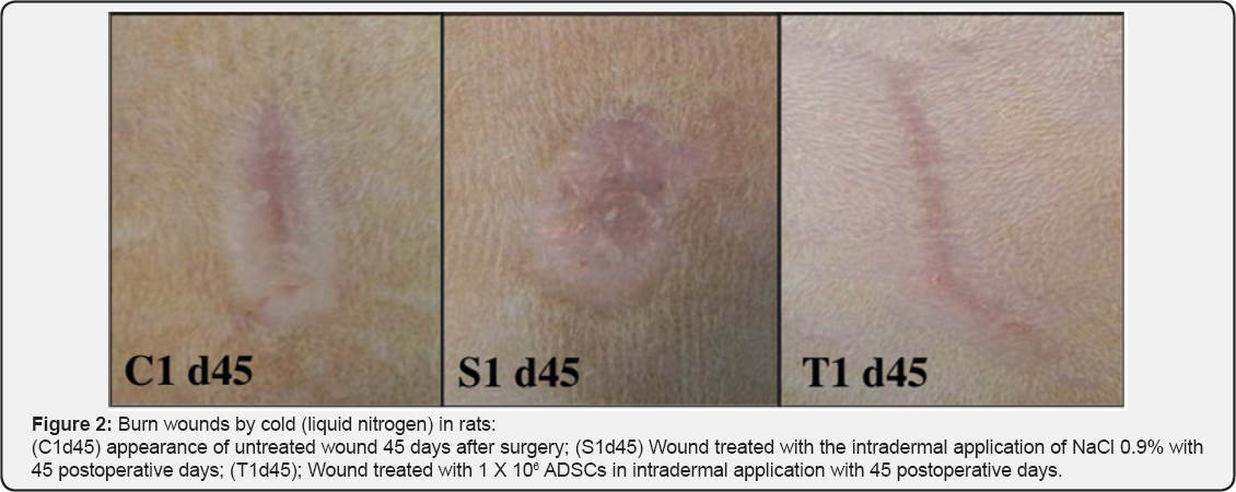
MSCs have a great potential to the tissue repairment and regeneration in reconstructive plastic surgery, preventing ischemic lesions in skin flaps [7-14] and in skin grafts [15,16]. MSCs are capable of reducing severe atrophy, retraction, fibrosis and ulceration skin signals, caused by radiotherapy [17,18]. Combined with the lipoinjection in face and body defects and in rejuvenating esthetic treatments, MSCs raise in 35% the survival rate and the microvasculature from the injected fat [18]. Also, diabetic patients with ischemic wounds show greater survival rates and lower number of amputations in affected limbs, when treated with Adipose derived stem cells (ADSCs) [19]. The MSCs improved the skin healing at induced burns in rats [20,21]. In dermatology, the MSCs are being clinically used for inflammatory diseases [22-28], Graft-Versus-Host Disease [23,25], Chrown Disease [29], Systemic Sclerosis, Lupus or dermatomyositis [24,28,30].
When compared with the usage of Growing Factors, that like any drug has a limited half-life, the cellular therapy using MSCs, specially in its two subtypes - the Bone-Marrow Mesenchymal Stem Cells (BM-MSCs) and ADSCs - can bring greater benefits because of: their expansion capacity in cellular culture; stable phenotypic expression; and possibility of differentiating in specialized cells, that secrete and suppress cytokines and growing factors in the injury [13,31-33]. However, long expansion periods of the cellular culture, to achieve effective therapeutic doses, raise the possibility of infection from the host. Because of that, the ADSCs still have to be heavily studied to the application in the clinic routine [2]. This way, this study has as objective briefly review the literature about the usage of MSCs and its subtypes, the ADSCs, to improve the skin healing.

Discussion

The MSCs are capable to answer and adjust their functions, when exposed to cells or typical factors of the lesion environment [33,34]. They have an attraction capacity to the inflammation areas [33-35] or tumors, because of the molecules CCL21 or HMGB1, known as homing. In case of lacking of this signalization, they are attracted in order of preference to the lungs, liver and spleen [35]. The MSCs show chemotaxis in vitro by the Stromal Cell-Derived Factor (SDF1), Platelet-Derived Growth Factor (PDGF), Insulin-Like Growth Factor-1, Interleukin-8 (1L-8) and Tumor Necrosis Factor-α (TNFα). In murine models, the MSCs used in the systemic administration are capable of getting into the damaged tissues [14,34,36].
The MSCs act, in many levels, in the three healing phases: inflammatory, proliferative and remodeling phase. Current studies indicate that the differentiation of the MSCs, that contributed to the tissue regeneration, is limited by the small survival rate of these cells in the damaged area. However, the paracrine signaling to the cells from the injury is the main mechanism of the MSCs, reducing the inflammation, stimulating the angiogenesis and inducing the cell migration and proliferation [33,37]. Nevertheless, other authors suggest that the differentiation of the MSCs in keratinocytes and endothelial cells have the same function as the paracrine signaling to accelerate the neovascularization and the reepithelialization of wounds [32] (Figure 1 & 2).

Some studies suggest that immunosuppressive potential is the same of both types of MSCs, ADSCs and BM-MSCs, in an inflammatory environment. Their local administration inhibited the T cells recruitment and proliferation [37-41] and decrease Interferon-γ (lFNγ),Tumor Necrosis Factor-α (TNFα), 1L-6 and IL-1β expression in the skin grafts [39-41]. In the inflammatory phase (one to three days), the expression of anti-inflammatory cytokines 1L-4 and 1L-10 is increased while 1L-2 pro- inflammatory cytokines, TNF-α and 1FN-γ are decreased, causing less local inflammatory reaction [37,42]. MSCs can also regulate macrophage activation and convert phenotype expression of M1 macrophages to M2 anti-inflammatory macrophages [39-41], which accelerates the wound healing. Additionally, MSCs act over the innate immunity to prevent the rejection of transplanted allografts, inhibiting the complement system activation [39]. The decrease of TFN-α also helps the reepithelialization after the proliferation phase [43]. The antimicrobial action from the MSCs are also important to limit the infection occurrence [37].
With the utilization of cell therapy in the proliferative phase (14 days) the production of important growth factors is increased: Vascular Endothelial Growth Factor (VEGF), Basic Fibroblast Growth Factor (bFGF) [32,37,42,43], Epidermal Growth Factor (EGF), Keratinocyte Growth Factor (KGF), Hepatocyte Growth Factor (HGF), Platelet-Derived Growth Factor (PDGF) and Transforming Growth Factor-beta (TGF-β). Through their paracrine action, the MSCs raise the migration and proliferation of keratinocytes, endothelial and epithelial cells. The fibroblasts proliferation is also enhanced, as well as the blood vessels' formation. This way, there is a better growing of a more vigorous granulation tissue [33,37,42,43]. The angiogenesis is a very important factor to the wound's healing, and the VEGF and the Fibroblast Growth Factor (FGF) are very important angiogenic factors [32].
Through the paracrine signaling, from the MSCs in the remodeling phase (21 days to one year), the fibroblasts produce collagen fibers in greater quantity and density, generating an increase in wound tensile strength [37,42,43]. At the same time, there is a smaller wound contraction and increased matrix-metalloproteinases production, that control the collagen deposition exacerbation, improving the aesthetic result and keeping the skin's function [33,42,43]. MSCs also contribute to the scar's appropriate remodeling with the increased secretion of VEGF and HGF, the adequate balance between TGF-β1 and TGF-β3 [37] and the increase of bFGF and TGF-β which inhibit the Alpha-Smooth Muscle Actin (α-SMA) expression, responsible for myofibroblast phenotype [32].
Studies indicate that there is also the possibility of using BM-MSCs and ADSCs to protect the damaged tissues caused by the 1schemia-Reperfusion 1njury (1R1) and to decrease the dysfunction of organs undergoing ischemia. The ADSCs are able to protect axial flaps from the 1R1 with results as satisfactory as the ones obtained in procedures without ischemia, because of the improvement in the angiogenic response and the blood perfusion elevation [10]. Many studies use the ADSCs to prevent ischemic injuries in skin flaps [7-14], as others [15,16] used them to improve the quality of skin grafts.

Conclusion

The MSCs - both BM-MSCs and ADSCs - are capable to answer and module their functions, when exposed to cells or typical factors of the lesion environment, attracted by the cell signaling to the inflammation areas. The researches developed till the moment point the paracrine signaling as the main mechanism from the MSCs, decreasing the inflammation, benefiting the angiogenesis and inducing the cell migration and proliferation in the damaged area. The progress on the researches about the association of MSCs with reconstructive surgery practices indicates good and productive results, which can benefit the clinical practice in this field.


To Know More About Open Access Journals Please click on: ttps://juniperpublishers.com/index.php

Monday, November 25, 2019

Proposition of a General Scoring System to the Back PEI- Juniper Publishers

Journal of Head Neck & Spine Surgery

Questionnaires are important tools to assessment due its easy application, low charge and possibility of self-report of the assessed. The Back Pain and Body Posture Evaluation Instrument (BackPEI) and the Back Pain and Body Posture Evaluation Instrument for Adults (BackPEI-A) are questionnaires to assess the back-pain presence and associated risk factors. The BackPEI assess only school children while BackPEI-A assess only adults and has a score system which indicates the subject risk factors exposition level to back pain development. The aim is proposing a score system specific to BackPEI. The score system uses only those questions involving risk factors to back pain development, such as: physical exercise practice; time spent watching television and using computer; number of hours sleeping and habit of reading/studying on bed; occurrence of back pain among parents; postures adopted to sleep, to sit, to write, to use computer and to lift an object from the ground; school bag used to transport material and mode of transportation school bag; and parents’ schooling. As higher as the score, lower is the risk factors exposition to back pain. From this communication, BackPEI has a general score system which expresses the level of risk factor exposition to back pain development among school children.
Keywords: Back pain; Posture; Adolescent; Development; Questionnaire
Abbreviations: BackPEI: Back Pain and Body Posture Evaluation Instrument; BackPEI-A: Back Pain and Body Posture Evaluation Instrument for Adults

Introduction

It’s been increasing the interesting to investigate the risk factors related to spine. This is due to high prevalence of postural changes and of back pain. These problems affect not only the adults [1-5], but also children and adolescents [6-10]. Most epidemiologic studies which investigate back pain and associated risk factors use questionnaires, due the easy application, low charge and possibility of self-report of assessed [11-15].
The BackPEI and BackPEI-A are questionnaires developed to assess both back pain presence and the associated risk factors, presenting validity and reliability proven. The BackPEI have 21 multiple choice questions [16] which assess only school children (from 11 to 16 years old). The BackPEI-A assesses exclusively adults and is composed by 20 multiple choice. Only the BackPEI-A presents a score system indicating the exposition level of risk factors to back pain development [17]. In this perspective, the aim of this short communication is proposing a score system specific to BackPEI.

Methods

The general score system proposed to BackPEI involves only the questions related to risk factors to back pain development among children and adolescents, such as: physical exercise practice (questions 1 and 3) [11,18]; time spent watching television (question 4) and using computer (question 5); number of hours sleeping (question 8) and habit of reading/studying on bed (question 6) [10,19,20]; occurrence of back pain among parents (question 17) [20,21]; posture adopted to sleep (question 7); to sit on a chair (question 10), to write (question 9), to use computer (question 11) and to lift an object from the ground (question 12) [22,23]; school bag used to transport material and mode of transportation school bag (questions 13 and 14) [24,25]; and parents’ schooling (questions 15 and 16) [21].
On question 1, affirmative answer scores 1, while negative answer scores 0. The question 2 does not score. On questions 3, 6 and 17, affirmative answer scores 0 and negative answer scores 1. On questions 4 and 5, the options containing up to 3 hours per day score 1 and the options from 4 hours per day score 0. On question 7, the option “face down (on my stomach)” scores 0, while the other options score 1. On question 8, options containing less than 7 hours spent sleeping per night score 0 and the other options score 1. On questions 9 – 12 and 14, the suitable postures score 1, and the unsuitable postures score 0. On these questions, only one option is considered suitable. On question 13, the options containing backpack with 2 straps, backpack with 1 strap and wheeled backpack score 1, while the others options score 0. On questions 15 and 16, the options “higher education (University)” and “secondary school (1st to 3rd grade)” score 1, and the other options score 0. The final score is resulting from sum of all scores (maximum of 16 points). As higher as the score, lower is the risk factor exposition to back pain development.

Conclusion

From this short communication, the BackPEI questionnaire now has available a general score system, like the BackPEI-A has. This score aims to express how much the school children assessed is exposed to risk factors related to back pain development.

To Know More About Journal of Head Neck & Spine Surgery Please click on:
 
To Know More About Open Access Journals Please click on: ttps://juniperpublishers.com/index.php

Tuesday, November 5, 2019

Vertebral Body Changes in the Lower Spine of Basketball Players-Juniper Publishers

Journal of Head Neck & Spine Surgery

Abstract

Professional sport activities or work may exert considerable stress on the spine. We aimed to assess the effects of loading on the deformation of the vertebral body of the twelfth thoracic vertebra (T12) and the lumbar vertebrae (L1-L5), which are subjected to considerable stress in basketball players. A total of 20 basketball players were assigned to one of two groups, based on their experience with playing basketball: under 5 years, junior group; over 5 years, senior group. All participants underwent magnetic resonance imaging; the relevant geometric variables were measured using specialized software, and the outcome measures were calculated using specific formulas; the outcome measures were compared between the junior and senior groups. The results indicated a significant difference between the two groups with respect to the compression deformity ratio at four levels (T12, L1, L2, and L3), and to the biconcave deformity at one level (L3). Our data suggest that the mechanical loading on lower spine plays an important role in the development of degenerative changes of the vertebral body, which may be considered a risk factor for future injury and low back pain in basketball players.
Keywords: Vertebral body changes; Magnetic resonance imaging; Basketball
Abbrevations: T12: Thoracic Vertebra; L1-L5: Lumbar Vertebrae; Ha: Anterior Margin; Hp: Posterior Margin; Hm: Halfway Between these Margins; Dap: Anterior-Posterior Diameter; BMI: Body Mass Index

Introduction

During sports activities, several important musculoskeletal structures are required to perform complex spinal movement patterns that involve bending and twisting at high speeds. Therefore, the spine is subject to the action of many forces, due to both physiological motion (flexion, extension, rotation, lateral flexion) and accessory motion (shearing tension, compression) [1]. Depending on the direction, magnitude, and point of application of the forces, the spine may suffer deformation and injury. Abnormalities of the spine may also cause injury and pain among athletes because of the unique demands related to each specific sports activity. Lumbar spine injuries represent a significant concern to the athletes, coaches, and physicians.
During athletic endeavors, the lumbar spine is subjected to considerable stress due to unfavorable biomechanical situations typically occurring during such activities [2]. The thoracolumbar and lumbar spine are particularly susceptible to injury due to the large forces exerted in these regions, which are related to: body weight; loads created by motions such as flexion, extension, and rotation [3]; and loads created by accelerating motions especially in sports demanding high speed movements.
In athletes, independent variables that contribute, individually or in combination, to lumbar spine injury include poor technique, poor conditioning, and abnormal anatomy; thus, young athletes may have a spinal deformity that is incidentally or potentially related to their sports activities [4].
Heavy physical work can lead to degenerative changes in the spine [5,6], with body position being a factor that can dramatically affect the load on the lumbar spine. It was shown that the vertebral body undergoes a gradual change in shape under the application of a constant load [7]. Compressive damage arising from repetitive loading is most likely a common event in life; damage to the vertebral body causes decompression of the adjacent disc, leading to internal disc disruption and further degenerative changes [8-10]. Compression fractures can occur with axial loading in a flexed or vertical position. The capacity of the spine to resist injury is decreased if the forces applied involve flexion and are of long duration [11].
It has been demonstrated that the load on the spine during physical activity can be measured based on changes in stature [12]. In analyses of force transmission and in model studies on the spine in high level athletes, it is necessary to estimate reference geometric parameters. It is particularly important to determine the size of the intervertebral discs, the size and shape of the vertebrae, and the overall shape of the spine [13].
Besides the loads carried or lifted by the upper extremities, loads caused by specific, frequent movement during sports activities cause significant loading stress to the vertebral bodies, inducing continuous remodeling of the vertebrae. This leads to degeneration and deformities of the vertebral body, which is associated with increased risk of injury. In the case of the lumbar spine, loads due to flexion, extension, rotation, and acceleration are typically associated with deformity of the vertebral body.
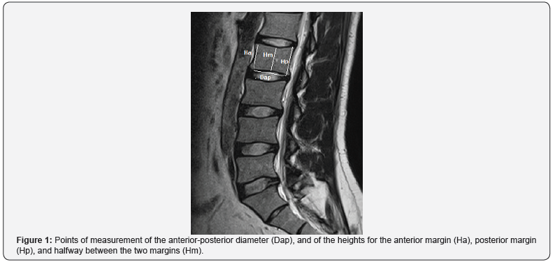
To assess the potential deformation, the height of the vertebral body is measured in three places: at the anterior margin (Ha), at the posterior margin (Hp), and at halfway between these margins (Hm). Three types of vertebral deformities can then be defined based on these heights and on the anterior-posterior diameter (Dap) of the vertebral body. Thus, an anterior wedge deformity is characterized by a low Ha/Hp ratio; a biconcave deformity is characterized by a low Hm/Hp ratio; and a compression deformity is characterized by a low Hp/Dap ratio [14].
The present study attempted to assess the effect of sportsrelated loading on the remodeling of the twelfth thoracic vertebra and the five lumbar vertebrae in basketball players, as predictive markers for future injury or lower back pain.

Material and Methods

Participants

For this study, 20 basketball players (mean age = 20.90, standard deviation = 2.84) with and without symptoms of lumbar pain underwent a case history and a physical examination. The participants were classified in two groups: senior players, with ≥ 5 years of experience; and junior players, with < 5 years of experience. The two groups were matched for sex and age. All participants provided informed consent before commencing the experiment. Players who underwent previous operation in their spine, and players with history of smoking habit were excluded from the study. The study was approved by the Institutional Review Board of our university.

Measures

The pain description for each player was identified by a selfreported questionnaire and a physical examination conducted by an orthopedic specialist. The age, body height, mass, and the body mass index (BMI) of all the subjects were documented. No participant was medicated during the study. A dedicated 1.5 T MRI was performed by technicians for lateral views of the lumbar spine and sacral region of all participants (Figure 1).
The whole body of the vertebra was estimated on each image using Kinovea version 0.8.15 (Kinovea, France) and syngo fast View version 1.0 (Siemens, Munich, Germany), and all measurements were performed blinded to any other information or measurement regarding the subject.

Procedures

After completing the imaging process, geometric variables were selected using the two referred software, including: lumbar body index (Hp/Ha ratio) (Smith et al., 1996), anterior wedge deformity (Ha/Hp ratio), biconcave deformity (Hm/Hp ratio), and compression deformity (Hp/Dap ratio) [14].

Statistical analysis

Data of outcome variables were tested using independent t-test for differences between the two selected groups. Data analysis was performed using SPSS version 17.0 (IBM, Chicago, Illinois, US), and level of significance was set as 0.05.

Results

Our findings showed that, between the senior and junior groups, there were no significant differences regarding age, weight, height, or BMI. This result suggests that these factors did not affect the results of the study.
The height of the vertebral body was measured in three places: at the anterior margin (Ha), at the posterior margin (Hp), and at halfway between these margins (Hm). Three types of vertebral deformities can be defined based on these heights and on the anterior-posterior diameter (Dap): anterior wedge deformity (low Ha/Hp ratio); biconcave deformity (low Hm/Hp ratio); and compression deformity (low Hp/Dap ratio). SD: standard deviation; N: number of participants in the group.
The height of the vertebral body was measured in three places: at the anterior margin (Ha), at the posterior margin (Hp), and at halfway between these margins (Hm). Three types of vertebral deformities can be defined based on these heights and on the anterior-posterior diameter (Dap): anterior wedge deformity (low Ha/Hp ratio); biconcave deformity (low Hm/Hp ratio); and compression deformity (low Hp/Dap ratio). P-values lower than 0.05 were considered significant.
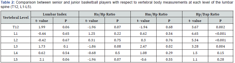
The ratios, for both groups, of Hm/Hp, Ha/Hp ratio, and Hp/ Dap for each level of the lumbar spine, and for the 12th thoracic vertebral body are shown in Table 1. Further, we observed significant differences between the senior and junior groups regarding Hp/Dap ratio at the spine levels T12 (t = 3.67, p = 0.002), L1 (t = 6.65, p ≤ 0.001), L2 (5.34, p ≤ 0.001), and L3 (t = 3.28, p = 0.004) (Table 2), revealing that senior basketball players exhibit greater compression deformity in these levels of the spine when compared to junior basketball players.
Accordingly, we also observed greater compression deformities in the L4 and L5 levels of the senior basketball players when compared with junior players; however, those results were not statistically significant.
Moreover, the senior basketball players had significantly higher Hm/Hp ratio at L3 level (t = 2.47, p = 0.02) when compared with the junior basketball players, revealing that senior basketball players exhibit greater biconcave deformities in L3 when compared to junior basketball players.
On MRI examination, no significant difference between the two groups was observed regarding anterior wedge deformity, or lumbar body index.

Discussion

High level sports participation in adolescents and young adults is associated with a greater incidence of low back pain and structural abnormalities, as revealed in imaging studies [15]. Back pain has significant effects on the athletes’ performance and is estimated to occur in 1.1% to 30% of athletes, with some variation depending on the type of sports activity. Among basketball players, low back pain is a common problem [16-20]. Pain in the spine is often difficult to diagnose. Loading appears to play an important role in the development of radiographic changes of the lumbar spine, and one factor that can dramatically affect the load on the lumbar spine is body position [21]. In the present study, we used MRI, which is considered the most accurate imaging modality for assessing the spine [22]. However, it is important to note that abnormal imaging findings are not always related to the source of the pain, as pathology can exist without pain, and vice versa. In the present study, we assessed degenerative changes related to vertebral body size and shape, which may be considered predictive markers for future lower back pain or injury.
Basketball is popular worldwide and creates unique physiological and physical demands on the players. For example, playing and dribbling the ball are usually executed in a position of spinal flexion, Repetitive jumping, landing from height, and abrupt changes of direction create significant forces acting on the spine. Such repetitive loading can create microscopic damage within a material or tissue, which gradually builds up until gross failure occurs. In living tissues, the process of damage accumulation is opposed by the process of adaptive remodeling [1,14]. According to Alexander [3], the type of injury that occurs in the lumbar spine, which is under significant stress in basketball players, is dependent on the direction, magnitude, and the point of application of the forces in the spine. Ruyssen-Witrand et al. [23] proposed that vertebral size should be considered a potential independent risk factor for vertebral fracture.
Several studies have compared lumbar spine abnormalities of elite athletes with those in non-athletic groups with respect to various sports such as wrestling, soccer, tennis, track and field, and gymnastics [24-26].
In this study, we focused on two groups with the same sex, range of age, and range of BMI, but with different experience with respect to playing basketball. We hypothesized that, independently of other factors, more years of playing in basketball-specific body positions and acceleration motions may cause more abnormalities in the lumbar spine. Indeed, we found that the vertebrae T12, L1, L2, and L3 showed a significantly higher compression deformity in senior basketball players. Likewise, L3 had more biconcave deformities (lower Hm/Hp ratio) in senior basketball players. Thus, playing basketball for more years appears to cause more loading on the lumbar spine, and deformity of the vertebral body.
Our result suggests that, in basketball players, T12 and L1-L3 are at higher risk of degenerative deformities and fractures, as they may be affected by compression and biconcave deformities to a higher extent. Schmitt et al. [26] reported that the concavity index at all levels of the lumbar spine was similar in different groups of track and field athletes. On the other hand, Reilly & Seaton [27] observed an average spinal shrinkage rate of 0.4mm/ min in players dribbling a hockey ball. Predisposing factors in basketball include repetitive spinal flexion, extension, twisting, and loading. Poor development of abdominal musculature in conjunction with strong paraspinal muscles may increase the stress on the lumbar spine during hyperextension, while repetitive jumping and changing of direction create significant forces throughout the spine [1]. In basketball, athletes must bend and rotate their bodies. The specific patterns of loading experienced by the basketball players during dribbling, frequent flexion, and hyperextension may explain the higher rate of radiographic changes we found for the vertebral body of T12, L1, L2, and L3 in more experienced players.
No other significant differences were observed in our MRI examination of the lumbar spine with respect to lumbar index at any level, or compression and biconcave deformity of vertebrae L4 and L5. We did find slight differences with respect to anterior wedge deformity, but this observation did not reach statistical significance, and thus requires more attention before it can be hypothesized as a risk factor for stress fractures.
The present study is limited due to its small population sample, and the fact that it did not account for factors like diet, quality of sleep, or the physical and mental condition of the participants.

Conclusion

In this study, we focused on loading on the lumbar spine caused by basketball-specific body positions, which may translate into vertebral deformities and thus increase the risk of pain and spinal injury, and ultimately prevent the player from participating in sport events. Athletes with minor fractures in the lumbar vertebral body but without neurologic involvement may be considered to return to the sport competition, but under the supervision of the coach and physician; however, they should be made aware of the risks of future injury caused by the deformities. Coaches and athletes may consider these risk factors and apply proper training and conditioning to avoid the development of vertebral body deformities.
In general, athletes and coaches in sports that have an increased risk of lumbar spine injury should be educated in preventive techniques. Specific sport training may be considered to support this part of the spine, with the understanding that significant forces are transferred to the lumbar vertebrae, and with knowledge of the sport-specific deformation likely to occur at various spinal levels.

Acknowledgment

The authors would like to thank the athletes who participated in this study, as well as the employees of the Orthopedics Department and the technicians of the MRI Centre at our hospital for their support with data collection.

To Know More About Journal of Head Neck & Spine Surgery Please click on:
 
To Know More About Open Access Journals Please click on: ttps://juniperpublishers.com/index.php

Tuesday, October 29, 2019

Bandcizer - A Tool to Assess Dosage of Elastic Band Exercises-Juniper Publishers

Journal of Head Neck & Spine Surgery

Introduction

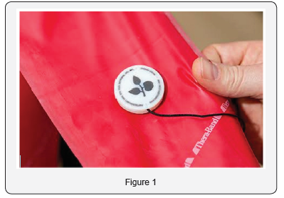
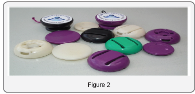
Home based treatment or rehabilitation is one the most blooming and evolving trend among budding and established physiotherapists as it gives improvement in pain intensity, quality of life and muscle strength at the comfort of being at home. Home based exercises are often carried out by patients with help of home-based exercise equipment’s. Bandcizer is a sensor for resistance Training. It helps to assess the dosage of Thera band resistance and Intensity Figure 1.

What is the use of Bandcizer?

Elastic band or resistive exercise ropes are used by many now a day as it provides the sufficient resistance for doing strengthening exercises. The duration, method, repetition and technique of using such exercises are a matter of concern for the physios who recommend home based exercises. Recent studies are suggesting the fact that secondary injuries following the practice of home-based exercises have increased drastically. It’s unclear why such injuries happen and it’s a serious which has to be looked into at the earliest.

Founder

BandCizer ApS is founded by Anders S. Sørensen & Tim Bang. A new commercially available sensor has been developed which automatically records and stores exercise data into its memory card or directly send into the mobile phone. The University Southern Denmark and the national Danish jointly developed “BANDCIZER™”. This new sensor may help clinicians as well as researchers to monitor the Home-based exercise program with ease. The BandCizer is a small device (slightly larger than a quarter) that attaches to TheraBand resistance bands.
BandCizer is capable of automatically storing the data such as day, time and quantifying important aspects of exercise such as ROM and important details such as TUT (Time under tension). It measures the changes in thickness of the elastic band. It is equipped with a LSM3330 3D digital gyroscope with a sample rate of 20Hz. The data recorded by the sensor is sent to a computer directly in form of text files via Bluetooth.

How it Works

BandCizer ‘s 3D gyroscope measures rotation and angular velocity occurring around three axes: x, y and z. The positions of these axes are constant in the BandCizer and cannot be altered. For example: if rotation movement occurs around a vertical axis, the x-axis of the gyroscope will register and record this movement, under the condition that the x-axis of the gyroscope is always positioned on thera-band to point vertically.
BandCizer App guides you towards optimal performance, compliance and progression your training and also provide interactive supervision with live bio-feedback, tailored for the individual [1-5].

Advantages and Usage

a) To increase quality in rehabilitation, sport and fitness.
b) To boost motivation and provide interactive supervision.
c) To document compliance and visualise performance.
d) To share result with therapist (Figure 2).

 
To Know More About Journal of Head Neck & Spine Surgery Please click on:
 
To Know More About Open Access Journals Please click on: ttps://juniperpublishers.com/index.php

Friday, October 18, 2019

Intra-Thyroidal Thymic Tissue May Display Radiological Features Suggestive of Malignancy - A Case Report-Juniper Publishers

Journal of Head Neck & Spine Surgery

Abstract

Introduction: Thyroid “incidentalomas” are increasing in frequency, thought mainly to be a result of the enhanced sensitivity of ultrasonography and the increasing use of this imaging technique. We present two cases of thyroid nodules in paediatric patients which were found to be intrathyroidal thymic tissue.
Case report: The first case displayed ultrasonographic features that were highly suspicious for papillary thyroid cancer with microcalcification, however following diagnostic hemithyroidectomy the lesion was diagnosed as intra-thyroidal ectopic thymic tissue. Histologically, calcified Hassall’s corpuscles would account for the calcified appearance of the nodule on ultrasound scan. We describe a further case of intra-thyroidal ectopic thymic tissue in another paediatric patient who presented with a thyroid mass.
Discussion:It is important to consider thymic remnant tissue as a differential diagnosis for incidental thyroid nodules.
Keywords: Paediatric; Otolaryngology; Incidental finding; Endocrine gland neoplasms; Thyroid malignancy

Introduction

Thyroid nodules are less commonly found in children, affecting 0.2-1.5%, when compared to adult patients [1]. Paediatric thyroid lesions are however more likely to be malignant and must be carefully investigated [1,2]. There are sporadic reports of intra-thyroidal thymic tissue in the literature, however increasing ultrasound scan use and enhanced sensitivity of ultrasonographic images has resulted in a rise in incidentally found thyroid lesions, which is likely to increase further [3,4]. This has created a diagnostic dilemma for clinicians and lead to an increase in diagnostic thyroid surgeries and an increase in detection of microcarcinomas. Thymic tissue has a distinctive appearance on ultrasound with features that may be misinterpreted as a malignant thyroid nodule [3].
We report two cases of intra-thyroidal ectopic thymic tissue in paediatric patients, both presenting with a thyroid mass. One patient displayed radiological features that were suspicious of malignancy. Both patients underwent diagnostic hemi-thyroidectomy.

Case Report

Case 1

A 4-year-old girl with no co-morbidities presented with cervical lymphadenopathy following an upper respiratory tract infection. An ultrasound noted cervical lymphadenopathy and a solitary thyroid nodule with appearances suspicious of papillary thyroid cancer. There was no family history or other risk factors for thyroid malignancy. Blood tests revealed normal thyroid function, calcitonin and thyroid auto-antibodies.
A repeat ultrasound scan of the neck revealed a hypo-echoic nodule within the left lobe of the thyroid, displaying micro-calcification and was taller than wide leading to a U5 (malignant) diagnostic ultrasound grading (Figure 1). Following diagnostic hemi-thyroidectomy, the nodule was histologically found to be ectopic thymic tissue within the thyroid lobe, with calcification of Hassall’s corpuscles, which would account for the calcified appearance on ultrasound scan.

Case 2

A 12-year-old boy with no co-morbidities presented with a midline neck swelling that had appeared 6 weeks previously. Clinically he was found to have a 4.0 x 5.0cm, non-tender neck mass in the midline with no movement on tongue protrusion. Thyroid function, calcitonin and thyroid auto-antibodies were normal.
Ultrasound revealed a 4.0cm hyper-echoic thyroid lesion arising from the isthmus. Fine needle aspiration cytology (FNAC) was reassuring with morphological features consistent with a benign colloid nodule and classified as Thy 2, however due to the size of the mass the child underwent hemi-thyroidectomy which revealed nodular hyperplasia; ectopic intrathyroidal thymic tissue was also identified throughout the sample.

Discussion

Intra-thyroidal thymic tissue is an uncommon cause of neck swellings and has rarely been reported in the literature with published data from a small number of case reports [3,5]. The thymus is involved in adaptive immunity and T-cell function; it proliferates in the neonatal period and first decade of life and is vital for development of a mature immune system [6]. Embryologically, the thymus is derived from the endoderm of the third and fourth pharyngeal pouch during the 6th gestational week and as the thymo-pharyngeal duct elongates during the 7th week the thymus migrates inferiorly and medially towards the superior mediastinum [4,6]. Ectopic thyroid tissue can therefore be found anywhere from the angle of mouth to the superior mediastinum [5], meaning ectopic thymic tissue can easily be misinterpreted as a pathological neck lump, particularly in the paediatric population.
There are various reports of ectopic thymic tissue. Most commonly it is found as aberrant tissue within the neck, known as ectopic cervical thymic tissue [5]. There are only sporadic reports of in thyroidal thymic tissue, however it has previously been misdiagnosed as papillary thyroid cancer on ultrasound, similar to the case presented [3,5]. The exact epidemiology of intra-thyroidal thymic tissue remains unclear, however one study from Japan suggests that it is present in 1% of children [7]. Similar numbers were found in a study of perinatal thyroid glands [8]. With the increasing use and enhanced sensitivity of ultrasound, higher numbers of ectopic thymic tissue located within the thyroid gland may be detected and potentially misdiagnosed as malignancy, leading to diagnostic surgery, as in the two cases presented.
We present the cases of two children with intra-thyroidal thymic tissue who were referred to our paediatric head and neck tertiary referral service with thyroid masses. One child had a thyroid lesion that was radiologically suspicious for a papillary thyroid cancer with micro-calcifications and a mass that was taller than wide. Micro-calcifications are considered a highly suspicious feature on ultrasound, suggestive of papillary thyroid cancer and would result in a U5 grading on ultrasound according to the British Thyroid Association guidelines [9,10]. Hyperechoic foci within ectopic thymic tissue may be misinterpreted as micro-calcifications, which could mimic a malignant thyroid lesion. The other child had incidental thymic tissue identified throughout the thyroid gland in association with nodular hyperplasia.
The cases presented highlight the complexities of preoperative diagnosis of intra-thyroidal thymic tissue. However, with increasing usage of ultrasound imaging it is important to consider thymic remnant tissue as a differential diagnosis for thyroid nodules, especially as the imaging findings may mimic a malignant thyroid lesion. Unfortunately, due to the suspicious ultrasound features diagnostic thyroid surgery may still be inevitable.

To Know More About Journal of Head Neck & Spine Surgery  Please click on:
 
To Know More About Open Access Journals Please click on: ttps://juniperpublishers.com/index.php

How Does the Multi-Disciplinary Team Impact Chronic Kidney Disease Management? - Juniper Publishers

  Urology & Nephrology - Juniper Publishers Abstract Chronic kidney disease (CKD) is a condition where a gradual kidney function loss le...