Showing posts with label More About Juniper Publishers. Show all posts

Thursday, August 22, 2019

Preparation of Novel Morphinan Derivative as a Result of Grewe Cyclization_Juniper Publishers


Peer Reviewed Chemistry Journals Juniper Publisher


Authored by Sureshbabu Jayachandra*

Abstract

The acid catalyzed cyclization of Benzyl substituted Octahydroisoquinoline results in the formation of a major product, N-Formyl Dextromethorphan and a minor product, one of the morphinan derivatives, which is formed via the mechanism of Super electrophiles. Excess Phosphoric acid acts as an electrophile and leads to the formation of a novel minor product but has a significant existence and can be isolated using Column Chromatography at the later stages.
Keywords: Morphinan derivatives; Alkaloids; Grewe cyclization; Super electrophiles

Introduction

Morphinan is the model chemical structure of a huge family of psychoactive drugs, containing opiate analgesics, antitussives and dissociative hallucinogens. It has a phenanthrene fundamental structure with A ring aromatic, the B and C rings being saturated, and an additional nitrogen-containing, six- membered, saturated ring, the D ring, attached to two carbons of the core, and with the nitrogen (Figure 1) [1]. If the D ring is “below” the core (dextro), the analgesic and euphoric properties are eliminated or are dramatically reduced, but the cough- suppressant property is retained, as in dextromethorphan [1].
Ofthe main naturally occurring opiates ofthe morphinan type- morphine, codeine and thebaine-thebaine has no therapeutic properties (it causes seizures in mammals), but it provides a low-cost feedstock for the industrial manufacture of at least four semi-synthetic opiates, counting hydrocodone, hydromorphone, oxycodone and oxymorphone, and, possibly more knowingly, the opioid antagonist naloxone [1]. The Benzyl substituted octahydroisoquinolines with a double bond common to the two fused rings served as a starting material in the synthesis of many morphinan derivatives. The synthesis of isoquinoline ring system is usually achieved by one of the traditional methods, such as Bischler- Napieralski reaction, Pictet- Spengler reaction or the Pomeranz- Fritsch reaction (Figure 2) [2].
So, Benzyl substituted octahydroisoquinolines (N-Formyl Octabase) is a raw material for the usual synthesis of Dextromethorphan Hydrobromide and one of the important intermediate is N-Formyl Dextromethorphan. The formation of this intermediate involves a well-known naming reaction, Grewe cyclisation [3]. The cyclisation has been performed using 85% phosphoric acid and toluene as a solvent (Scheme 1).
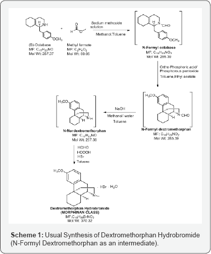
N-Formyl Dextromethorphan is further deformylated to N-Nor dextromethorphan and then methylated to form Dextromethorphan Hydro bromide salt (Scheme 1). In this manuscript, only the preparation of N-Formyl Dextromethorphan from N-Formyl octa base is projected and the formation of a significant Morphinan derivative.

Results and Discussion

The reaction of N-Formyl Octa base with phosphoric acid present in excess act as a catalyst, to form cyclized derivative, N-Formyl Dextromethorphan, using a well- known Grewe cyclization. Grewe cyclization is a synthetic methodology involving the formation of superelectrophiles. Superelectrophiles are multiply charged cationic species (dicationic, tricationic etc.) which are usually characterized by their reactions with weak nucleophiles like arenes, alkenes or alkanes. This mechanism involves chargely densed species formed because of super acidic media or excess acid catalyzed. Due to this, it leads to the double protonation of the molecule resulting in the reactive dicationic intermediate [4].
The two basic categories of superelectrophiles are:
    a) Gitionic (close) Superelectrophiles - where charge centres are separated by no more than one carbon atom or heteroatom. i.e. 1,2-dicationic or 1,3- dicationic.
    b) Distonic (distant) Superelectrophiles- where charge centres are seperated by 2 or more carbon atoms i.e. 1,4- dicationic or 1,5- dicationic.
Amongst the reactions involving superelectrophiles, a significant number involve the migration of carbon atoms, as in the case of our lab molecule that undergo ring expansion. N-Formyl Octa base when treated with excess amount of acid, so, the olefinic group in isoquinoline ring being a weak nucleophile, gets protonated along with the protonation of nitrogen atom. Here, protonation of nitrogen atom is the key element explaining the mechanism and the success of this reaction. Thus, the resulting molecule is 1,4- dication. The notable feature in this intermediate is that, tertiary carbocation is formed, therefore, a stable intermediate. Due to charge electrostatic repulsive effects, protonation at the double bond is regioselective forming 1,4-dication opposing the formation of 1,3-dication generating Distonic super electrophiles (more charge separation) (Scheme 2) [5].
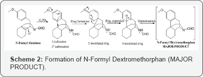
Now, again the electron rich arene will attack the carbocation to form 5-membered ring which is expanded to form fused 6 membered rings. This step can be explained that superelectrophiles undergo structural rearrangements that lead to favorable deprotonation step and this gives molecule with least or no positive charges. Thus, we achieve our major product and is formed because of most favorable mechanism. The mechanism adopted by superelectrophiles concluded that deprotonation- reprotonation is one of the most common path by which charge migrates in superelectrophiles. Rapid deprotonation in the strongest superacids is because of these reactive intermediates exhibiting extreme level of carbon acidity. Phosphoric acid (85%) in toluene is distilled using Dean- Stark apparatus to remove excess water azeotropically. The obtained phosphoric acid is approximately 97% and then phosphorus pentoxide is added under nitrogen atmosphere to obtain 100% phosphoric acid. Further, a solution of N-Formyl Octa base in toluene is added dropwise and the reaction is heated and stirred at 65-70oC for 12 hours to obtain the desired product (N-Formyl Dextromethorphan). The completion of reaction is analyzed by HPLC monitoring.
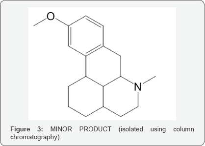
This N-Formyl Dextromethorphan is further insitu reduced to N-Nor Dextromethorphan using sodium hydroxide and methanol/ water mixture and then reacted with formic acid, formaldehyde and hydro bromic acid to form Dextromethorphan Hydro bromide (Scheme 1). During isolation of Dextromethorphan hydro bromide and LC-MS data, showed the formation of an unknown compound with same mass as Dextromethorphan but at different Retention time. The structure of this compound is analyzed and characterized using techniques like HPLC, 1H NMR, 13C NMR and DEPT (Figure 3).
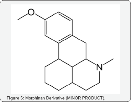
Considering all the insitu reaction stages and the possible by products, it is worthwhile to propose the formation of this compound from its precursor, originated in the step of Grewe cyclisation of N-Formyl Octabase although in minor quantity (Morphinan derivative) (Scheme 3).
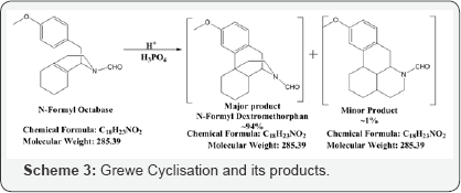
As all the 4 stages are carried forward in situ, and isolation is done only at the Dextromethorphan Hydro Bromide stage, the preparation of this minor product from its N-Formyl precursor takes place in the same way as N-Formyl Dextromethorphan to Dextromethorphan Hydrobromide. It has been in situ reduced to N-Nor using sodium hydroxide and methanol, and on treatment with with formaldehyde, formic acid gives Dextromethorphan Hydrobromide (~95%) along with this minor morphinan derivative (~1-2%) as per monographs of European Pharmacopoeia (EP) [6], United States Pharmacopoeia (USP) [7] and International Pharmacopoeia (IP) [8]. This minor product is isolated (at this stage) by column chromatography (MDC: MeOH :: 9:1) followed by Preparative HPLC (Scheme 4).
The formation of this intermediate can be explained with the possibility of formation of 1,3-dication over 1,4-dication (which is less probable). Another difference lies in the arene attack which attacks the carbocation from the comparitively less electron rich position and only reason to justify is the resulting separation of charges and formation of 1,5-dication resulting in 5-membered product which is expanded to form fused 6 membered rings with carbon migration, giving us the minor product (Scheme 5).
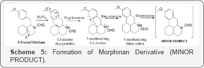
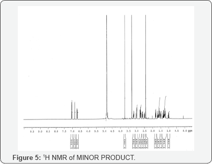
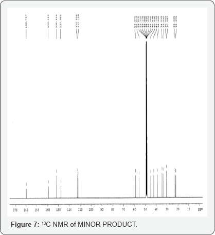
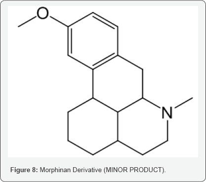
This isolated minor product is analyzed and characterized using Mass spectrometry, HPLC and NMR (1H and 13C NMR). DEPT- analysis showed the presence of 6 -CH2 groups instead of 7 -CH2 groups in the major product. All the characterization and analysis confirmed the formation and structure of the isolated minor product (Figures 4-9).

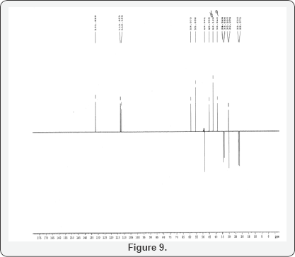
Dept of MINOR Product
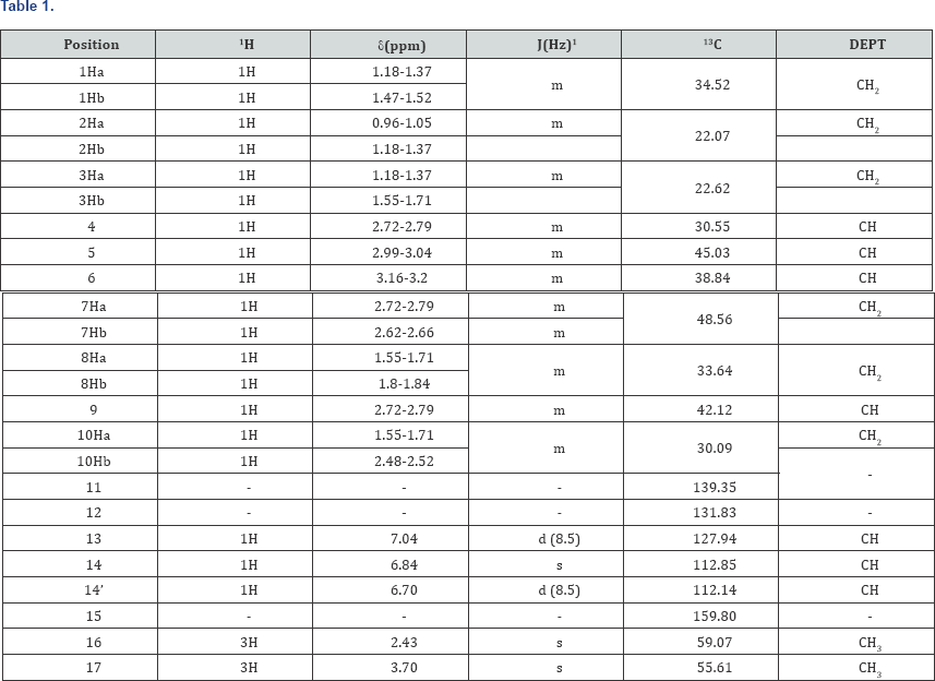
MINOR Product: Purity: 97%, Mass: 272 (m+1) (Figure 10) (Table 1).

s- singlet, d- doublet, m-multiplet, br-broad, 1H-1H coupling constants.

Experimental Section

Preparation of N-Formyl Dextromethorphan (MAJOR) and Morphinan Derivative (MINOR)

In a flask, charge Ortho phosphoric acid (~85.0 % w/w). Charge Toluene and Raise the temperature of the reaction mass. Reflux and maintain over Dean stark apparatus to remove water azeotropically. Cool the reaction mass under nitrogen atmosphere and Charge Phosphorus pentoxide under nitrogen atmosphere. Slowly add N-formyl octa base organic layer and Raise the temperature of the reaction mass under nitrogen atmosphere. Stir and maintain the reaction mass at 65-70oC under nitrogen atmosphere till reaction complies by reaction monitoring by HPLC after 10-12 hrs maintenance. Cool the reaction mass under nitrogen atmosphere. Concentrate the reaction mass under vacuum to remove toluene. To the concentrated mass charge ethyl acetate under nitrogen atmosphere and stir. In another flask, charge water, Cool. Charge ethyl acetate reaction mixture reaction mass in to chilled water. Stir, settle and separate the layers. Wash the organic layer with water again and then a wash of 7% sodium bicarbonate solution is given. Concentrate the organic layer u/v till almost no solvent distills.

Preparation and Isolation of MINOR Product

N-Formyl Minor product obtained in Grewe cyclisation, is treated with Sodium hydroxide in the mixture of methanol and water for 12 hours at 65-70oC to give N-Nor which is further treated with formaldehyde and formic acid in the presence of toluene for two hours at 60-65oC to give N-Methylated minor product which is further purified and isolated using Column chromatography and Preparative HPLC.

Acknowledgements

Our group would like to thank the Department of Scientific and Industrial Research India, Dr. Hari Babu (COO Mylan), Sanjeev Sethi (Chief Scientific Office Mylan Inc ) ; Dr Abhijit Deshmukh (Head of Global OSD Scientific Affairs); Dr Yasir Rawjee {Head - Global API (Active Pharmaceutical Ingredients)}, Dr Sureshbabu Jayachandra (Head of Chemical Research) Mr Manoj Pananchukunnath (Head of Global Injectables Scientific Affairs, Product Development) Dr. Suryanarayana Mulukutla (Head Analytical Dept MLL API R & D) as well as analytical development team of Mylan Laboratories Limited for their encouragement and support. We would also like to thank Dr Narahari Ambati (AGC- India IP) & his Intellectual property team for their support.

To Know More About Peer Reviewed Chemistry Journals Please Click on:
https://juniperpublishers.com/omcij/index.php

To Know More About Open Access Journals Publishers Please Click on: Juniper Publishers

Understanding the Relationship of Chronic Toxoplasma Gondii Infection and Schizophrenia_Juniper Publishers

Advances in Biotechnology & Microbiology Juniper Publishers


Authored by Taibur Rahman


Abstract

Toxoplasma gondii is a widely distributed neurotropic protozoan parasite that causes Toxoplasmosis in humans and animals. Approximately, 30-50% populations are infected by this parasite world-wide. After acute infection, T. gondii undergoes developmental switching from its highly replicating tachyzoite stage to slowly replicating dormant bradyzoite stage preferentially in brain and skeletal muscle. Therefore, brain and skeletal muscle may act as a reservoir of T. gondii for persistent infection. In immunocompromised individual and developing fetus, T. gondii bradyzoite can reactivate and thereby can cause severe neurological disease for instance schizophrenia. In this mini-review, we have linked the association of chronic T. gondii infection with schizophrenia. This study would help scientist to perform in-depth research on discovering the mechanism of T. gondii infection in schizophrenia.
Keywords: Toxoplasma gondii; Brain; Schizophrenia; Immunocompromised patients; Fetus

Introduction

Toxoplasma gondii is an important neurotropic parasite that can infect any warm-blooded animals including humans and causes Toxoplasmosis. The infection rate varies from 10-90 % world-wide depending on environmental or socioeconomic factors and geographic locations [1]. Due to its wide-spread nature and infection capacity, T. gondii has both medical and veterinary importance. After acute infection in human and warm-blooded livestock animals, T. gondii accomplishes its asexual stages life cycle through stage conversion of the parasite from fast replicating tachyzoite to slow replicating dormant bradyzoite particularly in brain and skeletal muscle [2]. This stage differentiation of the parasite brain and skeletal muscle permit them for establishing lifelong persistent infection. It has to be stressed that human can become infected primarily by ingesting undercooked or raw meats of infected livestock animals or eating contaminated foods and water [3].
European multicenter case control study showed that 30-63 % of acute infections of pregnant women are due to consumption of infected undercooked meats [4]. In immunocompetent host, primary infection is generally mild with flu-like symptoms whereas T. gondii infection can cause life threatening problems in immunocompromised patients for instance AIDS, transplant and cancer [5]. Furthermore, congenital toxoplasmosis can interfere brain development in fetus [6] which may provide significant impact on severe neurological damage or even death of the fetus. Neurological disorder like schizophrenia is present in ~1% people and ninth most common cause of disability over the world. The symptoms of the disease start from late teens to early adulthood, although the psychotic episodes can persist throughout the entire life of the patient. Till now, no single contributory driving force has been discovered; therefore, it is realistic only to explain some factors that were shown to be positively associated with schizophrenia. The well-known risk factors include: genetic predisposition, neurodevelopmental instability and environmental factors, including infectious agents [7,8]. In this study, we have deciphered the relationship of T. gondii infection and common neurological mental disorder schizophrenia.

Impact of T. gondii on Schizophrenia

Schizophrenia is a chronic and severe neurological disease that affect nearly one percent of adult population world-wide [9]. People with this disease suffer from hallucination (false perception), delusion (beliefs that conflict with reality), depression, apathy, poor social activities and lack of speech [10]. The exact etiological factor for Schizophrenia has not been identified yet but it is widely accepted that mostly genetic and environmental factors are prominent cause for pathogenesis of the disease [11]. Recent evidence suggests that infectious agents can act as a high-risk factor for onset of schizophrenia [12]. Previous studies have shown that infection with virus e. g influenza, herpex simplex, rubella, polio and varicella zoster may contribute the development of schizophrenia [13]. Recently, T. gondii, an important neurotrophic protozoan parasite, has been identified as an important risk factor for developing symptoms of schizophrenia [14]. Thereafter a number studies has investigated the association of T. gondii, and schizophrenia. Torry et al. [15] has summarized 38 studies of T. gondii, and schizophrenia and confirmed the increased prevalence of anti-T. gondii, antibodies in schizophrenia patients (odd ratio 2.73, 95% CI). For instance, Hamidinejat and colleagues has shown that 57 % patients with schizophrenia carry anti-T. gondii, IgG antibodies compared to healthy controls [16]. Another study conducted by Mortensen and colleagues suggest that early infection of T. gondii, in newborn may develop schizophrenia in later stage of life [17]. Contrary, some other studies has failed to reveal the association of T. gondii, infection with schizophrenia [18-20]. Although the role of T. gondii,infection in developing schizophrenia is controversial in very few studies, the above majority studies nevertheless, confirm the positive correlation of T. gondii, infection and development of psychiatric disease like schizophrenia. However, the mechanism of how the parasite develops the disease is remaining unknown.

Conclusion

Despite having few controversial findings, it can be said that there is strong correlation of T. gondii, infection and schizophrenia. The correlation between T. gondii, and schizophrenia might be explained in the way as T. gondii, develop bradyzoite tissue cysts and persist in brain of infected patient’s life time, thereby the parasite may somehow modulate neurological parameter which may directly or indirectly involved in that process. Further research is needed to find out the exact factors that determine the relationship of T. gondii,infection in schizophrenic patients.

Acknowledgment

We want to thank ministry of education, Bangladesh and department of Biochemistry and Molecular Biology, University of Dhaka for her cooperation.


To Know More About Advances in Biotechnology & Microbiology Please Click on:
https://juniperpublishers.com/aibm/index.php

To Know More About Open Access Journals Publishers Please Click on: Juniper Publishers

Monday, August 19, 2019

Helicobacter Pylori Strains Isolated from Iraqi Subjects with Gastritis, Peptic Ulcer, and Carcinoma of Stomach Genetic Sequences: A New Variants

GastroenterologyOpen Access Journals Juniper Publishers


Authored by Abdulghani Mohamed Alsamarai

Abstract

Background: Genetic variations of the infectious agents play an important role on the influence of natural disease severity, response to treatment and morbidity.
Aim: To clarify if there are genetic variations in H. pylori clinical isolates from cases with gastritis, peptic ulcer and carcinoma of stomach.
Materials and Methods:Eight PCR product sample were sent for sequence analysis of H.pylori isolated from biopsy of stomach and 20μl (10pmol) from the Forward primer sequence is 5′-GGTTCAAATCGGCTCACACT-3′ Reverse primer sequence is 5′-CTCCTTAATTGTTTTTACATAG-3′ They are made by primer 3PLUS software. The result of the sequence analysis was analyzed by blastn in the National Centre for Biotechnology Information (NCBI) and many software used to detect polymorphism in H. pylori.
Results: The present study H.pylori isolates were separated on the phylogenetic tree and belong to a population cluster different from previously reported H.pylori sequences. H.pylori variant 1 which was isolated from Iraqi patients with gastric cancer was most closely to strain Helicobacter pylori R037c isolated in Canada. While variants 2 and 3, which were isolated from peptic ulcer and variant 4, which was isolated from patient with gastritis were more closely to Helicobacter pylori 26695 isolated from patient with gastritis in UK. The present study data suggest that sub lineages evolution within the Helicobacter pylori species may be associated with the development of less or more pathogenic potential. In addition, the relation of the 4 variants to the American [Hp R037c] and European [Hp 26695] suggest that these variants diverged from American and European lineage ancestors as reported previously for other geographical areas. This study is the first one in Iraq that sequenced H.pylori isolates from patients with gastritis, peptic ulcer and gastric cancer and the gene diversity of the bacteria may be used as biomarker that may be used for follow up and prognosis in the above disease for prevention of development of cancer. Therefore, called GhaFa.1. Iraq for first isolate, GhaFa.2. Iraq for second isolate, GhaFa.3. Iraq for third isolate and GhaFa.4. Iraq for fourth isolate.
Conclusion: This study indicated that genetic variation in H. pylori was correlated to gastritis severity and was more in gastric cancer cases as compared to gastritis and peptic ulcer.
Keywords: H. pylori; Gastritis; Peptic ulcer; Carcinoma of stomach; Genetic variation; Sequencing
Abbrevations: NCBI: National Center for Biotechnology Information; PCR: Polymerase Chain Reaction; SNP: Single Nucleotide Polymorphism

Introduction

H. pylori infections are worldwide and affected many systems beyond the stomach [1]. However, the disease prevalence varied between different studies [2]. The infection was with a range of 28.3% to 100% in general population and it was more in subjects with gastritis and gastric carcinoma [2]. H. pylori infections was linked with gastric carcinoma [3], however, not all H. pylori infected individuals will develop carcinoma of the stomach. Thus, development of gastric carcinoma and its association with H. pylori infections may interfere with risk factors which lead to potentiate cancer in certain subjects and not the others [3,4]. Previous studies reported abnormality in biomarkers in individuals with gastritis, peptic ulcer and gastric carcinoma, and the abnormality was more prominent in subject with cancer of stomach [5,6].
Genetic variations of the infectious agents play an important role on the influence of natural disease severity, response to treatment and morbidity [7,8]. Thus, this study was conducted to clarify if there are genetic variations in H. pylori clinical isolates from cases with gastritis, peptic ulcer and carcinoma of stomach.

Materials and Methods

Eight PCR product sample were sent for sequence analysis of H.pylori isolated from biopsy of stomach and 20μl (10pmol) from the Forward primer sequence is 5′-GGTTCAAATCGGCTCACACT-3′ Reverse primer sequence is 5′-CTCCTTAATTGTTTTTACATAG-3′ they are made by primer 3PLUS software. The result of the sequence analysis was analyzed by blastn in the National Centre for Biotechnology Information (NCBI). Online at (http:// www.ncbi.nlm.nil.gov) and many software used to detect polymorphism in H. pylori.

Results and Discussion

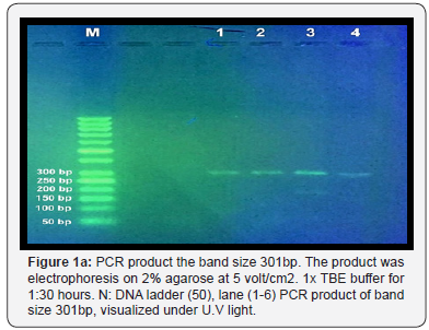
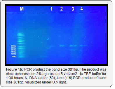
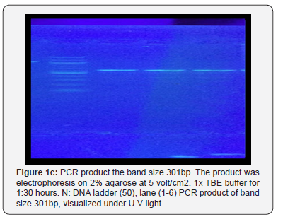
The result shown in Figure 1 A-C indicated that a yield of single band of the desired product with a molecular weight of 301bp for urease gene of H. pylori was obtained from 8 samples. Eight PCR product sample were sent for sequence analysis of H.pylori isolated from biopsy of stomach and 20μl (10pmol) from the Forward primer sequence is 5′-GGTTCAAATCGGCTCACACT-3′ Reverse primer sequence is 5′-CTCCTTAATTGTTTTTACATAG-3′
They are made by primer 3PLUS software. The result of the sequence analysis was analyzed by blastn in the National Centre for Biotechnology Information (NCBI). Online at (http:// www.ncbi.nlm.nil.gov) and many software used to detect polymorphism in H. pylori found variation include Transversion (refers to the substitution of a (two ring) purine or a (one ring) pyrimidine and transition a (point mutation) that changes a purine to another purine or pyrimidine to another pyrimidine
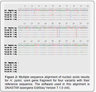
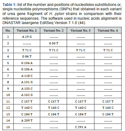
The polymorphisms of Helicobacter urease subunit alpha gene were detected by PCR / direct DNA sequencing method. Undoubtedly, too small sized of the studied groups is still a shortcoming of our research. Nonetheless, the observed sequencing variations nucleic acids indicate the detection of four different genetic variations with variable number of single nucleotide polymorphism [SNPs] (Figure 2). Sequencing results observed that variant No. 1 possesses very high SNPs variability compared with the reference as well as other variants nucleic acid sequences. The pattern and nature of each SNP that detected by sequencing may indicate that these SNPs are novel in their positions and consequences. The variant No. 1 has 13 SNPs (A 19 G, T 71 C, G 86 T, G 104 A, G 106 A, A 118 C, A 131 G, A 133 C, A 152 G, C 157 T, T 160 C, C 184 T, and A 209 T), and only four (T 71 C, C 157 T, T 160 C, and C 184 T) are in common with other three variants (Table 1). The variant No. 2 has 5 SNPs (G 50 T, T 71 C, C 157 T, T 160 C, and C 184 T), and four (T 71 C, C 157 T, T 160 C, and C 184 T) of them are in common with other three variants. The variant No. 3 has 5 SNPs (T 71 C, C 157 T, T 160 C, C 184 T, and C 291A), and four (T 71 C, C 157 T, T 160 C, and C 184 T) of them are in common with other three variants. The variant No. 4 has 4 SNPs (T 71 C, C 157 T, T 160 C, and C 184 T), and all of them are in common with other three variants, and no one unique SNP was identified.
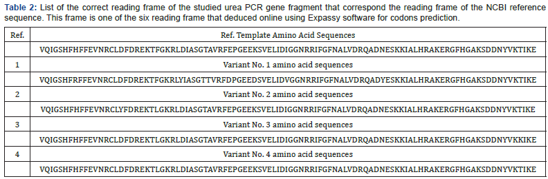
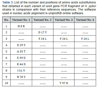
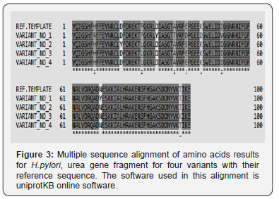
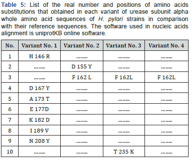
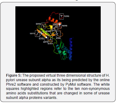
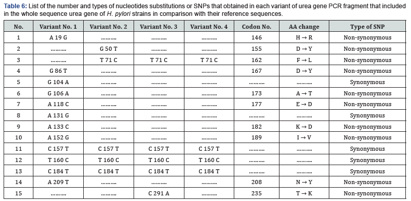
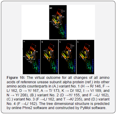
Since multiple reading frames can be extracted from each amino acid sequence, special software were used to predict the correct reading frame that suits the original reference reading frame in urease subunit alpha (Table 2). Once these suitable open reading frames were constructed, they compared with each other using uniprotKB software to see the final effect of the observed SNPs on the primary structure of proteins fragments Figure 3. Moreover, other data that found the high variability of urea gene isn’t only stated in the levels of DNA, but it extends into the proteins’ level too. According to Figure 3, it was found that variant No. 1 has seven amino acid changes (H 8 R, D 29 Y, A 35 T, E 39 D, K 44 D, I 51 V, and N 70 Y), and all of them aren’t in common with the other three variants (Table 3). Variant No. 2 has two amino acids changes (D 17 Y, and F 24 L), and only one of them (F 24 L) is in common with the other three variants. Variant No. 3 has two amino acids changes (F 24 L, and T 97 K), and only one of them (F 24 L) is in common with the other three variants. Whereas variant No. 4 has only one amino acid change (F 24 L), which is in common with the other three variants. According to previously mentioned data, it was found that the variant No. 1 has distinct and significant variations in comparison and the reference, as well as other three amino acids sequences of the other three variants Figure 4. However, to get a complete idea about the extent and nature of these amino acid’s changes, the whole sequence of urease subunit alpha was retrieved, and the accurate position of PCR products were localized. Table 4. Hence, the real positions of these changes were relocalized Table 5 and introduced in the whole sequence of this protein. Accordingly, only ten amino acid changes were represented in all of these four observed variants Table 6. Seven of non-synonymous mutations were represented in variant No. 1 (H 146 R, D 167 Y, A 173 T, E 177 D, K 182 D, I 189 V, and N 208 Y).
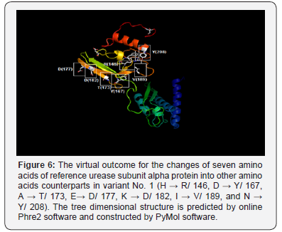
The non-synonym mutations that exist in variant No. 1 was shown seven amino acids changes. The conversions of histidine into argenine, lysine into aspartate, glutamate into aspartate, all polar charged amino acids, and isoleucine into valine, both hydrophobic aliphatic amino acids (in the positions 146, 177, 182, and 189 respectively), may not have a drastic effect on the protein structure since all of these converted amino acids residues have the same functional units Figure 5 & 6.
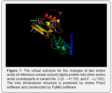
Whereas, the conversion of aspartic acid, the polar charged amino acid into tyrosine, the hydrophobic aromatic amino acid, alanine, the hydrophobic aliphatic amino acid into threonine, the polar uncharged amino acid, and asparagine, the polar uncharged amino acid, into tyrosine, the hydrophobic aromatic amino acid (in positions 167, 173, and 208 respectively) have potential drastic effect of on the final manifestation of the protein structure since all of these converted amino acids residues have different functional units Figure 6. The non-synonym mutations that exist in variant No. 2 was shown two amino acids changes. The conversions of aspartate, the polar charged amino acid into tyrosine, the hydrophobic aromatic amino acid, and the conversion of phenylalanine, the hydrophobic aromatic amino acid, into leucine, the hydrophobic aliphatic amino acid (in positions 155 and 162) may have dramatic effect on protein three-dimensional structure and function Figure 7..
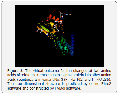
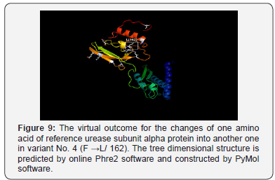
As well, the non-synonym mutations that exist in variant No. 3 was shown two amino acids changes. In addition to phenylalanine / leucine conversion that observed in variant No. 2, another conversion was observed from threonine, the polar uncharged amino acid into lysine, the polar charged amino acid (in position 235) These two changes may have dramatic effect on the three-dimensional structure too Figure 8. In variant No. 4, only one amino acid conversion was observed, which has been observed in both variant No. 2 and variant No. 3. Figure 9. It was obviously found in this study that variant No. 1 is genetically distinct from all other three observed variants as well as the reference sequences Figure 10. Thus, the variant No. 1 is highly polymorphic and may be regarded as a mutational hotspot, or the high variable site, leading to ligand diversity. The high genetic dissimilarity that we observed in this studied variant in comparison with its reference breed sequences might be due to several factors, such as the type, location, or other factors since the nature of polymorphisms were potentially highly dependable on several factors in the determination of its final genetic polymorphism.
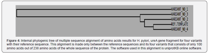
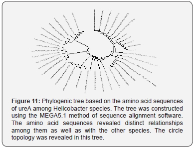
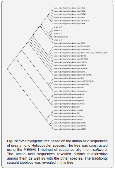
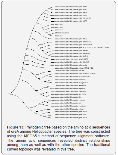
In addition to the closeness of the amino acid’s sequences of variants No. 2, 3, and 4, the phylogenetic tree that we construct has shown that the variant No. 1 has the most far genetic distance compared with the other observed three variants of this study as it was presented through several topological frames Figure 11- 13.
However, the observed variants of the current study are provided a beneficial tool for the geneticists and microbiologists to identify the unknown SNPs that may be useful for establishing a possible association with the genealogical relationships among these variations. The present study indicated that H.pylori isolated from patients with gastric cancer [variant 1] was highly polymorphic and was genetically distinct from H.pylori isolated from peptic ulcer [variants 2 and 3], and from patients with gastritis [variant 4]. These genetic diversity in our isolates was in line of previous studies performed in other geographical areas [8]. In addition, the variability was not confined to DNA only, but it extend to involve proteins and these variability may attribute to evolution of new H.pylori strains with high ability for gastric cancer induction. Cover [8], reported that there was presence of certain proteins in H.pylori with higher gastric cancer risk, which were absent in isolates with low gastric cancer risk. Thus genetic diversity and protein variability in H.pyloriisolates may influence the incidence of gastric cancer and development of gastrointestinal diseases.
The present study H.pylori isolates were separated on the phylogenetic tree and belong to a population cluster different from previously reported H.pylori sequences [9-12]. H.pylori variant 1 which was isolated from Iraqi patients with gastric cancer was most closely to strain Helicobacter pylori R037c isolated in Canada [11]. While variants 2 and 3, which were isolated from peptic ulcer and variant 4, which was isolated from patient with gastritis were more closely to Helicobacter pylori 26695 isolated from patient with gastritis in UK [9]. The present study data suggest that sub lineages evolution within the Helicobacter pylori species may be associated with the development of less or more pathogenic potential. In addition, the relation of the 4 variants to the American [Hp R037c] and European [Hp 26695] suggest that these variants diverged from American and European lineage ancestors as reported previously for other geographical areas [11,13].
This study is the first one in Iraq that sequenced H.pylori isolates from patients with gastritis, peptic ulcer and gastric cancer and the gene diversity of the bacteria may be used as biomarker that may be used for follow up and prognosis in the above disease for prevention of development of cancer. Therefore, called GhaFa.1. Iraq for first isolate, GhaFa.2. Iraq for second isolate, GhaFa.3. Iraq for third isolate and GhaFa.4. Iraq for fourth isolate.

Conclusion

This study indicated that genetic variation in H. pylori was correlated to gastritis severity and was more in gastric cancer cases as compared to gastritis and peptic ulcer. This finding suggest that organism gene diversity influence the outcome and prognosis of H. pylori infections.

Ethics Approval and Consent to Participate

The research protocol was approved by Tikrit University College of Science Ethical Committee and informed consent was taken from each participant before their enrolment in the study.

Consent for Publication

All the authors were agreed for manuscript publication

Authors Contributions

All the authors were contributed in the study design, samples and data collection, investigational work, data analysis, and manuscript writing.

To Know More About Gastroenterology Open Access Journals Please Click on:
https://juniperpublishers.com/argh/index.php

To Know More About Open Access Journals Publishers Please Click on: Juniper Publishers

Anthropology, Participatory Research and Development Cooperation: A Necessary Dialogue- Juniper Publishers
  Archaeology & Anthropology- Juniper Publishers Abstract Le domande sul “perché e per chi” if it generates conoscenza [1] sono da tempo...