Monday, December 19, 2022

Considerations for Designing and Implementing Phase II Oncology Clinical Trials - Juniper Publishers

Biostatistics and Biometrics - Juniper Publishers


Abstract

Oncology Phase II trials’ main objective is to collect preliminary efficacy data of an oncology treatment, that will be used to inform and decide whether to further investigate the treatment. Choosing a proper design is an important but not easy task in the treatment evaluation/FDA approval process. The objectives of this short article are to briefly summarize the different type of phase II clinical trial designs, including the traditional designs and the more recently developed adaptive designs; discuss the practical considerations in the selection of proper designs; provide recommendations for the implementation, especially some aspects to be considered for the adaptive designs.

Keywords: Oncology phase II clinical trials; Single arm designs; Randomized trials; Phase II screening trials; Adaptive designs

Introduction

Oncology clinical trials designed and implemented to test new treatments involve a series of phases. During the early phases (Phase I and II), researchers assess the safety, potential side effects, and the best dose of the new treatment. Specifically, Phase II clinical trials are designed to identify promising experimental treatments that can be studied and tested in the later phase (Phase III). Therefore, Phase II trials usually have small number of patients and homogeneous cohorts in terms of patient characteristics. These studies have relatively large Type I error (e.g., 0.10) and moderate power (e.g. 80%) compared to the confirmatory Phase III trials. In the later phase trials, researchers will then study whether the new/proposed treatment is superior to the current standard therapy using much larger sample sizes and more heterogeneous patient groups. Phase II single arm trials that used historical controls were predominantly designed and implemented in the last few decades. But in 2010, the National Cancer Institute’s (NCI) published guidelines for the design of Phase II clinical trials. These guidelines recommended and encouraged to use randomized Phase II trials instead of single arm trials. A barrier for the oncology trials is that number of available patients is typically very limited, especially for certain rare disease types. Therefore, Phase II-single arm trials with historical controls are still widely used [1]. So, the questions that researchers face when designing a Phase II clinical trial are: how to choose a proper design? A single arm with historical control or a randomized trial, traditional design or adaptive design? In addition, the statistical design of a trial should consider all the statisticians’ and physicians’ considerations:

a) disease type/patient population

b) primary endpoint(s)

c) available preliminary data of effect size

d) desired power and Type I error

e) adjustments for interim analysis

In this article, we review Phase II designs, both traditional and a few advanced adaptive designs for oncology trials. We also discuss key factors and practical considerations about designing Phase II trials based on our experience in an academic medical center research environment.

Types of Phase II Designs

Phase II oncology clinical trials designs can be divided into two categories: single arm trials and randomized trials. Single arm trials only include the experimental arm and the comparison of the primary endpoint(s) to a historical control. For single arm trials, the traditional statistical designs used are the Simon’s two stage design [2] and the Fleming one stage design [3]. Both designs use binary primary endpoints. For example, overall response rate and progression free survival (PFS) at a certain time are the most commonly used. The Simon’s two-stage design could either use optimal or minimax approaches. Minimax designs minimize the total number of sample size (patients) while optimal designs minimize the expected sample size. Under Simon’s two stage designs, the trial will be stopped at the end of the first stage only if the treatment appears ineffective (lack of efficacy). Otherwise, the study will continue. Simon’s two stage designs require a full stop of the trial to evaluate the futility boundary before starting enrollment of patients into the second stage. Therefore, if the primary endpoint requires longer evaluation period (e.g. PFS rate at 1 year), one-stage designs or the other alternative designs are better options as they will result in shorter study periods.

The availability of patient data based on evolving sequencing clustering/omics methods and progress in imaging technologies make comparisons against historical controls inaccurate. Therefore, randomized trials with inclusion of an “active” control group are preferred to avoid a high risk of “false positives”. Randomized trials have been extensively reviewed in the literature [4-6]. Sharma et al. [7] have discussed the evidence in favor of randomized Phase II trials and barriers to widespread the use of these designs [7]. Randomized trials protect against selection bias that may yield results that are more promising. Screening randomized trials [8] screen-in potentially effective drugs instead of screening out very ineffective drugs. For this situation, the false positive rate (Type I error) could be set up at a higher level for these types of trials. More advanced adaptive designs were developed in the last decades. Adaptive designs may increase the probability of success, shorten the decision timelines, and deliver the right treatment to the right patient. For single arm trials, Predicative Probability designs [9], which are based on Bayesian predictive probability, allow for continuing assessing futility and efficacy during the trial. Bayesian Optimum Predictive designs (BOP2) [10] can be used for simple or more complicated endpoints with a Dirichlet-multinomial model. For example, it could be toxicity and response, or two types of response (e.g., response rate, PFS rate, or surrogate biomarker response). Moreover, interim analyses can be set at a specific number of patients. These designs can have more than one interim analysis for futility. For randomized trials, the most commonly used adaptive design in Phase II/III studies [11], is the Group Sequential Design (GSD) [12-14]. GSD allows for premature termination of a trial, due to efficacy or futility, based on the results of interim analyses. GSD has fix randomization and total sample size, but can be stopped early for efficacy, or futility, or both. Early stopping ensures that a new drug can be investigated and approved sooner if it is beneficial, or it avoids wasting resources if a negative result is observed. Usually Lan and DeMets version of the O’Brien-Fleming spending function will be utilized for futility stopping boundaries [15]. Other different spending functions, like Alpha, Gamma, Rho spending functions could also be used [16]. More recently, Master protocol trials [17] could be the next-generation Phase II clinical trial design and FDA developed draft guidelines (September 2018). Master protocol trials include Basket trials, Umbrella trials, and Platform trials. Basket trials test the effect of one treatment on multiple diseases or multiple disease subtypes. Umbrella trials have many different treatment arms for at least one disease. Platform trials have several treatments for one disease type perpetually, and further accept additions or exclusions of new treatments during the trial. Master protocol trials can use either traditional frequentist designs or Bayesian adaptive designs. For example, Simon’s two-stage design can be used for each parallel sub-study (disease type or sub-disease type) in a basket trial; Bayesian response-adaptive randomization may also be used in Umbrella trials.

Primary Endpoint

Usually, the traditional primary endpoints for Phase II trials are response rate, progression free survival (PFS) rate at a certain time point, or time-to-event PFS. Overall survival (OS) usually is the primary endpoint for the large confirmatory Phase III trials. However, OS could also be a specific required primary endpoint for a certain disease in Phase II trials. Biomarkers (e.g., a classifier, prognostic, or predictive marker) also could be selected as the primary endpoints for Phase II trials. As we know, the number of patients (sample size) required to enroll into a Phase II trial depends on the targeted effect size (difference) of the primary endpoint. It is critical to correctly choose the primary endpoint(s) to have robust and successful trials. There are several considerations when choosing primary endpoints:

a. Is there any disease specific/standard primary endpoint? As we mentioned earlier, some diseases have specific primary endpoint requirements. For example, OS is the standard primary outcome for determining Glioblastoma treatment efficacy.

b. How long is the follow-up time? Usually, evaluation of response rate and surrogate markers could be relative quick while time-to event PFS/OS could have longer follow-ups.

c. Will the study evaluate response rate or PFS? If the treatment does not improve response but can have more stable disease (SD) and maintain the SD for a longer time, then PFS is preferred over response rate.

d. Have the surrogate biomarkers been verified yet? Biomarkers must be validated before they can be used as primary endpoints. Otherwise, it could lead to a dramatic inflation of the Type I error rates.

Power and Type I Error

Early phase clinical trials serve as the gatekeeper to rule out the inefficacious treatments as fast as possible. Therefore, Phase II trials have small sample size with slightly larger Type I error rate (false positive rate) and moderate power (e.g. 80%). For single arm trials, it is common to use 90% power and 10% Type I error rate, or 80% power and 5% Type I error rate. For randomized trials, 80% power is commonly used. Screening randomized trials could have Type I error rates as large as 20%.

Preliminary Data

The preliminary data on primary endpoints, for example the historical control and targeted response rate, are one of the key information used in Phase II designs to determine the effect size. Information on clinically relevant effect sizes is likely provided by the physician or principal investigator (PI) based on current clinical practice. Effect sizes can be also estimated from the literature on similar drugs, preliminary data from early phase trials on the study drug(s), large historical data banks from patients previously treated on similar trials. While multiple statistical approaches have been discussed, the design of Phase II trials should always be planned with the clinician’s input, having a comprehensive understanding and interpretation of the current literature, as well as based on preliminary data. A systematic review of these sources can provide more reliable data to inform the design of the trial and conduct comprehensive simulations. A close collaboration between the clinician and the biostatistician could provide unique opportunity to conduct literature review before planning and designing trials. A systematic literature review could be time consuming. Some bioinformatics tools, like text mining, could be used to efficiently collect preliminary information from existing large databases.

Implementation

Phase II trials, especially adaptive designs, require close collaborations among all staff in the study team. The study team includes the physician/PI, biostatistician, data manager/coordinator, research nurses and IT support. The communication among all staff is key to have a robust design and implementation of the trial. While many practical considerations are like traditional designs and advanced adaptive designs, additional considerations may be required for particular types of adaptive designs [18]. First, these more advanced designs require additional planning time to select the proper endpoint(s) and obtaining preliminary data. Adaptive designs are usually more complicated than the traditional designs. Simulations are needed to help interpreting and understanding the operation characteristics curve of the adaptive design. Second, prior to the trial initiation, all team members need training on the logistical aspects of the trial, and the team should know the communication matrix during the trial. The training is essential to minimize errors and ensure the successful implementation of the trial. Third, interim analyses or adaptive procedures, which should be clearly stated in the protocol, need to be conducted timely at the designated time. This requires consistent data monitoring and quality checking throughout the trial duration, and the statistician needs to be readily prepared to work on all the analyses. These additional efforts should be considered in the study budget and the planning process. Prior to obtaining funding to run the trial, institutional support should be provided to support investigators team on the pre-planning process. Each adaptive trial design could be implemented slightly differently depending on the available resources/infrastructure of each institution.

Conclusion

As we have discussed, several aspects need to be considered to choose a proper statistical design: single arm versus randomized trial, traditional design versus adaptive design. The background of the disease type and patient population as well as the preliminary data are all critical elements for Phase II clinical trial designs. While more advanced adaptive designs have been proposed to improve the operation characteristics over the traditional designs, PIs should be provided with the opportunity to learn about these designs and build trust with the biostatistician and the rest of the research team on the implementation process. Institutional support and infrastructure are the most critical aspects for the successful designing and implementation of investigator initiated clinical trials in academic medical centers.

Click here: https://juniperpublishers.com/index.php 

Thursday, December 15, 2022

Standing on A Beach / Staring at The Sea: How the Sleeve Art Made for The Singles of The Band the Cure - Juniper Publishers

 Archaeology & Anthropology - Juniper Publishers

Abstract

Graphic design as a visual design discipline provides pre-print solutions by organizing the main two elements: type and image. Every graphic design presents a product or a service, and an album cover as a graphic design product presents the music and the musician besides protecting the recorded medium. Music as an art form based on sound and silence has been recording since the end of the 19th century for collecting and distributing the music to the masses. The primitive sleeves created for titling and protection turned into tools of labeling, offering and presenting as form they got and the materials they have been made of. Among its similar the 1986 released compilation album of the English rock band The Cure, Standing on a Beach / Staring at the Sea certificated by five times with platinum and six times with gold and critically acclaimed as the one of the finest albums of the „80s.The aim of this paper is to put forth the making of the sleeve art of the singles of the band The Cure with its unique attitude, which also shows the tight bonds among various graphic design elements like line, color, tone, light and shades and music. Does the sea as black and white in the background, the Figure old and thorn and the lettering in far behind have a relation of the music the band The Cure is made?

Keywords: Graphic design; Sleeve art; The Cure; Sea; Design elements

Introduction

Music as an artistic expression and cultural outcome [1] has been packaged with different kind of sleeves since it has been recorded for re-listening. Initially these recordings made for professional purposes only like record studies, collecting and archiving. On primitive sleeves it is fair enough to have the written information only like the date, place and names of the performance and the performers. But when it comes to the times of mass production and selling the music, the circumstances have changed. From that time on there is a new component: the buyer, someone who is both a listener and a collector, a fond of music so a fan of the performer / performers. As a conclusion the primitive forms of sleeves created for titling and protection only turned into tools of labeling, presenting and advertising surfaces for graphic design to organize. Graphic design as a two-dimensional design discipline organizes type and images according to the product’s needs, usage and functions [2]. Graphic design serves to sectors like food industry, every kind of media, pharmaceutical industry, fashion and many others with its own elements shape these types and images under the guidance of its principles. In this paper, music industry has been chosen as the sector graphic design serves. Among its contemporaries the 1986 released compilation album of the English rock band The Cure, standing on a Beach / Staring at the Sea chosen to examine [3]. Because it has been certificated by five times with platinum, six times with gold and critically acclaimed as the one of the finest albums of the „80s.

The sleeve art of the album with its visual elements organized according to visual principles of graphic design accepted as indicators of the auditory experience the album promised to. So, the aim of this paper is to put forth the making of the sleeve art of the singles of the band The Cure with its unique attitude among various visual elements like line, color, tone, light and shades; the visual principles like balance, hierarchy, unity / harmony, dominance / emphasis, similarity and contrast and the band The Cure’s music especially in this singles album [4] (Figure 1).

The Study

The study has begun with the examination of the album’s musical content; has continued with the visual examination of the sleeve art; resulted with a comparison of the two and concluded by finding out the similarities and contrasts. If the similarities are more than the contrasts, it can be said that the sleeve art is consistent as physical form and content. If the contrasts are more than the similarities, it can be said that the sleeve art isn’t consistent as physical form and content, or it has another aim or meaning. Besides being a musical album, standing on a Beach / Staring at the Sea album labeled as genre post-punk, new wave and gothic rock [5] Post-punk as a style inspired by punk’s energy, but left the rock clichés behind. Drew influences from art rock’s tendency towards sonic experimentation, linking radical content to radical form; the lyrical surreal imagery and soundscape are used to evoke and signify one mediated phantasy [6]. Transformed music into an epic of timelessness [7] combined with electronic styles like dub, funk and disco accepted as black, post-punk is under the effect of art and politics. On the other hand, the second one, new wave was more complex as music and lyrics also different than blues and rock & roll; but under the influence of many other styles. And finally the third one, gothic rock with its darker sound than the post-punk and new wave with minor or bass chords, reverbs, dark arrangements or dramatic and melancholic melodies in a combination with themes like sadness, nihilism, dark romanticism, tragedy and melancholy [8-10].

The thirteen songs combined for this album are Killing an Arab, Boys Don’t Cry, Jumping Someone Else’s Train, A Forest, Primary, Charlotte Sometimes, The Hanging Garden, Let’s Go to Bed, The Walk, The Love Cats, The Caterpillar, In Between Days and Close to Me. While emotional sensitivity is obvious on some of the titles of the songs, the same emotional sensitivity easily can be seen when reading / listening the lyrics of the rest. As a single compilation album it marks the first decade of the band The Cure from increasingly dark and tormented music to dominant, melodic bass lines, whiny, strangulated vocals, and a lyric obsession with existential and almost literary despair: Gothic new wave [11]. Songs based on the limited number of the musical instruments, which are six-strings bass, acoustic guitar, Smith‟s voice and the string sound of Solina – the synthesizer [12- 16]. Taylor in his 2006 dated study titled The A to X of Alternative Music defined The Cure music as initially began alongside postpunk bands that were driven by punk spirit, but soon withdrew into themselves and developed a stillness which begat a sense of epic power and serve them well for three albums, conforming them as leaders of the goth scene. Unwilling to be typecast, Smith explored the possibilities of pop music and over the course of another four albums reconciled the frivolous and bleak aspects of their music in a way which might have inspired Pixies amongst others. On the front cover of the album in left hand side of the Figure 2, a black and white realist photograph seen. It is visually clear and plain; the viewer can easily perceive what captured. An old man face seen in a beach, which is almost empty in front of an open sky. He is looking directly in the eye of the viewer closely. He is old, thorn and melancholic in his eyes. His face with the wrinkles and grey striped hair creates a textural contrast with the plain black jacked of him and the sky but creates a textural harmony with the grains of the sand and the rocks. Besides its lack of colors, the photograph‟s dominant tone is light grey in a combination with pitch black, which increase the emptiness effect creates the feeling of loneliness and melancholy. While the horizon on the photograph placed a little above of the center point of the cover, the head of the figure divide the surface into two equal halves vertically. But the eyes are centered horizontally for a better eye contact, which makes the photograph much more realistic and effective. As seen on the compositional examination on right hand side in Figure 2, there is a grid system consists of equal units both horizontally and vertically. Because there is a diagonal organization an asymmetrical balance established to create inequality concluded in tension.

Two amorphous rocks easily can be seen on soft plain light grey sand with their black grounds and white tops. These two rocks have two functions: First they are leading the eye on and send it to the back to the horizon, then to the thin grey line and finally above to the tiny typographic elements, which are the title of the album and the name of the band. The tiny sizes of the letters both suites the thin lines and textures all over the photograph. The typography organized as a one line sentence in same size, type and manner suits both the horizon, the rocks, the wrinkled forehead, the eye & the eyebrows and closed thin lips. So all the visual elements mentioned suit each other as structure and distributed all over the surface not equally but similarly. On Figure 3 the back of the cover seen as a lighter real life photograph without any figures but another interestingly amorphous rock. With its low saturated coloring, emptiness, amorphous rock, grained textures, semi-diagonal composition and tiny lettering this back cover suits the front cover very well, so with the content very well. Approximately same leveled horizon in a combination with the semi-diagonal seaside matches the front cover. The giant amorphous rock / s create / s a heavy weight balanced with the dark stripes of the barcode and tiny lettering of song list centered vertically. Tiny lettering’s second function is to lead the eye to the endless horizon to nourish the emptiness effect. Inside the booklet there are three different approaches seen both suits the same design attitude with some exceptions inside the general loop like low saturated tones, diagonal compositions and texture the most dominant element: On Figure 4 the details of the songs placed as tiny and as white as they can be on a dark grey submarine. Very darker but diagonal composition created by degrading tones and textures; very lighter but centered and equally distributed list, getting wider at the bottom like the lighten tones of the sands, and finally another type of information than being the A & B side songs signed the bottom of the surface. The second approach can be seen on Figure 5 as a black and white submarine photograph. On a flat darker surface fishes perceived with their reduced tones equal in size but unequally distributed in an open composition diagonally, which contrasting and dynamic. Although the photograph consists of three tones and a plain ground the attributes mentioned prevent the photograph to become dull. The third approach can be seen on Figure 6 as the middle two pages of the booklet in form of another sandy photograph. After many suspicions about the previous photographs whether they are black & white or very low saturated now it is very obvious. A semisaturated realistic photograph placed with the covers of all the single albums of the band in this compilation. Like the sand waves distributed unevenly the covers placed like creating two uneven loops. With the black leaves surrounding from three different directions the covers mainly create a diagonal composition, which keeps the dynamism and tension in high levels.

The semi-saturated coloring as a definition isn’t enough to explain the interesting colour distribution seen. A special fluorescent illuminating effect circulates all over the sand but the covers. The fluorescent pink, purple, yellow and blue touches of colours on mainly black and white single covers are contrasting the interesting tones of the sand cooling up, warming down and degrading horizontally wave by wave. On the Figure 7 the two CDs designs seen under the titles of A and B. Because of the high amount of the lettering a plain grey background has been chosen. To let the design plain and clear white are the letters, and same as fonts, but different in size. A hierarchy established among the information and this hierarchy leads the sizes and the placements of the words. Like in every other pages, the front and back covers of the sleeve the typographic elements aligned centred, evenly and symmetrically distributed. There is only one typeface used, which is straight, plain, and sans serif with unchanging thicknesses, which balance the textures, asymmetry and fluidity, and be legible enough.

Results

Musically darker sound with minor or bass chords, reverbs, dark arrangements or dramatic and melancholic melodies in a combination with themes like sadness, nihilism, dark romanticism, tragedy and melancholy. Visually all the faces of the sleeve art are black & white or low saturated can create hopelessness, sadness and being in bad mood. All compositions made are diagonal or semi-diagonal, so asymmetric and asymmetrically balanced can create tension, chaos and tragedy. The horizon and vertical center used to calm all the designs can create emptiness – loneliness effect. Many kinds of contrasts created by size, tone and texture concluded in dynamism can be the reflection of the rebellion to the mainstream genres and the power to change it.

Conclusion

The study has begun with the examination of the album’s musical content; has continued with the visual examination of the sleeve art; resulted with a comparison of the two and concluded by finding out the similarities and contrasts. Because the similarities are more than the contrasts, they are harmonious, it can be said that the sleeve art is consistent as physical form and content. 

To Know more about Archaeology & Anthropology 

Click here:  https://juniperpublishers.com/gjaa/index.php

To Know more about our Juniper Publishers

Click here: https://juniperpublishers.com/index.php

Tuesday, December 13, 2022

Prolonged Survival in a Liveborn Male Prenatally Diagnosed Non-Mosaic (Complete) Trisomy 22: A Case Report - Juniper Publishers

 Pediatrics & Neonatology - Juniper Publishers

Abstract

Trisomy 22 is a chromosomal disorder rarely associated with the survival of live-born infants secondary to severe, congenital anomalies. Most affected children die before one year of age. Typical malformations include microcephaly, congenital heart disease, and renal malformations, along with other atypical features. This case presents a complete, non-mosaic trisomy 22 male with prolonged survival to-day of life 112 (four months of age). Trisomy 22 was confirmed via peripheral blood cytogenic studies showing complete, non-mosaic additional chromosome 22 in all metaphases. The patient had multiple organ malformations consistent with previously documented cases of trisomy 22, with the addition of intrahepatic cholestasis, congenital hypothyroidism, absent gallbladder, and early onset seizures. To date, only 29 liveborn cases have been reported and none have reported liver and gallbladder disease in association with Trisomy 22.

Keywords: Trisomy 22, chromosomal abnormality, mosaicism, liveborn

Introduction

Trisomy 22 is a chromosomal disorder, which is one of the most common autosomal anomalies leading to early, spontaneous abortions. Greater than 96% of errors leading to trisomy 22 occur during oogenesis [1]. Non-mosaic trisomy is rarely associated with live-born infants, with few documented cases reported in the literature. Infants born with non-mosaic trisomy 22 have a poor prognosis, with prolonged survival being unusual [2]. It is important to differentiate non-mosaic from mosaic trisomy 22, as mosaic trisomy 22 is compatible with life and prolonged survival. Of the documented cases, survival of non-mosaic trisomy 22 has varied from a few minutes after birth to three years, with median survival of four days [3]. In utero, the most common feature associated with trisomy 22 is intrauterine growth restriction and oligohydramnios. Multiple organ malformations and fetal structural anomalies are typically seen but presentation can be variable [4] [5] [6].

Case Presentation

A single-live born, African-American, male was born to a 35-year-old G2P1001 at 38 and 3/7 weeks via spontaneous vaginal delivery (SVD). Mother was induced secondary to intrauterine growth restriction of the baby. Mother previously delivered a healthy term child. Mother had no major medical conditions and denied any use of tobacco, drugs, or alcohol. Mother’s only known medications were albuterol, escitalopram and loratadine. There was no consanguinity. Family history was unremarkable for previous genetic disorders.

Fetal trisomy 22 without mosaicism was confirmed via amniocentesis at 22 weeks’ gestation. Amniocentesis showed 47, XY, + 22 in all cells. Prenatal ultrasound identified multiple congenital defects, including congenital cardiac disease and genitourinary anomalies, along with oligohydramnios. Ultrasounds prior to birth were significant for estimated fetal weight (EFW) less than the second percentile, elevated dopplers, two vessel cord and decreased amniotic fluid, with an Amniotic Fluid Index of 11.5. The infant was also expected to have bilateral cleft lip with possible palatal involvement, questionable ambiguous genitalia, absent vs. left pelvic kidney and cardiac anomalies. Neonatal Intensive Care Unit (NICU) consultation and counseling was done prior to delivery in regards to the poor prognosis; however, parents wished to proceed with the pregnancy.

Infant was born at 38 and 3/7 weeks via SVD secondary to induction of labor and intrauterine growth restriction. APGAR at one and five minutes of life were three and five, respectively, with infant requiring brief positive pressure ventilation (PPV) before transitioning to continuous positive airway pressure (CPAP). Birth weight was 1570 grams, less than the third percentile, length was 41.5 cm, less than the third percentile, and head circumference was 27.5 cm, less than the third percentile. Infant transitioned to noninvasive positive pressure ventilation secondary to respiratory acidosis shortly after arrival to the NICU.

On exam, infant noted to have contractures of all extremities, microcephaly, orbital hypertelorism, down-slanting palpebral fissures, low-set ears with pits bilaterally, bilateral cleft lip involving nasal septum (palate intact), flattened nasal bridge, webbed neck, clinodactyly of fifth digit on bilateral hands, severe hypospadias, and imperforate anus. Echocardiogram was performed and significant for a large inlet ventricular septal defect, with anterior extension, a large, bidirectional patent ductus arteriosus, and a thickened, tri-leaflet and doming aortic valve. Left and right ventricular systolic function was mildly diminished. There was no surgical intervention for his cardiac defects due to his poor prognosis. Renal ultrasound was significant for a left ectopic pelvic kidney, with bilateral kidneys small for age. Patient had colostomy placed secondary to imperforate anus. Thyroid studies were consistent with hypothyroidism and patient was started on levothyroxine.

The infant was noted to develop an increasing direct hyperbilirubinemia, without transaminitis, on day of life (DOL) three. A formal gastroenterology and hepatology consult was placed on DOL four. Abdominal ultrasound was obtained and showed normal liver parenchyma, with concerns for biliary atresia versus neonatal hepatitis. Timing of biliary of atresia was too early in this infant and in the setting of normal liver parenchyma, neonatal hepatitis was less likely. HIDA scan was subsequently performed that showed intrahepatic cholestasis. Patient was started on Ursodiol on DOL seven.

Infant was discharged home on DOL 55 with home hospice. Shortly after discharge, the infant had new seizure activity while at home and had to be started on Levetiracetam. An EEG was later obtained and significant for the presence of left cortical dysfunction with nonspecific etiology.

Infant was re-admitted to the hospital following cardiac arrest requiring CPR at home on DOL 99. Patient was stabilized and discharged home on DOL 109, but readmitted three days later for acute, hypoxic respiratory failure. Due to patient’s worsening clinical status and poor prognosis, family opted for palliative care and infant died on DOL 112. Autopsy not performed as parents declined.

Cytogenic findings

After birth, Fluorescence in situ hybridization (FISH), with a panel of probes specific for detection of chromosomal trisomy: 13, 18, and 21, was performed on peripheral blood leukocytes of the patient. No evidence of aneuploidy was seen for these chromosomes. Peripheral blood leukocytes were then sent for full chromosomal analysis, with 20 metaphase spreads counted. GTGbanding was performed on the metaphase spreads, revealing 47, XY, +22 in all cells analyzed (see Figure 1). There was no evidence of mosaicism.

Discussion

Trisomy 22 (“complete” or “non-mosaic” Trisomy 22) is a rare chromosomal disorder. In contrast to mosaic trisomy 22, complete trisomy 22 is incompatible with life. Survival beyond the first trimester is rare. To date there are only 29 documented survivors and mean survival has been about four days. Our infant discussed here is now another documented survivor, with survival to four months of age. Typically, survived children have typical features of midface hypoplasia with flat/broad nasal bridge, ear malformation with pits or tags, cleft palate, increased distance between organs or bodily parts, unusually small head, congenital heart disease, genital abnormalities, and IUGR (NORD, 2021). Trisomy 16 occurs most frequently (1% of pregnancies) followed by trisomy 21 (0.4%) and trisomy 22 (0.5%) [7] [8].

In this report we describe the clinical presentation of a live born complete trisomy 22 diagnosed at 20 weeks’ gestation with confirmation of diagnosis postnatally. This infant is one of the rare cases known to survive up to four months of age. Presentation prenatally was typical according to previous reports of findings.

However, the infant presented with a direct hyperbilirubinemia as early as three days of life. The infant had a direct bilirubin of 1.6 on DOL one and total bilirubin of 6.1 that proceeded to increase to a direct bilirubin of 5.4 on DOL four with total bilirubin of 8.6. Infant initially had direct hyperbilirubinemia without elevated transaminases; however, at around two weeks of life, he developed elevated transaminases with ALT as high as 251 and AST 519. Infant was worked up to assess for the cause of the direct hyperbilirubinemia and a Gastroenterology and Hepatology consult was placed on DOL three.

Ultrasound findings presented normal liver parenchyma (see Figure 2A and 2B); however, this imaging could not rule out biliary atresia vs. Neonatal hepatitis. Timing was too early for the possibility of biliary atresia and infant had pigmented stools [9]. Neonatal hepatitis was initially excluded as a differential as initial transaminases were low; however as discussed earlier these peaked at around two weeks of life. Thyroid studies were obtained and significant for the diagnosis of congenital hypothyroidism. Infant was subsequently started on Levothyroxine, which may have been a possible etiology for his conjugated hyperbilirubinemia.

A HIDA (see Figure 2C) scan performed showed evidence of intrahepatic cholestasis; gallbladder was unable to be visualized on both abdominal ultrasound and HIDA scan. Interestingly, we found only one individual report of a complete trisomy 22 with thyroid isthmus agenesis and absent gallbladder; however, this infant was a feticide at 27 week’s gestation [10]. While it seems that congenital hypothyroidism presents in Mosaic Trisomy 22, there were no reports found in non-mosaic trisomy infants. Congenital hypothyroidism was seen in our case, along with report of absent gallbladder and development of direct hyperbilirubinemia, with findings of intrahepatic cholestasis.

Further workup included a newborn screen reported with elevated cystic fibrosis (CF) IRT, but with no CF mutations, as well as an amino acid profile above normal limits but with low risk of maple syrup urine disease. While Total Parenteral Nutrition (TPN) can be a differential for TPN associated cholestasis, the infant in our case developed direct hyperbilirubinemia prior to starting TPN [11]. Liver biopsy may have been an approach to provide a definitive explanation for the infant’s findings; however, after infant’s demise, autopsy was declined by parents.

Another rare development in our case is the development of seizure activity, requiring the patient to be started on Levetiracetam for control of his seizures. One previous case report identifies the development of seizures between the ages of three to five years in two trisomy 22 patients [12]. However, no other case reports have reported seizures in a complete trisomy 22 at an early age. Our patient developed seizures at around two months of age at home, which were described as bilateral upper extremity flexion that started and stopped over 15 minutes. Seizures resolved following administration of lorazepam. Infant was subsequently started on Levetiracetam by the palliative medicine physician.

Infant had a coding event at home requiring chest compressions and subsequently admitted to the PICU. With background of seizures, an EEG was obtained while infant was admitted that showed abnormalities due to the presence of left temporal asymmetry sharp and slow activity, which was suggestive of left cortical dysfunction with nonspecific etiology. At the time of admission infant was on palliative management and due to decompensation events through his hospital course, a MRI of the brain was never completed.

Conclusion

The infant presented in this clinical report provides new findings that may be associated with the diagnosis of Trisomy 22. Due to the rare survival of infants with non-mosaic Trisomy 22, most documented abnormalities are limited to findings seen prenatally via ultrasound or findings seen in the few documented live-born infants [13]. The prolonged survival of the patient in this case allowed for the discovery of congenital hypothyroidism, gallbladder agenesis, intrahepatic cholestasis, and early onset seizures. These findings in a non-mosaic trisomy 22 patient are uncommon or non-existent in the current literature. The severity of these anomalies, as well as the known severe congenital malformations are important factors to consider in the medical management of patients with non-mosaic trisomy 22. These factors also play a significant role when counseling families prenatally and postnatally [14] [15].

To Know more about Pediatrics & Neonatology 

Click here:  https://juniperpublishers.com/ajpn/index.php

To Know more about our Juniper Publishers

Click here: https://juniperpublishers.com/index.php 


Monday, December 12, 2022

The Needs and Applications of Delivery Systems to Fortify Food with Active Ingredients - Juniper Publishers

 Nutrition and Food Science - Juniper Publishers


Abstract

Fortifying food products with bioactive agents is a major initiative within the food industry for human health. However, many agents are water insoluble with low bioavailability; some are unstable and easily degrade during storage. Thus, it is necessary to overcome these obstacles before adding them to the final food products. Entrapping bioactive agents into suitable delivery systems such as liposomes, emulsions, etc., has been proven to be a useful method. This review simply describes the reasons for bioactive agents needed to be entrapped in delivery systems, the common formulations used, and their applications. This would provide a general understanding of how active ingredients generally enrich food.

Keywords: Active ingredients; Delivery systems; Emulsion; Liposome

Introduction

Consumers are increasingly demanding a healthy diet rich in active ingredients such as flavors, vitamins, minerals, antioxidants, lipids and so on [1-3]. However, on the one hand, the compositions of active ingredients in natural food are usually limited; on the other hand, some agents may be high in raw food, but their bioavailability is very low [4-6]. For example, β-carotene has received increasing attention during the past 30 years due to its beneficial health effects such as antioxidant and anticancer activities as well as its ability to reduce the risk of heart disease and certain chronic disease [7]. However, humans cannot synthesize β-carotene in their bodies, and they must obtain it from the foods they consume. Many vegetables and fruits (peppers, tomatoes, carrots, mangoes, and kale) are rich in β-carotene, but literatures show that only a minor part (<10%) of the carotenoids in these raw foods is absorbed [8,9]. In addition, many active ingredients including β-carotene also have many other disadvantages such as low water-solubility, physical or chemical instability, undesirable flavor, and so on [10-12]. All of them make active ingredients difficult to directly incorporate into final food products. Therefore, development of strategies to incorporate active ingredients is necessary if such foods are to keep successful and initiative in the marketplace.

Luckily, these challenges can be overcome by entrapping active ingredients into suitable delivery systems. During the past decades, delivery systems (such as liposomes, emulsions, lipid particles, microcapsules, beads, etc.) have been effectively designed and utilized in food industry to encapsulate, protect, and deliver functional components before introduced into final food products [13-16].

Among of them, emulsions especially for oil-in-water emulsion have attracted the interest of many research groups in food and pharmaceutical fields due to their favorable properties such as good biocompatibility, easy design and preparation [3,17]. To obtain uniform emulsion, the oil phase dissolved with bioactive agents is mixed with emulsifier water solution, blended and then passed through a homogenizer. In this system, emulsifiers play an important role in the formation of emulsion and its stability thereafter. To be an effective emulsifier, ingredients should exhibit perfect emulsifying activity [18]. That means it should quickly adsorb to the surfaces of small oil droplet form an interfacial coating and appreciably decrease the interfacial tension during homogenization [3]. Synthetic emulsifiers such as Tween 20, tween 80 are the traditional emulsifiers for many products. With the increasing demand for food and beverage with natural ingredients in recent years, many food manufacturers are trying to replace synthetic emulsifiers with natural and sustainable alternatives. To date, researchers have found a lot of natural emulsifiers (such as protein, polysaccharides, phospholipids, and bio surfactants), which are able to form and stabilize oil droplets [18-21]. Chung et al., [18] found that emulsion prepared by using quillaja saponin as a natural emulsifier could produce whitish milk similar to a commercial liquid creamer. Lin et al., [22] have prepared β-carotene loaded emulsion by using modified starch. He et al., [4] have fabricated curcumin emulsions containing Konjac glucomannan stabilized with whey protein isolate and achieved a controlled and sustainable release of bioactive compounds from emulsions. Lv et al., [20] have compared the effectiveness of a number of natural emulsifiers (whey protein, gum arabic and quillaja saponin) on the production and stability of vitamin E fortified emulsions and found that whey protein isolate and quillaja saponin were more effective at forming emulsions containing fine droplets than gum arabic, while quillaja saponin based and gum arabic based emulsions exhibited better resistance to pH change. Due to the above-mentioned advantages, emulsions are already widely utilized in the food industry, e.g., in dressings, sauces, soups, beverages, dips, creams, and desserts [6,19,22].

Liposome, as one of the most common used formulations, also has great potential to embed bioactive agents. A liposome is a self-assembling and cell-resembling delivery system with concentric bilayer structure. The size of liposome is typically about 10 to 1000nm with the structure varying from a balloonlike unilamellar to an onion-like multiple structures [3,12]. There are different hydrophilic and hydrophobic regions in liposomes separated by surfactant substances (such as phospholipids) [3]. Therefore, both hydrophilic and lipophilic molecules and even amphiphilic molecules can all be entrapped in the special bilayer. Since liposomes were first described in the 1960s by Alec Bangham, they have been beneficial in medical, cosmetic, and agricultural fields, and have become integral to food research. More recently, liposomes have been widely used to encapsulate proteins/peptides/enzymes, polyphenols/flavones, essential oils/fatty acids, vitamins, energetic substrates, and minerals [3,23-25]. Active agents in the form of liposomes could then be effectively added into different food products: dairy products (milk, cheese, and yogurt), drinks (juices and milk drinks), meat (pork, beef, and rabbit), and other products (chocolate, tofu, etc.) [12,27-32].

Delivery systems containing bioactive agents could be directly added into food material with simple blending or homogenization and bring a number of benefits to food industry: supplying good protection for bioactive agents against environment stress, masking off-flavor (bitterness, astringency), improving storage and handling characteristics, extending shelf life, besides increasing bioavailability. However, the food system microenvironment (pH, intrinsic component, and ionic strength etc.) and emulsifier properties used are critical factors needed to take into account if appropriate final food is wanted [3,6]. For example, whey proteinbased emulsion could effectively inhibit oxidation of fish oil in milk, but is less stable in yoghurt and dressing [18].

Conclusion

A number of challenges associated with active ingredient need to be addressed when incorporated into food. Suitable delivery systems such as emulsions, liposomes, etc. could be designed to encapsulate active agents followed by simple blending or homogenization. However, whether appropriate final food could be obtained is dependent on food and emulsifier properties.

Click here: https://juniperpublishers.com/index.php 

Friday, December 9, 2022

The Multisystem Disease COVID 19: proBNP at ICU-Admission Might Help in The Prognostication of a Patient’s Hospital-Survival - Juniper Publishers

 Anesthesia & Intensive Care Medicine - Juniper Publishers

Abstract

Background: COVID-19 presents a wide spectrum of clinical manifestations from asymptomatic infection to severe pneumonia accompanied by ARDS and multisystemic failure. N-terminal-pro-B-type natriuretic peptide (NT-proBNP) has been proven to be a good predictor of outcomes in patients with ARDS and might be an indicator of severity for SARS-CoV-2 infection, too.

Methods: We conducted a single-centre, retrospective cohort study of critically ill adult patients at the intensive care unit (ICU) with confirmed COVID-19 infection at the clinical centre of Hanau, Germany. Patients were admitted from March 19th to May 25th and were followed-up until June 25th, 2020.

Results: Of 34 patients admitted for COVID-19 to the ICU, 18 (52.9%) survived and 16 (47.1%) died. The majority was male 27 (79.4%). Many patients had cardiovascular diseases 23 (67.6%), chronic kidney diseases 12 (35.3%) and thrombo-embolic events 11 (32.4%). Non-survivors were older than survivors (76.0±7.3 vs. 58.5±14.9 years; p=0.003). In non-survivors we found significant higher levels of d-dimers, creatinine, urea and lactate at ICU-admission compared to survivors. In our population, NT-pro-BNP-Level at ICU-admission could partially help in the prognostication of patients’ hospital survival

Conclusion NT-proBNP may help to categorize the severity of the multisystem COVID-19 disease at admission to the ICU.

Keywords: COVID-19, pro-BNP, ARDS, multi-organ failure, cardiac insufficiency

Abbreviations: COVID-19: Coronavirus disease 2019; pro-BNP: Pro–B-Type Natriuretic Peptide, Acute Respiratory Distress Syndrome: ARDS

Introduction

Coronavirus disease 2019 (COVID-19) has spread rapidly throughout China (Wuhan, Hubei) and almost all countries around the world. Its etiological agent is the coronavirus SARS-CoV-2 [1]. On March 11th, 2020 the World Health Organization (WHO) declared a pandemic. The number of fatalities owing to COVID-19 is escalating and estimated more than 3.1 million patients died world-wide [2].

Patients with COVID-19 present with clinical symptoms of variable severity which might range from detection of virus RNA without symptoms to multiorgan failure and acute respiratory distress syndrome (ARDS) in up to 15% of patients [3,4]. It is difficult to estimate the clinical course in advance and some patients may detoriate rapidly [3].

The immune response to SARS-CoV-2 is known to involve all the components of the immune system that together appear responsible for viral elimination and recovery from the infection [5]. In the late stage of the disease, severe cases suffer from ARDS, metabolic disorders, multiple organ dysfunction (MODS) and coagulation disorders.

Up to now the exact pathophysiological mechanisms responsible for the different clinical courses of COVID-19 patients are not clear. In some patients a severe inflammatory response might lead to a decrease and functional impairment of CD4+ T cells and CD8+ T cells, extraordinarily increased neutrophils, disseminated intravascular coagulation and finally even death [6].

In patients with ARDS of other origin Lai et al. concluded 2017 that N-terminal proB-type natriuretic peptide (NT-proBNP) is a good predictor of outcomes. B-type natriuretic peptide (BNP) was first described in the porcine brain, but BNP in humans originate primarily from the heart’s ventricular myocardium. The secretion of BNP is mediated by the ventricles of the heart in response to excessive stretching of heart muscle cells. Several studies have reported that BNP or NT-proBNP was elevated in patients with ARDS. But only Determann et al. and Park et al. focused on NTproBNP. Accordingly, this peptide may theoretically be used as an indicator of clinical severity for SARS-CoV-2 infection. In a metaanalysis including 13 observational studies and a total of 2248 patients, Sorrentino et al. [4] demonstrated that an elevated NTproBNP level on admission is associated with a worse prognosis in COVID-19 patients [4].

Therefore, we investigated during the first wave of the pandemic in Germany whether the level of NT-proBNP is a possible predictor of mortality in patients with COVID-19.

Materials and Methods

Trial design

We conducted a single-center, retrospective cohort study of consecutive adult patients hospitalized and admitted to ICU with confirmed COVID-19 infection by positive reverse transcription polymerase chain reaction at the clinical center of Hanau, Germany. The hospital is a designated hospital to treat patients with COVID-19 and teaching hospital of the University of Frankfurt, Germany. Patients were admitted from March 19th, 2020 to May 25th, 2020, and they were followed-up until June 25th, 2020. One patient was included despite a negative SARS-CoV-2 PCR due to the typical clinical course and radiological findings in the thorax CT typical for a COVID-19 pneumonia. Clinical information was collected on admission and during ICU stay by attending physicians. This project was performed in accordance with the Declaration of Helsinki and after approval of the local Ethics Committee of the Landesärztekammer Hessen, Frankfurt, Germany (2020-1795-evBO; 28.08.2020).

Data collection

The medical records of the patients were reviewed by a trained team of physicians working in the Hospital of Hanau, Germany, during the epidemic period. Patient data including demographics, medical history, laboratory examinations, comorbidities, complications, procedures, and outcomes (death, need for intensive care unit {ICU}, intubation, mechanical ventilation, renal replacement therapy, ICU- and hospital length of stay {LOS}, and hospital discharge) were collected and analyzed.

Statistical analysis

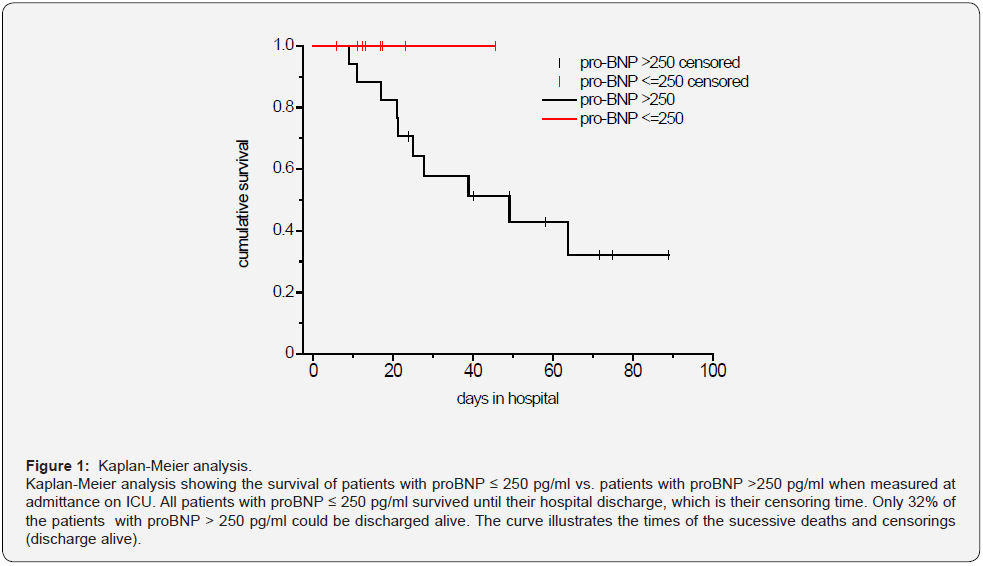
Continuous variables are presented as mean±SD and median (25%, 75% quartil). Categorical variables are expressed as absolute number of patients and percentage. The mean values for continuous variables were compared using independent group t tests when the data were normally distributed, otherwise, the Mann-Whitney test was used. For pro-BNP we calculated the area under the receiver operating characteristic {ROC} in respect of survival and explored the optimal cutoff value. By means of Kaplan- Meier curves, the survival of patients with pro-BNP below / above the cutoff is illustrated. A p-value less than 0.05 was considered statistically significant. Because of the explorative nature of the study, we did not perform an α-correction for multiple testing, therefore the p-values must be interpreted carefully. All statistical analyses were performed with IBM® SPSS®, version 27 for Windows.

Results

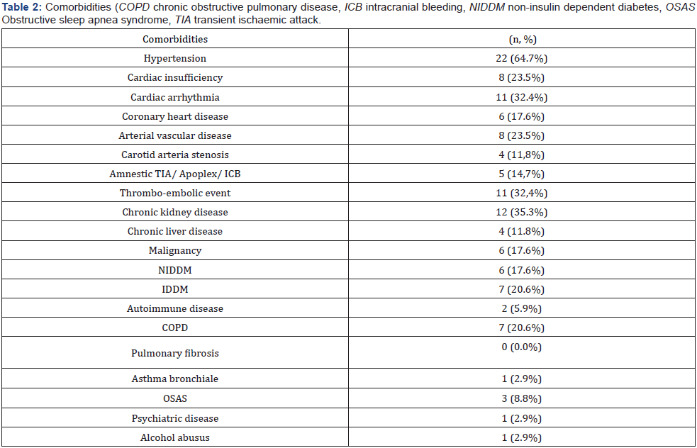
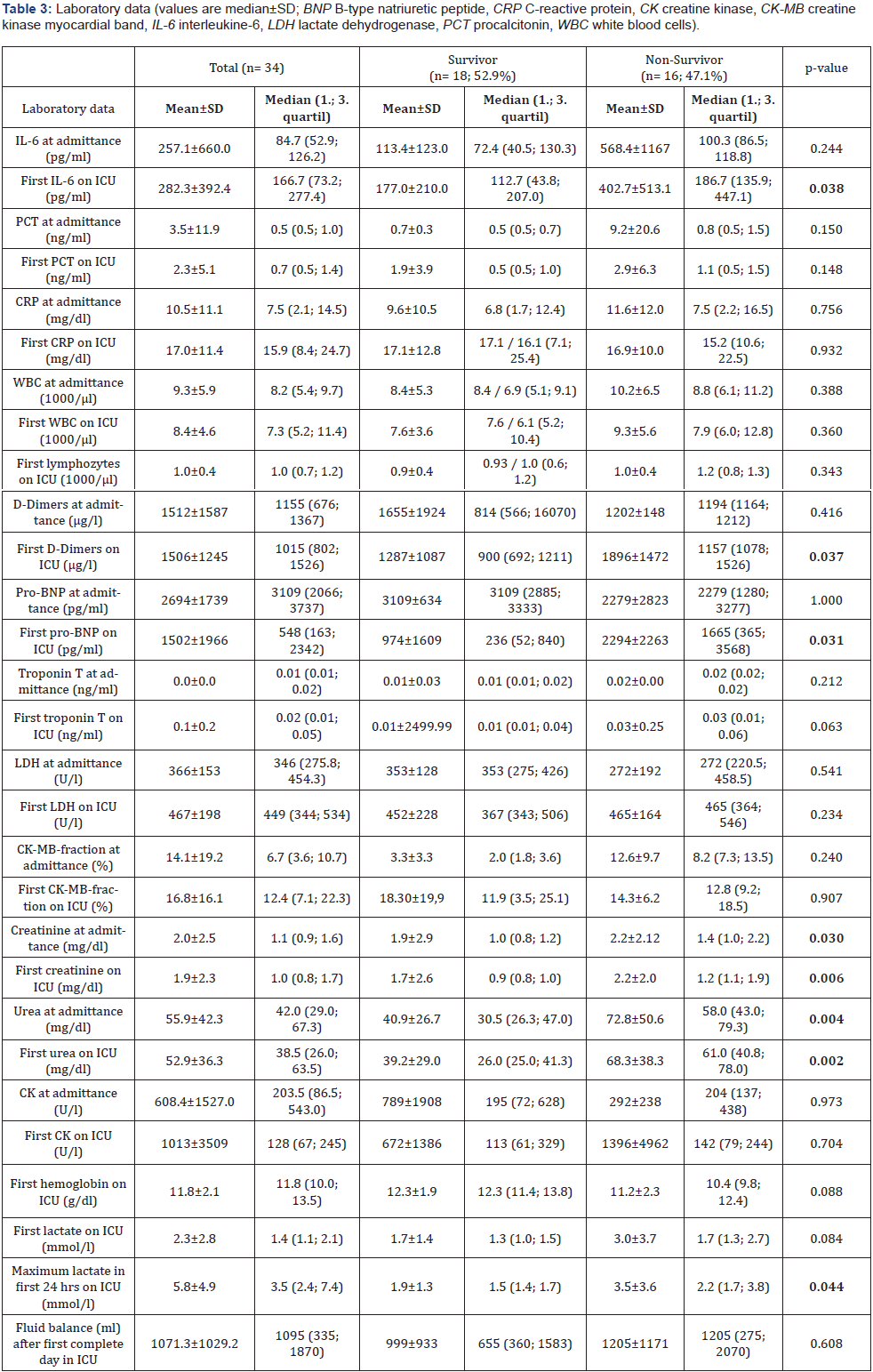
A total of 34 patients were admitted to ICU for COVID-19 during this study period. We had 18 (52.9%) survivors and 16 (47.1%) non-survivors. 27 of them were male patients (79,4%). The age of the total cohort was 67.8±13.9 years. Basic clinical characteristics and respiratory parameters before intubation and extreme values during the first 24 hours of ventilation on the ICU are shown in Table 1. The comorbidities of our patients are demonstrated in Table 2. Most of the patients had cardiovascular diseases 23 (67.6%), chronic kidney diseases 12 (35.3%) and thromboembolic events 11 (32,4%). In the absence of contraindications our patients were anticoagulated slightly elevated to reach a level between prophylactic and therapeutic anticoagulation. Laboratory data are shown at hospital admittance and the first value measured on intensive care unit (ICU) (Table 3).

All 34 patients were admitted to the ICU due to progressive respiratory failure. We treated 28 (82.4%) patients with highflow oxygen therapy, 24 (70.6%) patients were intubated and invasively ventilated, 22 (64.7%) were proned and only 3 patients (8.8%) were non-invasively ventilated. Many patients (28, 82.4%) showed laboratory or clinical signs of kidney injury, 15 (44.1%) patients developed a new AKI, 9 (26.5%) an acute on chronic kidney injury and only 4 (11.8%) patients chronic kidney injury. 19 patients (55.9%) needed renal replacement therapy at any time during their ICU stay whereof only 4 (11.8%) had pre-existing end-stage renal disease.

From our 34 ICU patients, almost half (16, 47.1%) died during their hospital stay. Non-survivors were older than the survivors (75.5±7.3 vs. 60.9±14.9; p=0.003); the youngest non-survivor was 64 years old; the oldest survivor was 88 years old. The laboratory data demonstrated for the non-survivor group at admission on ICU increased levels of IL-6, abnormal levels for d-dimers, pro-BNP, creatinine, urea and lactate compared to survivors. The survivors had a higher paO2 level and Horovitz index before intubation. The total duration of high-flow nasal cannula oxygenation therapy (NFHC) was longer for the survivors.

Because of the described elevation of pro-BNP in ARDS and its potential use as a prognostic marker in ARDS we looked in more detail at the pro-BNP level at admittance on ICU and survival. We calculated the receiver operating characteristic (ROC) curve for pro-BNP in respect of survival, which resulted in an AUC of 0.760 (p = 0.007). This is a hint, that pro-BNP is a significant predictor of the survival. We received a pro-BNP value of >244 pg/ml as the optimal cut-off value for our sample. With this cutoff value the sensitivity is 1 and the specifity 0.533. As the adjacent cutoff-value is 252, we determined 250 as optimal cutoff.

The Kaplan-Meier analysis (Figure 1) shows the survival of patients with proBNP ≤ 250 pg/ml vs. patients with proBNP > 250 pg/ml. All patients with proBNP ≤ 250 pg/ml survived until their hospital discharge, which is their censoring time. Only 32% of the patients with proBNP > 250 pg/ml could be discharged alive. The curve illustrates the times of the successive deaths and censorings (discharge alive).

Discussion

In this retrospective analysis of 34 ICU patients with respiratory insufficiency due to a COVID-19 infection during the first part of the pandemic in a municipal hospital in Germany we found clinical courses similar to those in other studies [7,8]. Severe respiratory failure in patients with SARS-CoV-2 infection is only one sign of the multisystem inflammatory syndrome [9-11], which is consistent with the high rate of patients with renal failure and the need for renal replacement therapy in more than half of the patients [12,13]. The high rate of multiorgan failure together with the increased age of our patients may at least partially explain the high mortality of our patients which is comparable to data from other studies with COVID-19 ICU patients [14].

More than two third of our patients were invasively ventilated and only a minority was non-invasively ventilated because at that time - during the “first wave” in Germany - we worried about an increased risk of transmission for the ICU staff with the use of noninvasive ventilation. Accordingly, we suggested a reduced risk of transmission of SARS-CoV-2 when patients were intubated timely. In the meantime, it has been demonstrated that is reasonable, safe and recommended in the guideline to try nasal-high flow oxygen and non-invasive ventilation in patients with respiratory insufficiency due to COVID-19 when patients are closely monitored [2,15-18].

Very soon after the first patients with COVID-19 had been treated it was recognized that there is an increased risk for thrombo-embolic events in these patients due to an inflammatory alteration of the endothelium and an inflammatory pro-coagulatory state [19,20]. It was difficult and potentially misleading to count the number of thrombo-embolic events in our patients because we did not screen systematically for these events as other groups did [20].

The major finding in this - small group - of patients with COVID-19 is that an elevated pro-BNP level on admission to the ICU shows the tendency of a worse prognosis.

Many clinical data described a cardiovascular manifestation induced by this viral infection especially in critical ill patients. Acute myocardial injury manifested mainly by elevated levels of high-sensitive troponin I, and arrhythmias have been detected [9,21]. Guo et al. [3] reported in their study among 187 patients with COVID-19, 52 (27.8%) exhibited myocardial injury as demonstrated by elevation of troponin T (TnT) levels, and the mortality was markedly higher in patients with elevated TnT levels than in patients with normal TnT levels (59.6% vs 8.9%). The authors suggest that myocardial injury due to the inflammation might play a major role in the clinical detoriation of COVID-19 patients and that those patients with elevated troponin T levels (27.8% of patients) had more malignant arrhythmias and a higher mortality [3]. Consistent with our results of increased levels of pro-BNP in non-survivors they found a correlation between elevated pro-BNP and troponin T levels [3].

In our group of non-survivors TnT levels were a bit higher at admission to the ICU (0.03 ± 0.25) and proBNP was significantly higher in the non-survivor group (2294±2263; p =0.031) compared to the survivors. The comorbidities of our patients represented mostly cardiovascular diseases, hypertension on top of all (64.7%). Alvarez-Garcia et al. [5] pointed out that patients with a history of heart failure (HF) hospitalized for COVID-19 face nearly 3 times the risk of mechanical ventilation and twice the risk of mortality compared with patients without HF [5]. Similar to our results Sorrentino et al. demonstrated a correlation between increased pro-BNP levels and severity of COVID-19 disease but in contrast in their study the pro-BNP levels in non-survivors were already increased at admittance to hospital compared to survivors [4]. In our investigation patients with COVID-19 had also elevated levels of proBNP at admission to the hospital. Thus, we learned from several investigations during the pandemic that predictors of a fatal outcome in COVID- 19 cases included age, the presence of underlying diseases, the presence of secondary infection and elevated inflammatory indicators in the blood [5,14,22,23]. Although the high accuracy of NT-proBNP is already established in the diagnosis of acute heart failure, the prognostic value of this marker for patients with COVID-19 remains uncertain [4].

Our study has several limitations. Only 34 patients with COVID-19 were included during the first wave in Germany, and a larger randomized cohort study is needed to verify our conclusions. Unfortunately, we could not provide more information regarding cardiovascular complications as e.g., cardiovascular ultrasound or detailed hemodynamic monitoring. Due to the restricted options and the increased efforts in the isolation ward data was not complete in all patients.

Last but not least, the data of this retrospective study permit a preliminary assessment of the clinical course and outcomes of patients with COVID-19.

The causes of death may involve multiple organ dysfunction in most cases, and it is difficult to differentiate the myocardial injury as the main and direct cause in an individual case. Long-term observation and prospective study design on the effectiveness of treatments are needed. We still have to wait for long-term results after surviving Covid-19 disease [24].

Conclusion

Many critically ill patients with COVID-19 pneumonia suffer from multi-organ dysfunction including cardiac insufficiency. We concluded the measurement of specific laboratory data as NT-proBNP may help to categorize the severity of the COVID-19 disease at admission to the ICU.

Click here: https://juniperpublishers.com/index.php 

Thursday, December 8, 2022

Photografted Chitosan as Antibacterial Agent for Textiles - Juniper Publishers

 Organic & Medicinal Chemistry - Juniper Publishers

Abstract

The natural biopolymer chitosan was applied to textile substrates by ultraviolet photo grafting to confer antibacterial activity against a wide range of microorganisms. In this way an efficient and solid textile finishing was ensured by a low environmental impact process. The use of an environmentally friendly product is thus coupled to the adoption of an eco-sustainable process, that can guarantee the protection of both health and environment and produce fabrics complying with current regulations. Fabrics treated in this way can find application in all cases where it is necessary to counteract the proliferation of bacteria: medical devices, hospital and hygienic textiles, technical and furniture fabrics, but also in the field of water purification, as active biological filters.

Keywords: Chitosan; Antibacterial agent; Textiles; Cotton; UV-grafting; Water purification; Escherichia coli; Staphylococcus aureus; Klebsiella

Introduction

In last decades, globalization and even more stringent ecological regulations push textile companies to search for product quality improvements by environmentally friendly processes. Among targets, the use of processes with low water and energy consumption and the replacement of polluting or toxic chemicals with others of natural origin, with low or no environmental impact, are prominent. Furthermore, the frequent spread of diseases and infections highlights the importance of health and hygiene, promoting the production of antibacterial fabrics not only to avoid the degradation of the fabrics themselves, but also to effectively prevent the propagation of pathogens.

Fibers and fabrics of natural origin, hydrophilic and biodegradable, provide an optimal and favorable environment for the growth and proliferation of bacterial colonies; therefore, an antibacterial finishing, through appropriate chemical modification of the fibers surface, is particularly useful on these textiles. Metal ions (Ag+), quaternary ammonium salts, phosphonium salts or Triclosan are the chemical biocides commonly used today, but they were proved to be toxic or carcinogenic, so application on textiles, especially if in direct contact with the human skin, is not recommended considering the possible release of the antibacterial agent during use.

Biopolymers with intrinsic antibacterial activity, of natural origin from renewable sources, applied by an economic and ecological finishing process, is an interesting alternative for the development of bioactive textiles by eco-sustainable treatments. The proposed solution focuses on the finishing of textile fabrics by chitosan, grafted on fibers by photocuring [1]. Moreover, the application of a chitosan treated gauze for water filtration was also studied, aimed to disinfection and elimination of the same pathogenic microorganisms [2]. Water sanitation is an important issue worldwide: every year, more than 3 million people die in the world from diseases related to unhealthy water, responsible for 88% of diarrhea cases. The study has therefore a high application potential, with positive impacts in both scientific and socio-economic fields.

Chitosan UV grafting

Chitosan is a natural biopolymer mainly obtained from the exoskeleton of crustaceans. It is an abundant, low-cost by-product of the food industry, with well-known antibacterial properties against both Gram positive and negative bacteria, due to its combined bactericidal and bacteriostatic action, against fungi and viruses. Chitosan has no toxicity, and it is not a strong oxidant, avoiding the release of dangerous by-products during use; moreover, it is inert in water. For all these reasons chitosan can be considered as a sustainable alternative to other antimicrobial agents. Chitosan is already used in some textile finishing, applied by wet thermal curing with energy consumption, costs, and possible fabric degradation. Moreover, the addition of toxic reagents, such as glutaraldehyde [3], or expensive, such as genipin, is required as crosslinking agents to impart the required durability to repeated washings [4].

Therefore UV-curing of chitosan on fibers to confer antimicrobial activity was proposed as an alternative process to thermal curing or chemical crosslinking, obtaining a good effectiveness and fastness of the treatment. In UV-curing, radical species are generated by the interaction of UV light with a suitable photo initiator, which induces grafting reactions of reactive monomers and oligomers at low temperature and quickly, avoiding the emission of VOC, with lower environmental impact and lower cost than thermal process. Both chitosan and fibers, mainly cellulosic fibers, take part to this photochemical process. If a chitosan and initiator mixture is adsorbed onto the fibers and subsequently UV-cured, the polymeric chains can form inside the textile structure, which can also establish graft bonds, making the treatment solid and resistant. Moreover, the interpenetration of components and homogeneous distribution of monomers, even at a low concentration, contribute to obtain textile materials with modified surface properties, without high polymer add-on [5]. Consequently, the fabrics may retain their original properties of hand and breathability. Finally, the technology is easily implemented in the textile supply chain.

Chitosan as antibacterial finishing for textiles

Chitosan was applied by photograflting to several natural and synthetic fabrics, namely cotton, silk [5], wool [6], polyester and polyamide. The process was optimized, in terms of polymer add-on, ultraviolet exposure, impregnation and drying modality. The antimicrobial activity of treated samples was tested against Escherichia Coli, Staphylococcus Aureus and Klebsiella pneumonia, and compared with same samples treated by a commercial antibacterial agent based on Triclosan and quaternary ammonium salts.

The study confirmed the efficiency of chitosan against the tested bacteria and an excellent solidity of the treatment. A process yields equal to 100% was obtained on cotton and silk, while the hand and color of the treated fabrics were unaffected up to 2% add-on. However, the bacterial colonies were totally removed on fabrics treated with chitosan add-ons equal to just 0.3% owf, a very low amount unaffecting the peculiar properties of natural fibers. This strong antibacterial activity was unchanged after 30cycles washing, confirming the fastness of the treatment and the effectiveness of ultraviolet radiation to form strong radical bonds between chitosan and fibers.

The comparative test with the commercial product showed that it was less performing than chitosan, since after 30 washing cycles the antibacterial activity decreased to 98%, due to a certain release or inactivation of the antibacterial agent by surfactant action, while chitosan treated fabrics keep 99,999% of colony reduction [7]. Similar results were obtained, in terms of antibacterial activity, also on wool and synthetic fibers, but in this case a surface pretreatment was required to increase hydrophilicity and promote the absorption of the chitosan solution, due to hydrophobicity of this kind of fibers [6].

Chitosan for water purification

Water filtration tests were carried out on a cotton gauze treated with chitosan. The process for chitosan grafting was the same as for fabrics, but the add-on was increased till 25% owf. The as treated gauze was tested for continuous filtration of water contaminated by Escherichia, Staphylococcus or Klebsiella. The functionalized gauzes were tested in dynamic conditions, with bacteria inoculum continuously flowed through the filter several times. Results showed that a contact time, between the gauze and the contaminated water, of 4seconds was enough for the total elimination of Escherichia and Staphylococcus from the water, while after 8sec the Klebsiella was reduced by 98%. It is highly interesting since this fast speed of action guarantees total disinfection with a rapid flow of water, making the chitosan treated gauze an excellent candidate as biological filter. The comparison was made in this case with a cationized cotton gauze, but the material was not suitable for filtration, reaching only 40% of bacterial reduction after a contact time of 20 seconds [2]. Considering water treatments, it is noteworthy that in previous studies the same chitosan treated gauze was found active also against other water pollutants: for the capture of heavy metals, like copper or chrome, present in water [8], and for sequestration of dyes from textile dyeing wastewaters [9].

Conclusion

Chitosan UV-grafting on both natural and synthetic fibers yields strong antimicrobial properties against different classes of microorganisms. The coupling of a natural, biocompatible, and renewable biopolymer with an ecofriendly photo grafting process makes it competitive and interesting versus the traditional toxic antibacterial agents applied by thermal processes, energy and water consuming. Therefore, the possibility to replace the established antibacterial finishing processes, based on hard and energy demanding chemical treatments, with an ecofriendly low temperature process using ultraviolet light and a natural and safe biopolymer such as chitosan, is undoubtedly an interesting result which can contribute to change the production of antibacterial fabrics toward a sustainable development.

It was confirmed by a semi-industrial scale-up and a deep cost analysis of the whole process. On the other hand, chitosan shows also promising applications in the field of water purification. In fact, it can totally remove pathogenic microorganisms from contaminated water in few times, allowing the continuous treatment of great amount of water with a little amount of filtering material. Some literature [10] suggests that chitosan and its derivatives can also suppress viral infections in various biological systems. However, the mechanism of this antiviral activity is poorly understood. Future studies could be focused on the investigation about antiviral properties conferred to textiles treated by chitosan photo grafting. If it will be confirmed, the potentialities for chitosan applications would be even more interesting.

Click here: https://juniperpublishers.com/index.php  

Unveiling the Intersection of Age and Gender in the Emotional Well-being of Older Students: A Pilot Study at the Permanent University for Adults in Alicante (Spain) - Juniper Publishers

 Gerontology & Geriatric Medicine - Juniper Publishers Abstract This study assesses the emotional impact and well-being of older stud...