Wednesday, September 30, 2020

Forensic Evidence of a Surgical Fetus- A Rare Case Report - Juniper Publishers

Forensic Sciences & Criminal Investigation -                            Juniper Publishers 


Abstract

Rape is the fasted growing crime in the country compared to other crime against women. It is also seen minor girl age between (7-12year) is found rape-victim and sometimes reported pregnant being raped. In such cases, pregnancy is terminated by doctors using different methods to save the life of minor innocent girl, protect from Human Rights and social stigma. In one case, a girl child, age about 12 year, was staying with single parent family. She used to visit and play with her friend being the neighbour. Taking advantage of age, the girl was lured and repeatedly raped by her friend’s uncle, an elderly man age about 57 year. One day, the victim girl complained abdominal pain and the doctor confirmed her pregnant but then she was about 20 weeks. The mother took up the matter to legal court to save her daughter being unable to access to medical termination of pregnancy (MTP). Considering the girls age/health, medical board was held under judge’s order and decided to end the pregnancy. The unborn fetus of the victim girl was removed and received in forensic science laboratory for DNA evidence to link the accused and establish the crime. The minor girl was saved to carry the pregnancy further. The details have been discussed in this rarest of the rare case.

Keywords: Minor girl; Illegal sex; Fetus, Surgery; DNA evidence

Introduction

In the life cycle of women, they are subjected to different violence in the society. The crime against women includes mostly sexual assault, foeticide, infanticide, dowry death, bride burning forced prostitution/sterilization etc. [1-3]. But rape/sexual assault is one of the most common crime reported against women of different age groups even kids and aged women are not spared. The sex with minor girl is an evil scenario in the society and reported for various reasons [4] According to National Crime Records Bureau (NCRB), MHA, Govt. of India, 2013 Annual Report, 24,923 rape cases were reported across India in 2012, out of which 24,470 committed by someone known to the victim [5] The statistical figure of rape cases is available but pregnant being raped is not much as such statistics under such crime head is not found in literature. The following are the reasons for increase of sexual offence cases in the country [6,7].

a) Man dominated society

b) Improper respect for women

c) Girls are not properly educated to react

d) Use of mobile blue film

e) No exemplary punishment and trials get unusually delay

f) Want of quality investigation

g) Media and dress code

h) Sadistic lifestyle

i) All want girl friend

j) Use of internet-based child sexual exploitation networking (Dark net market).

There is law to permit termination of pregnancy when there is threat to life or abnormal fetus. In this case, the victim was minor and under order of Hon’ble court unborn fetus was surgically removed by a team of doctors to terminate the pregnancy.

Case Report

A girl child age about 12-year was staying with single parent and used to visit her friend’s house being neighbor. Taking advantage of frequent visit, her friend’s uncle an elderly man age about 57 year lured the girl and developed physical relation. Subsequently, the girl got pregnant. One day having severe abdominal pain the girl child (victim) told her mother and on medical checkup, she was confirmed to be pregnant. Thereafter, the child revealed the hidden truth to her mother about sexual abuse by the uncle of her friend. She (victim) was then twenty weeks of pregnancy and risky on her age/health to continue. Hence took help of Hon’ble court for termination of pregnancy to save the life of minor girl child and lodged a FIR in the local police station for justice. The case was registered u/s 376(2)(1)(a)/506 Indian Penal Code (IPC) and sec 6 of Protection of Children from sexual offences (POCSO) Act.

Materials and Methods

In this critical case, as per order of the Hon’ble Court the fetus of the victim girl was removed by surgical method by a team of gynecological experts. Subsequently biological fluid (blood) from the offender and the victim girl was collected as per blood sample authentication form. The illegal surgical fetus after removal was also received for DNA evidence.

Medical examination and result

Potency test of the offender

A. General Physical Examination: Height 157cm, weight 51kg, pulse 78bpm, beard and moustache adult type, axillary and pubic hair adult type.

B. Local Examination: Penis length 8cm and circumference 9cm in flaccid condition, both the testes descended into the scrotal sac, scrotal skin is healthy; no genital, extra-genital injury was noted.

C. Based on the above findings the medico-legal expert opined that there was nothing to suggest that the offender is not capable of performing sexual intercourse

Surgical method used to remove unborn fetus

It was a case of late mid-trimester abortion. As per protocol, medical termination was attempted first by using prostaglandins and oxytocin. But as it failed it was decided to do surgical induction by transabdominal anterior hysterotomy under general anesthesia. In this procedure, after opening the abdomen low transverse incision was made on lower segment of the uterus. Abortus and placenta were removed completely; uterine wall was sutured in layers. Complete hemostasis was secured followed by closure of abdomen. Postoperative period was monitored meticulously by intravenous fluid, antibiotics, sedation and oxytocin.

Forensic DNA Evidence

Samples received from three different sources were properly preserved in the laboratory. The DNA Typing from three sources was made by using Genetic Analyzer AB 3130, Life Technologies Holding Pvt. Ltd., USA, following standard procedure available in literature [8]. The DNA profiling of all the three different exhibits are shown in (Table 1). The allelic distribution of fetus, victim and offender shown conclusively confirms that the victim and the offender are the biological parents of the fetus (Figure 1-3).

Conclusion

In this case the minor innocent girl was easily lured by the elderly man and repeatedly raped to satisfy his sex desire. But the offender could not think of the consequence as a result the girl got pregnant being raped. Moreover, the innocent girl could not understand what is being done on her and going to happen. She was also not taught to protest from such inhuman activity by her parents. As a child, she (victim) totally surrendered without any resistance to the offender and became the rape-victim of the devil. The fear of threatening in the mind of the girl being killed by the offender also kept her silent. As such, she was not at fault, abuser to blame. The criminal profile of the accused in this case, appears to be psychopath and mentally perverted and could not understand the barbaric act with a girl 12-year age almost like a daughter. The offender also did not care about stringent punishment prescribed under Protection of Children from Sexual Offences (POCSO) Act, leading to life imprisonment and even death. The offender is to be further examined by the psychiatric professional to know such unusual aggressive behavior. This is humiliating event in women’s life. The incident reminds a quote:

“Surgery during life is painful and benefit individual.

Surgery after death is not painful but benefits mankind”

The forensic result showing DNA evidence supported by the statement of the victim girl and medical report and circumstantial evidence could conclusively identified the offender & establish the crime.

To Know more about Forensic Sciences & Criminal Investigation   

To Know more about our Juniper Publishers

Click here: https://juniperpublishers.com/index.php 

Tuesday, September 29, 2020

Prevalence of Antiplatelet and Anticoagulant Medications in Rural Emergency General Surgery and Impact on Clinical Practice, Morbidity and Mortality - Juniper Publishers

 Open Access Journal of Surgery - Juniper Publishers  

Abstract

Background: An increasing number of general surgical patients are being prescribed antiplatelet and anticoagulant medications (APAC) for various cardiovascular conditions. We aimed to characterise APAC usage in emergency general surgical patients admitted to a rural hospital and assess the impact of APAC usage on patients.

Methods: A prospective study of emergency general surgical admissions at Northeast Health Wangaratta from July to October 2014 was conducted. A questionnaire was used to collect patient data. 
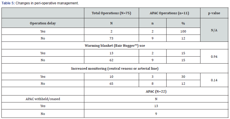
Results: 118 patients were classified into two groups: non-APAC (n=96, 81%) and APAC (n=22, 19%). The majority (91%) were antiplatelet medications. Patients older than 60 years were more likely to be on APAC (p<0.0001). Patients admitted with bleeding pathology were more likely to be on APAC (p<0.05). 15% of all emergency operations were on APAC. There was no difference in the use of central venous or arterial line for increased monitoring (p=0.14) or in the use of warming blanket (p=0.94). APAC patients were more likely to receive transfusion during admission (p=0.04). The recorded morbidities (n=2) and mortality (n=1) cases in this study were all in the APAC group.
Conclusion: The use of APAC is more prevalent in those aged 60 years and above. 91% of APAC are antiplatelet medications. Patients on APAC are more likely to be admitted for bleeding issues, receiving transfusion, and an increased risk of morbidity and mortality. There is an urgent need for well-established, recognised and more accessible clinical guidelines regarding the emergency APAC management, especially for antiplatelet medications, in rural emergency general surgical patients.

Keywords: Antiplatelet; Anticoagulant; Rural general surgery; Emergency general surgery; Morbidity; Mortality

Abbreviations: APAC: Antiplatelet and Anticoagulant medications

Introduction

Antiplatelet and anticoagulant medications (APAC) are being widely used in the prevention and treatment of various cardiovascular and thromboembolic diseases. With their ever-expanding indications in an aging Australian population, the management of APAC in elective and emergency surgical patients is becoming increasingly challenging, and the advent of novel oral anticoagulants is adding more complexity to this challenge. Patients on APAC present a clinical challenge for treating surgeons, physicians, anaesthetists and haematologists as there needs to be a careful balance between the bleeding and thromboembolic risks associated with cessation of these medications. Surgeons need to decide on the timing and type of operation performed, along with the judicious use of limited, often precious blood products and haemostatic agents.

Previously established guideline on perioperative management of APAC was mostly on the perioperative management and emergency reversal of warfarin [1]. However, the more recent guideline now includes the perioperative management of other APAC including the novel oral anticoagulants [2]. Although the guidelines are helpful, the APAC management in some patients still depends on the clinician’s judgement and individual patient’s clinical situation. The aim of our study was to characterise the prevalence of APAC medications in rural emergency general surgical patients, and to assess the impact that these APAC have on the clinical management of these patients and associated morbidity and mortality.

Materials and Methods

A prospective study of all general surgical admissions through the Emergency Department of Northeast Health Wangaratta (Victoria, Australia) from July to October 2014 was conducted. During these four months, a datasheet for each patient was completed by a member of the surgical team at the time of admission and during inpatient stay. Data collected included patient demographics (gender, age, length of stay, admission diagnosis), APAC usage (type, indication, duration of cessation), admission diagnosis, length of stay, types of operation, anaesthesia techniques, morbidity and mortality. Data was then statistically analysed using ANOVA (t-test) method (Microsoft Statistical Package). The ethics approval was obtained from the hospital ethics committee.

Results

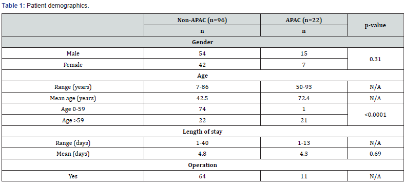
Patient demographics (Table 1)

A total of 118 consecutive general surgical patients were admitted through the Emergency Department. The patients were classified into two groups: non-APAC (n=96, 81%) and APAC (n=22, 19%). There was no difference of APAC usage based on gender (p=0.31). Patients in the APAC group were older compared to the non-APAC patients (mean age 72 vs 43 years old). Amongst patients younger than 60 years old, only one patient (1%) was on APAC. This contrasted with those aged 60 and above, in which 49% were on APAC (p<0.0001). The age groups were analysed in this way as there appeared to be a distinct increase in APAC usage from the age of 60 years old. The length of stay was not affected by APAC usage (non-APAC mean 4.8 days vs APAC mean 4.3 days) (p=0.69). 15% of all operated patients were on APAC medications.

Open Access Journal of Surgery

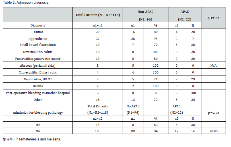
Admission diagnosis (Table 2)

There were 13 patients (out of 118) who were admitted for bleeding problems such as upper and lower gastrointestinal bleed, as well as bleeding complications following surgery at another hospital. In this group, the patients were more likely to be on APAC (p<0.05) with five patients (38%) being on APAC. Also, 20% (4 out of 20 patients) of all trauma patients were on APAC.

Open Access Journal of Surgery

APAC medications (Table 3)

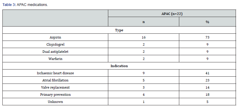
In this study, 19% (22 out of 118) of all emergency general surgical patients were on APAC. 82% of the patients (18 out of 22) were on single antiplatelet medication (aspirin or clopidogrel). two patients were on dual antiplatelet agents (aspirin, and clopidogrel or ticagrelor). No patient was on any of the novel oral anticoagulants (dabigatran, rivaroxaban or apixaban). The recorded primary indications for APAC medications include ischaemic heart disease (n=9, 41%), atrial fibrillation (n=5, 23%), valve replacement (n=3, 14%), and primary prevention (n=4, 18%).

Open Access Journal of Surgery

Operations and anaesthesia (Table 4)

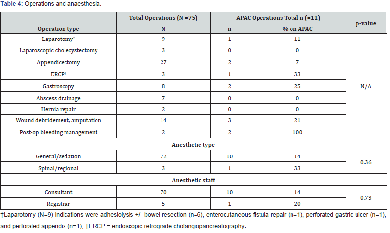
15% (11 out of 75 patients) of emergency general surgical patients requiring operations were on APAC. 11% of all laparotomies, and 25% of all gastroscopies were performed on patients taking APAC. The two patients who were operated for bleeding following surgery at another hospital were both in the APAC group. There was no statistically significant difference in the mode of anaesthesia chosen (general/sedation vs. spinal/regional) (p=0.36) nor seniority of staff performing the anaesthesia (p=0.73) between the two groups.

Open Access Journal of Surgery

Changes in peri-operative management (Table 5)

59% (13 out of 22) of APAC patients had interruption or cessation to their medications, on average by 3.5 days (range 1-13 days). Two out of 75 emergency operations were delayed due to APAC usage. In this study, there was no statistically significant difference in the use of central venous or arterial line for increased monitoring between the two groups (p=0.14). There was also no observed difference in the use of a warming blanket (Bair Hugger™) between the two groups (p=0.94).

Open Access Journal of Surgery

Transfusion, morbidity and mortality (Table 6)

The recorded morbidity (2 patients) and mortality (1 patient) in this study were all in the APAC group (p<0.01). The first complication was a 71-year old male on aspirin who had an appendicectomy and post-operatively developed coffee-ground vomiting. The second complication was a 63-year old female on aspirin who had a traumatic degloving lower limb injury. She was noted to have excessive blood loss at home shortly after discharge. There was no thromboembolic complication recorded. The single mortality in the study was of an 87-year old female on warfarin for mechanical mitral valve, who had a car accident and died shortly after in the Emergency Department from a cardiac arrest. Overall, six patients required transfusion, three from the non-APAC group and three from the APAC group (p=0.04).

Open Access Journal of Surgery

Discussion

An increasing number of general surgical patients are being prescribed antiplatelet and anticoagulant medications (APAC) for various cardiovascular and cerebrovascular conditions. This widespread usage is on the rise especially in the face of increasing prescription of novel oral anticoagulants (direct thrombin inhibitor, anti-factor Xa). In our study, 19% of all emergency general surgical admissions were on APAC, with antiplatelet medications being the most prevalent (91%). The absence of any patients on novel oral anticoagulants in this study may reflect the prescribing practice in rural areas like Wangaratta. However, we predict that such prescribing practice will change with more prescription of novel oral anticoagulants. We speculate that a similar study conducted in a tertiary metropolitan hospital may reveal different prescribing patterns. In our current study, patients on APAC tend to be older, with the majority of them being 60 years old or older.

An increasing number of general surgical patients are being prescribed antiplatelet and anticoagulant medications (APAC) for various cardiovascular and cerebrovascular conditions. This widespread usage is on the rise especially in the face of increasing prescription of novel oral anticoagulants (direct thrombin inhibitor, anti-factor Xa). In our study, 19% of all emergency general surgical admissions were on APAC, with antiplatelet medications being the most prevalent (91%). The absence of any patients on novel oral anticoagulants in this study may reflect the prescribing practice in rural areas like Wangaratta. However, we predict that such prescribing practice will change with more prescription of novel oral anticoagulants. We speculate that a similar study conducted in a tertiary metropolitan hospital may reveal different prescribing patterns. In our current study, patients on APAC tend to be older, with the majority of them being 60 years old or older.

Emergency surgery exposes patients on APAC to increased risk of peri-operative bleeding. Depending on the estimated risk of peri-operative bleeding due to APAC, some of the patients on APAC will have their APAC withheld or ceased, and the required operation may have to be delayed until the effects of their APAC are reversed. In our study, 15% (11 out of 75) of all patients requiring emergency operation were on APAC medications, and of these two patients had their operation delayed due to APAC. Among the APAC medications, a warfarin reversal protocol was the most readily available at institutional and national level [1]. However, a more recently developed guideline in December 2018 now incorporates the perioperative management of the novel oral anticoagulants, including emergency reversal of dabigatran with idarucizumab in case of life-threatening bleeding or when an urgent procedure is required [2]. However, as observed in this study, the use of antiplatelet medications was far more prevalent, owing to their frequent usage in the primary and secondary prevention of many cardiovascular and cerebrovascular diseases. Despite their prevalence, there is currently no well-established clinical guideline specific to the management of patients on antiplatelet medications who require emergency general surgery [4]. Current guidelines are limited to the management of elective surgical patients with ischaemic heart disease who are on antiplatelet medications [2,5].

If the cessation of antiplatelet medication is judged essential due to high risk of operative bleeding, then the recommendation is to discontinue aspirin for five days, ticagrelor for five days, and clopidogrel seven days prior to the procedure [2]. However, this recommendation poses danger for high-risk patients as a large meta-analysis involving 50,279 patients on aspirin revealed that its withdrawal could increase the risk of cardiovascular events 3-fold with the average time from aspirin withdrawal to the onset of acute coronary syndrome being 10.7 days [6]. Also, the discontinuation of clopidogrel within 4-6 weeks following bare-metal coronary stenting poses the risk of the development of stent thrombosis with a high perioperative mortality of 20- 40% [7]. Hence, discontinuation of antiplatelet agents should be advocated only after a careful risk-benefit analysis where bleeding risk clearly outweighs that of cardiovascular events. In the elective surgical setting, the usage of bridging tirofiban and intravenous heparin has been described in three cases of patients with drugeluting stents for elective surgery where authors attributed the achievement of minimal surgical bleeding and absence of stent thrombosis to their usage [8].

In life-threatening surgical conditions where urgent or immediate surgical intervention is necessitated, there is lack of universal evidence-based guidelines to mitigate the effects of the antiplatelet agents. Platelet transfusion has the potential to restore normal platelet functions and limit haemorrhage progression in patients on antiplatelet agents, however, its efficacy and indications have not been fully established and clinical data are variable [9-12]; hence, their effectiveness of prophylactic use of platelet transfusions in trauma and emergency surgery for patients on antiplatelet agents remains controversial. Despite this, emergency platelet transfusion is frequently used in neurosurgical patients with traumatic brain injury on antiplatelet medications as these patients have an high mortality rate, and many advocate platelet transfusion regardless of the platelet count [13,14]. However, some authors argue that platelet transfusion in this setting does not reduce mortality rate either [15]. Furthermore, another study suggested that platelet transfusion does not restore platelet function for patients on clopidogrel, and it may only be effective for some patients on aspirin [9]. Platelet function tests, such as Verify Now™ are available to help identify patients who may benefit from platelet transfusion [9,16]. However, these tests are time-consuming and are not performed routinely in most hospitals. In our study, the two patients on antiplatelet agents who required transfusion did not receive any platelet transfusion.

A large cohort study showed that in patients with coronary stents, interrupting antiplatelet treatment by more than five days pre-operatively significantly increased the risk of cardiovascular or cerebrovascular thrombosis [17]. 59% of our APAC patients who had interruption to their medications did not have any thromboembolic complications. The mean duration of APAC interruption in this study was 3.5 days (range 1-13 days). For two patients with dual antiplatelet agents, only one of their medications was temporarily ceased.

In regard to impact of APAC usage on anaesthetic management, we hypothesised that due to multiple medical comorbidities and anticipation of increased blood loss, APAC patients would receive increased anaesthetic monitoring and hypothermia prevention. However, our data showed no statistically-significant difference in the use of central venous or arterial line and warming blanket between the two groups. APAC usage is often noted as a contraindication for neuraxial anaesthesia [18,19] due to increased risk of spinal haematoma with potential devastating neurological sequelae. In our study, we observed no difference in the anaesthetic technique (general/sedation vs. spinal/regional) used between APAC and non-APAC groups. This can most likely be contributed to the small sample size as only one patient in the study had a spinal anaesthesia in the non-APAC group.

The limitations of the study include the small number of recruited patients and that the data that is now 5 years old. In particular, we expect that the usage of novel oral anticoagulants would be much more prevalent if the study is repeated in the current time. Despite such limitations, we believe the current study serves as a useful baseline study to demonstrate the prevalence and effects of APAC in emergency general surgery patients in a rural hospital context. The scope for future research includes recruitment of a greater number of patients to incorporate those on novel oral anticoagulants.

Conclusion

From the highlighted cases of bleeding in APAC patients, we identified that decisions in regard to APAC interruption, type and amount of blood products used were often left up to the individual surgeon’s clinical decision. Apart from emergency warfarin reversal protocol, and more recently for emergency reversal of dabigatran with idarucizumab, there is still a lack of well-established, easily accessible guidelines for emergency management of APAC in non-cardiac surgery, in particular for patients on antiplatelet agents. We propose that such an evidencebased clinical guideline should incorporate APAC pharmacologic data, available coagulation tests, and possible reversal agents or blood products. This would require collaboration of specialist groups at institutional and national level, ranging from surgeons, haematologists, cardiologists, neurologists and anaesthetists. This would not replace clinical judgement for each individual patient but provide the essential guidance for judicious and effective use of blood products, along with identifying opportunities for future research.

To Know more about Open Access Journal of Surgery

To Know more about our Juniper Publishers


Monday, September 28, 2020

SARS-CoV-2 Infection and Diabetes/strong - Juniper Publishers

 Current Research in Diabetes & Obesity - Juniper Publishers

Abstract

 The outbreak of severe acute respiratory infections is being one of the most serious risks to global health. In early December 2019, many pneumonia cases with unknown reason emerged in Wuhan, Hubei, China. Sequencing the samples from lower respiratory tract, scientists have revealed a novel coronavirus that was named 2019 novel coronavirus (SARS-CoV-2). The most common symptoms identified were: fever, dry cough and dyspnoea. Thus, doctors were concerned about the possibility that patients with cardiovascular disease, diabetes or other chronic diseases were more exposed to complications.

It is now well known that human pathogenic coronaviruses (severe acute respiratory syndrome coronavirus [SARS-CoV] and SARS-CoV-2) bind to their target cells through angiotensin-converting enzyme 2 (ACE2), which is expressed by the epithelial cells of the lung, intestine, kidney and blood vessels. The expression of ACE2 is mainly increased in patients with hypertension treated with ACE-inhibitors and in patients with diabetes. This could explain why these patients are more susceptible to the infection and its severe consequences, including death. Actually, current literature has shown a relation between metabolic comorbidities and a worse outcome for Covid-19 infection. The aim of this short report is to report the current knowledge on the relation between hyperglycaemia, diabetes and severity of SARS-CoV-2 infection.

Keywords: COVID-19; Diabetes; ACE2; Obesity; Hypertension; IL-6

Abbreviations: SARS-CoV-2: 2019 Novel Coronavirus; SARS-CoV: Severe Acute Respiratory Syndrome Coronavirus; ACE2: Angiotensin-Converting Enzyme 2; ROS: Reactive Oxygen Species; IL-6: Interleukin-6

Introduction

In December 2019, several cases of respiratory infections in humans were reported in Wuhan, China [1]. The recognized pathogen was a novel virus, named “2019 novel coronavirus (SARS-CoV-2)” and was first isolated on 7 January 2020. Since then, the virus has spread worldwide and has infected 1 914 916 patients globally, causing 123 010 deaths as of 15 April 2020 [2]. The SARS-CoV-2 is an enveloped, single-stranded RNA virus that can be transmitted from human to human through respiratory droplets [3]. Moreover, since SARS-CoV-2 RNA has been detected in the stool of some patients, faecal-oral transmission could be possible [4]. The phylogenetic analysis revealed that COVID-19 is potentially a zoonotic virus. According to the similarity of SARS-CoV-2 to bat SARS-CoV-like coronaviruses, it is likely that bats serve as reservoir hosts for its progenitor [3]. The most common symptoms at onset of COVID-19 disease are fever, cough and fatigue. Other symptoms include headache, haemoptysis, diarrhoea, dyspnoea. Older people and patients with pre-existing medical conditions such as high blood pressure, heart disease, lung disease, cancer and diabetes appear to develop serious illness more often than others [5].

Several investigations have demonstrated a higher susceptibility to some infectious diseases in patients with diabetes, probably because of a dysregulation of the immune system. In fact, diabetes is a multifactorial metabolic disease, characterized by insulin resistance, glucose intolerance and hyperglycaemia [6]. A recent study reported that the mortality rate of COVID-19 in patients with diabetes and without other comorbidities is about 16% [7]. Hence, we review the current clinical evidence of the correlation between diabetes and COVID-19 infection.

Diabetes and Inflammation

Nowadays it is well known that chronic hyperglycaemia accelerates the formation of advanced glycation end products and increased levels of free fatty acids and stimulates the production of inflammatory mediators and reactive oxygen species (ROS) [8]. It is proposed that systemic immune activation and pro-inflammatory cytokines are central to the development of micro- and macro-vascular complications associated to chronic hyperglycaemia, particularly in obese patients with type 2 diabetes [9]. In addition, the inflammation related to obesity is characterized by an increased activation of innate and adaptive immunity cells in adipose tissue with an increased release of inflammatory factors and chemokines locally and systemically [10].

Several studies on SARS-CoV showed that a known history of diabetes and an ambient hyperglycaemia, before any steroid therapy, are independent predictors of morbidity and mortality, due to the consequent inflammation and the hypoxia of the tissues. In this way, hyperglycaemia might reflect the multisystem involvement and underline the high risk of death among diabetic patients developing SARS [11]. More importantly, the normalization of blood glucose levels with the suppression of ketosis are able to reduce mortality and morbidity especially in diabetic patients [12].

Therefore, diabetes seems to worsen the outcome of viral infections as already happened with the 2003 severe acute respiratory syndrome due to SARS-CoV or the H1N1 infection. This seems to be the case also for patients affected by COVID-19. In fact, people with diabetes have a low grade of chronic inflammation that could facilitate the progression of the typical ‘cytokine storm’ that has been shown to be the cause of severe cases of COVID-19 infection [13]. Cytokine release syndrome is a systemic inflammatory response, which can be caused by infection, some drugs and other factors, characterized by a sharp increase in the level of a large number of pro-inflammatory cytokines [14]. A retrospective study in Wuhan has shown that among different markers of inflammation (C-reactive protein, fibrinogen, D-dimer), interleukin-6 (IL-6) seems to be the more represented in diabetic patients than in patients without diabetes [7].

The IL-6 has a pleiotropic activity on inflammation and immunity. Usually, it induces the synthesis of acute phase proteins whereas it inhibits the production of albumin. Moreover, IL-6 stimulates the acquired immune response and promotes the proliferation of several non-immune cells [15]. Therefore, the increased synthesis of IL-6 could play a relevant effect and lead to the ‘cytokine storm’ associated with the COVID-19 infection. During this inflammatory storm, the D-dimer increases significantly. This iper-inflammation can also lead to an overall hypercoagulable state or even disseminated intravascular coagulation. The presence of higher level of D-dimer and fibrinogen and higher production of pro-inflammatory cytokines in patients with diabetes could indicate that they are also more inclined to present a hypercoagulable state than patients without diabetes. These data show that COVID-19 patients with diabetes are at higher risk of excessive uncontrolled inflammation responses and hypercoagulable state, which may contribute to poorer prognosis of COVID-19 [16].

The Role of ACE-2 Receptor

COVID-19, like others coronaviruses (SARS-CoV in 2003), uses a specific ACE2 receptor to invade cells, particularly type II pneumocytes. Many studies also confirm, through biophysical and structural analysis, that the 2019-nCoV S protein binds angiotensin-converting enzyme 2 (ACE2) with higher affinity than severe acute respiratory syndrome (SARS-CoV) [17,18]. It is well known that ACE-2 receptor can be found in lung, kidney, heart and also in the pancreatic islets. In some studies, it has been described that the severity of the disease and damage to several organs (lung, kidney, liver) is related to organ expression of ACE2. The localization of ACE2 in the endocrine part of the pancreas suggests that SARS-CoV enters islets using ACE2 as its receptor and may potentially cause acute hyperglycaemia and diabetes [19]. Therefore, it is possible to speculate that SARS-CoV2 could act in a similar way and worsen diabetes control in patients with diabetes or induce hyperglycaemia in non-diabetic patients. Preliminary reports showed that hyperglycaemia may be present in more than 50% of patients with the novel COVID-19 infection [20]. Interestingly, previous studies on SARS-CoV in 2003, have demonstrated that hyperglycaemia is an independent predictor of death and sometimes diabetes have occurred during the course of SARS. Usually, hyperglycaemia has been transient [21]. These consequences could be explained by the fact that SARS-CoV and SARS-CoV2 can damage islets cells and reduce insulin production [22].

Conclusion

According to a retrospective study led to Wuhan, almost 20% of the patients affected by COVID-19 had diabetes as underlying disease with poorer prognosis [23]. Another study, of about 150 patients (68 deaths and 82 recovered patients) in Wuhan, showed that the number of comorbidities is a significant predictor of mortality [24]. The most distinctive comorbidities of 32 nonsurvivors from a group of 52 intensive care unit patients with novel coronavirus disease 2019 (COVID-19) were cerebrovascular diseases (22%) and diabetes (22%) [25]. A report of 72,314 cases of COVID-19 published by the Chinese Centre for Disease Control and Prevention showed increased mortality in people with diabetes (2.3%, overall and 7.3%, patients with diabetes) [26]. These data are important to define that an impairment of glucose metabolism may significantly affect the prognosis of COVID-19. In fact, these patients show a higher mortality rate, which further support the hypothesis that diabetes is a risk factor for the prognosis of COVID-19. It is possible that there is a one to one relationship between COVID-19 infection and diabetes due on the one hand to the chronic inflammation typical of the diabetic patient and on the other hand to the direct invasion of pancreatic islets by the virus. Further studies are needed to better understand the pathophysiological mechanism underlying this process.

To Know more about Current Research in Diabetes & Obesity

To Know more about our Juniper Publishers

Friday, September 25, 2020

Effect of Lupeol, A Triperpenoid Compound on the Drug Resistant Gene, MecA of Methicillin Resistant Staphylococcus Aureus - Juniper Publishers

 Biotechnology & Microbiology - Juniper Publishers

Abstract

mec A gene is found to be responsible for drug resistance in Methicillin Resistant Staphylococcus aureus. Lupeol obtained from aqueous extract of bark of Alstonia scholaris, a traditional medicinal plant inhibited the growth of MRSA. The aqueous extract obtained from the bark of the tree analyzed by LC-MS and found to contain lupeol. On treating with MRSA cultures, it is found to have an inhibitory action at concentration of 50μg/ml. On RT-PCR analysis and mec A expression protein analysis, it is revealed that lupeol inhibits cell wall synthesis at sub inhibitory concentration of 20μg/ml when added in the growth medium. It inhibits mec A gene.

Keywords: MRSA; Lupeol; Alstonia scholaris; RT-PCR, MRSA; MecA

Introduction

Emergence of drug resistance is an alarming threat to the medical field. Microbes develop resistance due the regular use antibiotics. Methicillin Resistant Staphylococcus aureus (MRSA) is one such organisms. There are different mechanisms which causes the development of drug resistance. mecA is the gene associated with drug resistance in MRSA [1,2]. Plant antimicrobials are a cheap alternative against MDR. The gene responsible for different drug resistance have been reported in S. aureus [3-5]. Lupeol is a triterpenoid compound found in vegetables like cabbage, tomato etc. It is an anti-inflammatory agent and can affect the pathways having nuclear factor kappa B (NFκB), cFLIP, Fas, Kras, phosphatidylinositol-3-kinase (PI3K)/Akt and Wnt/β-catenin in a variety of cells. The action of Lupeol obtained from the extract bark of Alstonia scholaris was tested against the MRSA in the present study [6].

Material and Methods

Plant material was selected locally and identified and deposited in the herbarium, Department of Botany, University of Calicut (Voucher no.6234). Extract was prepared as described earlier with some modifications. In brief the bark collected was air dried and extracted with sterile water [7].10 gm of the sample was mixed with 50 of sterile water and kept on the shaker at 200rpm for 10 hrs. The supernatant was collected and filtered and dried. The extract was subjected to bioassay guided fractionation with increasing order of polarity, starting from solvent with least polarity. Solvents such as hexane, chloroform, methanol and water were used for extraction. The final extract with water has subjected to dryness. The dry mass was then dissolved to get a extract of concentration 1mg/ml and stored under refrigeration as stock till further use. MRSA (ATTC 1109) strain was procured from the Jubilee hospital, Thrissur used for the study. The culture was screened with oxacillin at 5μg/ml concentration and screened for resistance using nutrient agar plates using Bauer Kirby method and as per the NCCLS, 2000.

For RT-PCR study MRSA broth were prepared in Muller-Hinton medium with the extract of concentration 25μg/ml. The experiment was done with PCR primers as reported by Mariana et al, [8] with some modifications [8]. Total RNA was extracted from cells in the logarithmic phase of growth and purified using Guanidium Isothiocyanate. Unless otherwise specified, cells were grown to mid logarithmic phase in NB (pH 6.6) at 36°C, conditions optimum for expression of the toxic proteins. 750μl culture was centrifuged and resuspended in 600μl Lysis buffer (freshly supplemented with 0.7% β-ME) and mixed well by vortex. 60μl of 2M Na-acetate (pH-4.0) was added and mixed with vortex. An equal volume of hot phenol (68°C) saturated with DEPC water (pH 4.0) was added and vortex vigorously for 5 minutes. The mixture was incubated at 68°C for 10 minutes, cooled, 120μl of chloroform was added and vortex vigorously for 15 minutes with intermittent incubation on ice.

The mixture was centrifuged, 150μl of the aqueous phase was transferred to a fresh micro-centrifuge tube and equal volume of isopropanol was added. The solutions were mixed well and incubated at -20C for 1-2 hours. RNA was precipitated by centrifugation at 13,000 rpm for 20 minutes and the pellet was dissolved in 500μl of Lysis solution. RNA was re-precipitated by adding an equal volume of isopropanol, kept at -20°C for 1-2 hrs. After centrifugation, the pellet was washed in 80% ethanol, dried at room temperature and dissolved in 10μl of DEPC treated water. RNA was quantified by measuring the absorbance at 260nm (A)*. For all experiments with RNA, extensive precautions against RNase contamination were taken.

Semi-quantitative RT-PCR

Reverse transcription of the isolated RNA was performed to synthesize the first strand of cDNA with reverse primer and then amplification of cDNA was done using specific Primer sets.

Procedure

Isolated RNA samples were subjected to DNase treatment to make them free from any contaminating DNA.

a. For cDNA synthesis a total of 200ng of RNA was taken, incubated at 70°C with specific anti sense primer for 10 minutes annealing in a thermal cycler.
b. Add 5X buffer, and then Superscript RT at 42°C, allow the reaction to carry in thermal cycler for successful reverse transcription.
c. Following cDNA synthesis, amplification of specific genes responsible for cell wall synthesis (mecA genes) was done using specific primer sets.
d. Amplification was for 35 cycles (each cycle consisted of 94°C for 30 seconds, 50°C for 30 seconds and 72°C for 30 seconds, followed by a seven-minute extension at 72°C)
e. Genomic DNA served as a positive control, and DNase treated RNA that had not been reverse transcribed was used as a negative control.
f. Aliquots removed at 25, 30 and 35cycles for each PCR product was electrophoresed, and the gels were analyzed with a Gel Doc System.
g. PCR products were normalized according to the amount of 16S rRNA detected in the same cDNA sample.16S rRNA is a housekeeping gene and is constitutively expressed.
h. Each set of experiments was repeated at least thrice.

DNase Experiment Set Up

DNase treatment of isolated RNA samples were done before cDNA preparation to remove any DNA contamination with RNA, so that Reverse Transcriptase can only reverse transcribes the mRNA to prepare the complementary DNA.

a. The procedure generally follows the given set up:

b. Autoclaved water -- As required to make the total volume 10μl.

c. RNase Inhibitor -- 0.5μl

d. RNA -- Desired volume in microliter to have total 1micogram

e. DNase Enzyme -- 1μl

f. (Total 10μl of reaction set up)

g. This reaction mixture was kept for 30 minutes at 37°C water bath. The reaction was stopped by adding 1micoliter EDTA to each microfuge tube, to chelate Mg2+ ions. Finally, the heat inactivation of DNase enzyme was done at 70°C water bath for 10 minutes.

LC-MS analysis of the extract
5μl of the sample was injected to the HPLC. The system was with dual pump, rhedyne injector SPD photodiode array detector and 6.12 sp5 integration software. The compound was identified as lupeol by LC MS analysis [9].

Discussion

LC-MS analysis showed the presence of lupeol in the extract (Figures 1-3). PCR results clearly indicate that lupeol inhibits the mecA synthesis in MRSA at mRNA level (Figure 4 & 5). Lupeol can be used to develop a new drug against MRSA infections. Use of antimicrobials against drug resistance was described earlier by different workers [10-12]. Earlier research showed that lupeol is a strong anticancer agent. The action mechanism against amino acid synthesis has to be studied for further verification of the result.

Thursday, September 24, 2020

Evaluation of the Gingival Health Status in Children Suffering from Some Renal Disorders - Juniper Publishers

 Dentistry & Oral Health  - Juniper Publishers

Abstract

Salivary secretory Ig A is characterized as a considerable indicator for assessment of the risk of pathological processes affecting periodontal tissues and structures. The initiation and progression of the excretory system disorders of pyelonephritis and nephrotic syndrome correlate to the necessity of frequent hospitalizations of patients in child’s age. The aim of the study is to be evaluated the gingival health status in children suffering from pyelonephritis and nephrotic syndrome. In the study are applied clinical, laboratory and statistical methods. Among the children with diagnosed nephrotic syndrome is registered moderate negative correlation between salivary sIg A and PLI, as well as moderate negative correlation between salivary sIg A and GI. The healthy representatives of the study are characterized with significant negative correlation between the indicators of salivary sIg A and PLI, as well as significant negative correlation between salivary sIg A and GI. The participants with nephrotic syndrome are characterized with moderate negative correlation between the clinical indicator of PLI and salivary pH level. In the group of children with pyelonephritis is recorded significant negative correlation between PLI and salivary pH. Among the healthy representatives is registered extremely great negative correlation between both of the indicators of PLI and salivary pH. Great negative correlation between the clinical indicator of GI and salivary pH value is ascertained among the representatives of the three groups included in the study. The lowest level of secretory sIg A is registered among the participants suffering from nephrotic syndrome. The increased concentration of salivary secretory Ig A correlates to the lower levels of the PLI and GI. The reduced level of sIg A in saliva predisposes to initiation and progression of inflammatory reactions of the gingival tissue.

Keywords: Gingival health; Children; Pyelonephritis; Nephrotic syndrome; Salivary Ig A

Introduction

In saliva of healthy people can be determined insignificant quantities of organic substrates, namely: bilirubin, creatinine, triglycerides, cholesterol [1,2]. Salivary concentrations of urea and uric acid are similar to these in plasm and can vary as a consequence of metabolic disturbances such as kidney disorders [3], gout [4] or metabolic syndrome [5]. Other essential organic molecules- ascorbic acid and vitamin E, ensure the functionality of salivary antioxidant protective system [6,7]. Salivary DNA is routinely applied in many clinical laboratories for the purposes of assessment of individual’s genetic predisposition towards some diseases. Saliva-based assays are efficiently implemented for identification of HIV infection [8], monitoring of the course of renal disorders [3], prevention of cardio-metabolic risk [9], detection and quantitative evaluation of viral nucleic acids [10], forensic medicine investigations [11], oral health-related researches [12,13], as well as in condition of monitoring of medicines’ abuse [14]. Ig A is detected in mucosa secretion, precisely as an ingredient of saliva, tears, sweat, milk, and secretion of respiratory, urinary, and gastro-intestinal tract. Ig A is produced by plasmocytes of the mucosa-associated lymphoid tissue. Secreted Ig A is connected to glandular epithelial cells which are supplied with corresponding receptors on their basal surface [15]. The main function of Ig A is to connect to the microorganisms trying to colonize mucosa and to prevent their adhesion upon epithelial cells [16]. As a marker of human immune response, secretory Ig A fluctuates in conditions of local or common status-related bacterial and viral infections. Based on profounf investigations, A. Ivanova, A. Krasteva-Panova, Z. Krastev, established that the referent values of secretory Ig A in mixed saliva of children vary in the interval between 30-130 mg/L [17]. The synergetic and additive interaction of secretory Ig A with other antibacterial compounds in saliva such as lysozyme, lactoferine, peroxidase and mucines have the potential to protect oral mucosa from penetration of various antigens [18]. Some researchers accentuate on the interrelation between low levels of salivary Ig A and enhanced susceptibility of the organism to periodontal disorders [19-26].

Salivary secretory Ig A is characterized as a considerable indicator for assessment of the risk of initiation and progression of pathological processes affecting periodontal tissues and structures [27,28]. Simultaneously, an investigation among patients suffering from differentiated forms of primary glomerulonephritis, respectively: Ig A-related mesangyal nephritis, idiopathic nephrotic syndrome, idiopathic membranous nephropathia, accentuates on the establishment of significantly increased concentration of immunoglobulins, and especially Ig A, in saliva [29]. Urinary tract infections are characterized as inflammatory processes which affect the topographic zone between the urethra and renal parenchyme, clinically manifested with the compulsatory symptom of bacteriuria. These excretory system disorders are diagnosed in different periods of childhood, including the breastfeeding period and early childhood. In condition of status febrilis and anaemia with non-identified etiology in the age between one and three years the diagnosis of pyelonephritis has to be taken into consideration. In child’s age bacterial infections of the urinary tract are ranged as second in distribution, following the infections of the upper segment of the respiratory tract [30,31].

Aim

The aim of the study is to be evaluated the gingival health status in children suffering from some renal disorders, namely pyelonephritis and nephrotic syndrome.

Study Design

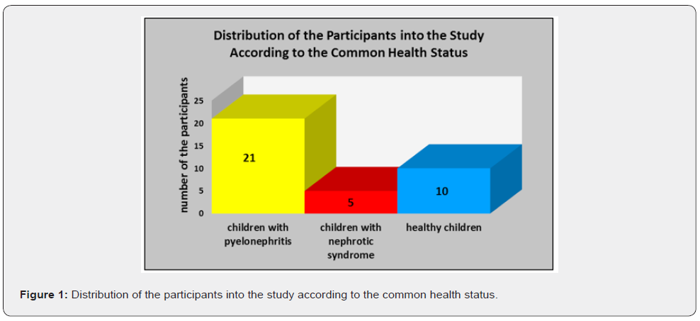
Subject of the study: A total number of thirty-six (36) participants are included in our study. All of them belong to the age interval of childhood, respectively up to the age of 18. These children are divided into three groups according to their common health status. The number of participants with diagnosed excretory system disorders equals to twenty-six (26). A control group of ten (10) healthy children also takes part in that investigation. A declaration of informative consent has been signed by a parent or guardian of each of the participants into the study (Figure 1).

Methods

In the context of that study are applied clinical methods, laboratory methods and statistical methods.

Clinical Methods

The gingival status of the examined participants is assessed by the clinical indices of Plaque index by Silness-Lȍe (PLI) and Gingival index by Lȍe-Silness (GI). By application of the plaque index PLI Silness-Lȍe is evaluated the level of accumulated dental plaque on teeth surfaces as an essential factor for initiation and progression not only of carious lesions, but also of gingival inflammation. The representative teeth which are included are. The level of dental plaque is assessed with figures in the interval from 0 to 3, with record of the medial, distal, vestibular and oral surfaces of all the applied ramfjord teeth. The sum of figures illustrating the plaque on ramjord teeth surfaces of each participant in the study is divided to the total number of teeth surfaces, respectively 24, which results in the average individual value of the plaque index PLI Silness-Lȍe. B the means of the gingival index GI Lȍe-Silness is ascertained the status of gingival inflammation. With the figures from 1 to 3 is registered the degree of gingivitis of the same teeth, respectively teeth surface as in the indicator of PLI Silness-Lȍe. The figure of 0 corresponds to healthy gingiva, without symptoms of edematization or bleeding. The figure of 1 associate to slight degree of gingival inflammation, clinically manifested by slight degree of edematization, without bleeding. The figure of 2 is equal to moderate degree of gingivitis. The last is characterized with edematized marginal gingiva and interdental papillae, combined with the symptom of provoked bleeding (bleeding on probing). The figure of 3 is related to severe degree of gingival inflammation, which correlates to pathological morphological substrate, interstitial edema in the zone of marginal gingiva and interdental papillas and spontaneous bleeding. The sum of all the recorded figures is divided to the total number of examined teeth surfaces, respectively 24. As a result, is obtained average individual value of the gingival index GI Lȍe-Silness for each of the participants included into the study.

Laboratory Method

The level of salivary secretory Ig A is evaluated by implementation of the method of radial immune diffusion. The individually collected samples of non-stimulated mixed saliva are collected in small containers with volume of 5 ml. The containers are stored at t = - 80 ○C. The salivary samples are being centrifuged for 15 minutes with 14000 revolutions per minute in condition of t = 4 ○C. According to the protocol, on a special plate supplied with outlined wells are deposited the centrifuged individual samples of non-stimulated mixed saliva. The plate with the samples is stored in its original package for providing proper level of humidity and hermetization for the period of incubation of the samples for 72 hours in room temperature (t = 22 ○C). After the fixed interval of 72 hours is made a record of the diameter of the circle around each well, measured in mm. Based on a monogram by the manufacturer of the product, each value of all of the registered diameters corresponds to a definite concentration of sIg A evaluated in mg/L.

Statistical Methods

Descriptive Analysis: The average value of n figures is determined when the total sum of all figures is divided in their whole number of n. The average value is the most widely applied parameter of central tendency. The median is the value which is in the middle position of the order of explored variables, or that is the value which separates the cases aligned according to the criterion of differentiation into two equal parts. Similar to the average value, the median is a definite characteristic of each statistical order and is unique for each sequence of data. The standard error is the quantitative expression of the uncertainty of the evaluation of the average value. Taking into consideration the variable of standard error and the rate of probability, the actual level of average value for the whole population is calculated in the range of average value plus/minus the standard error in terms of the fixed rate of probability. That is related to the variable of the interval of confidentiality. In the context of our investigation the interval of confidentiality equals to 95%.

Correlation Analysis: One of the main tasks of statistical analysis in the scope of medicine science is related to determination and establishment of interrelations between various events and phenomena, figured out at definite levels. The interrelation between the factorial characteristic and several or a great number of variations of the results-associated characteristic is defined as an interrelation of correlation. The coefficient of correlation by Pearson evaluates the significance of the linear interrelation between two variables. There is a five degreerelated scale of evaluation of correlation between two parameters based on definite intervals of variations of the coefficient of correlation by Pearson. In the interval between 0,00 and 0,25 is registered slight correlation. In the interval between 0,26 and 0,49 is evaluated moderate correlation. The interval of values of the coefficient by Pearson between 0,50 and 0,69 corresponds to significant correlation. The interval of values of the coefficient of correlation by Pearson between 0,70 and 0,89 regards to the condition of great correlation, and in the interval between 0,90 and 1,00 is established extremely great correlation. The positive interrelation of correlation is characterized with simultaneous increase or decrease of both of the investigated variables. The negative interrelation of correlation is marked out with elevation of the values of one of the explored parameters and reduction of the values of the other one (Figure 2).

Results

The lowest average value of the indicator of secretory salivary Ig A, namely 33,5920 mg/l, is established among the examined children with diagnosed nephrotic syndrome. Among the participants suffering from pyelonephritis and healthy children are registered similar values of this parameter of sIg A, respectively 58,7143 mg/l and 56,8800 mg/l. The lowest value of the median of secretory Ig A is recorded among the patients with nephrotic syndrome, namely 40,0000 mg/l. The healthy participants and children with established pyelonephritis are characterized with similar levels of that indicator, respectively 52,2000 mg/l and 52,1000 mg/l. The lowest minimal value of sIg A amounts to 4,06 mg/l and is recorded among the examined patients with nephrotic syndrome. Greater minimal value of secretory Ig A, equal to 12,80 mg/l, is registered for the healthy controls. The highest level of that indicator is related to the representatives of the study with the diagnosis of pyelonephritis, respectively 14,70 mg/l. The lowest maximal value of the variable of salivary sIg A, namely 57,2000 mg/l, characterizes the participants in the study with diagnosed nephrotic syndrome. Among the participants without common health disorders the maximal value of sIg A amounts to 123,0000 mg/l. In the group of patients suffering from pyelonephritis is ascertained the maximal value of sIg A equal to 130,0000 mg/l.

With the highest average value of the Plaque Index by Silness- Lȍe (PLI), respectively 1,8640, and the highest average value of the Gingival Index Lȍe-Silness (GI), respectively 1,6020, are characterized the children suffering from nephrotic syndrome. Among the patients with diagnosed pyelonephritis and healthy controls are recorded similar levels of the average value of PLI equal to 1,3957 and 1,3010, respectively. The average values of GI among the participants with pyelonephritis and children without common health disorders amount namely to 1,0390 and 1,0150 (Tables 1-3). Based on the calculated coefficient by Pearson there have been established definite interrelations. Among the patients suffering from pyelonephritis has been recorded slight negative correlation between salivary sIg A and the Plaque index PLI, as well as between salivary sIg A and the Gingival index GI. Among the children with diagnosed nephrotic syndrome has been registered moderate negative correlation between salivary sIg A and PLI, as well as moderate negative correlation between salivary sIg A and GI. The healthy representatives of the study have been characterized with significant negative correlation between the indicators of salivary sIg A and PLI Silness-Lȍe, as well as with significant negative correlation between salivary sIg A and GI Lȍe- Silness (Graph 1).

Among the participants with the diagnosis of nephrotic syndrome has been ascertained moderate positive correlation between the indicator of salivary sIg A and salivary pH. Children without common health disorders included in the investigation confirm similar level of moderate positive correlation between both of these variables of salivary pH and sIg A (Graph 2). Among the patients suffering from nephrotic syndrome there has been recorded great positive correlation between the clinical parameters of the Plaque Index PLI Silness-Lȍe and Gingival Index GI Lȍe-Silness. The children with diagnosed pyelonephritis and healthy controls in the study have been characterized with extremely great positive correlation between the indices of PLI and GI (Graph 3). The participants with the diagnosis of nephrotic syndrome have been characterized with moderate negative correlation between the clinical indicator of PLI Silness-Lȍe and salivary pH level. In the group of children with established excretory system disorder of pyelonephritis has been recorded significant negative correlation between the Plaque Index PLI and the para-clinical parameter of salivary pH. Among the healthy representatives in the investigation has been registered extremely great negative correlation between both of the indicators of PLI and salivary pH (Graph 4). Great negative correlation between the clinical indicator of GI Lȍe-Silness and salivary pH value has been ascertained among the representatives of the three groups included in the study, namely in the group of patients with pyelonephritis, in the group of participants with nephrotic syndrome and among the healthy controls (Graph 5).

Discussion

The results obtained in the context of our study are based on the explicitly manifested interrelations between the local immunity factor of salivary secretory Ig A and the specifics of common health status on individual and group level. There are definite, considerable correlations between local immunity into oral cavity and basic clinical parameters of the state of gingival tissue complex. A scientific investigation accentuates upon the interrelations between the salivary secretory Ig A in its role for physiological, functional and clinical manifestation of local acquired immunity in oral cavity, from one side, and effectors on systemic and local level, on the other side [37]. The secretory Ig A is characterized as a fundamental indicator for proper assessment of the level of risk of alteration of hard teeth tissues and periodontal structures in condition of pathological processes [27,28]. Among some of the participants in our study with the diagnosis of nephrotic syndrome is verified moderate negative correlation between sIg A and the Plaque Index PLI. Simultaneously, there is negative moderate correlation between the parameters of sIg A and the Gingival Index GI. Immunesuppressive activities of corticosteroids perform inhibiting effect upon the protective mechanisms of local immunity in oral cavity. This is related to suppression of the synthesis and secretion of salivary immunoglobulins, including immunoglobulin A (Ig A) [17,18,33]. In parallel, the common health disorder of renal failure is associated with a definite increase of blood and salivary urea level with risk of establishment of the pathological state of uremic stomatitis. The elevated rate of urea in blood and saliva is related to considerably lower distribution of non-cavitated and cavitated carious lesions, but to enhanced risk of gingival and periodontal disease [38-42]. We confirm the key role of secretory Ig A in non-stimulated mixed saliva as a plaque-inhibiting indicator, with suppressive activity against progression of the gingival inflammatory process.

In the context of a study performed by Rashkova et al. [26] a number of thirty (30) children without common health disorders and with clinically healthy gingiva are compared with thirty (30) children also without somatic deviations, but with clinically manifested plaque-induced gingivitis. In the group of 30 children with diagnosed gingivitis the average value of salivary sIg A amounts to sIgA= 41,07±32,14 μg/ml. The group of 30 healthy participants at child’s age with no symptoms of gingival inflammation are characterized with higher average value equal to sIgA=48,3±32,41 μg/ml. There has been established a statistically significant correlation between sIgA and the plaque index PLI Silness & Lȍe (p< 0,05). There has been ascertained no correlation between salivary sIg A and the degree of bleeding on probing (BoP). The parameter of sIgA is evaluated as a variable with considerable significance of assessment of the risk of pathological processes with impact upon oral cavity tissues. Secretory Ig A in the medium of non-stimulated mixed saliva performs plaqueinhibiting activity with key control function against advancing of the gingival inflammatory process [27]. In our study the children suffering from nephrotic syndrome are treated by application of corticosteroids [34-36]. In the specific therapy-related conditions of that excretory system disorder salivary sIg A is characterized with caries-protective activity against initial carious lesions and indirect impact upon the levels of plaque and gingival indices.

Based on the principles determined into the researches by Kiselova [17] and Dencheva et al. concerning the behavioral patterns of dental medicine doctors towards patients suffering from urinary tract infections, we accentuate on the explicit necessity of regular visits at dentist’ s office on each three months. The purpose of these appointments is associated to the performance of professional complex oral-hygiene procedures in combination with programs for motivation and re-motivation of patients [17]. The infections affecting hard teeth structures and periodontal tissues have to be adequately and radically treated [43-48].

The specifics of clinical manifestation and therapeutic protocols of the excretory system disease of pyelonephritis correlate to numerous factors with negative influence on the processes of formation of enamel, respectively on its qualitative and quantitative traits. There have been investigated the adverse effects of wide spectrum antibiotics [49-52], limitation of proteins’ consumption in condition of modified dietary regime, with prevailing frequency of intake of cariogenic foods [53], recurrent episodes of sub-febrile and febrile body temperature [54]. These variables combined with the need of application of steroid antiinflammatory drugs concern the disease of nephrotic syndrome. All of these factors serve as definite prerequisites for impingement of oral-dental health and disturbance of the periodontal status of the patients with excretory system disorders of pyelonephritis and nephrotic syndrome [55].

In the context of profound, thorough investigations of the salivary marker of pH is ascertained its essential role of a considerable factor for initiation and progression of the carious process [56-58]. In our study we accentuate on the interelations between dynamics of salivary pH levels and gingival health status in children with diagnosed nephrotic syndrome and pyelonephritis. In parallel, the researchers A. Ivanova, A. Krusteva and Z. Krustev evaluate the average value of pH in mixed saliva equal to 7,2 [17]. The limited consumption of foods rich in proteins, with respect to protection of the renal system from functional overload, is compensated with considerable amounts and high frequency of intake of sugar-containing products among the participants with established nephrotic syndrome and pyelonephritis. As a consequence of performance of this explicitly modified dietary regime there is a considerable number of episodes of reduction of salivary pH level, facilitating the inflammatory process of the gingival tissue [56,59].

Conclusion

1. The lowest level of secretory Ig A in saliva is registered among the participants suffering from nephrotic syndrome.
2. The increased concentration of salivary secretory Ig A correlates to the lower levels of the Plaque Index PLI and the Gingival Index GI.
3. The reduced level of secretory Ig A in saliva predisposes to initiation and progression of inflammatory reactions of the gingival tissue. 

To Know more about Dentistry & Oral Health  

To Know more about our Juniper Publishers


An Evidence Based Review of Vitamin D in COVID-19 Severity and Mortality - Juniper Publishers

 Complementary Medicine & Alternative Healthcare - Juniper Publishers Abstract Introduction: This evidence-based review aims to exp...