Oceanography & Fisheries - Juniper Publishers

Abstract
This work is devoted to the study of the features of biogenic migration of natural radionuclides in the conditions of Lake Issyk-Kul located in the north-eastern part of Kyrgyzstan. Lake Issyk-Kul is a high-mountainous, thermal, drain less reservoir with brackish water with a high content of natural uranium. This is due to climatic conditions, the geological structure of river valleys, and the peculiarities of the composition of river waters capable of extracting uranium from rocks. Calculations using the Erica tool 1.3 software showed that the natural uranium content in the water of Lake Issyk-Kul (0.809 Bq/l) is safe for living organisms of the aquatic ecosystem, the level of absorbed dose, accumulation and risk factors vary within the normal range. According to the Erica tool 1.3 database, low radiation doses (0-50 μGy/h) can stimulate the growth of aquatic plants. The species composition of the ichthyofauna of Lake Issyk-Kul consists of 22 species of fish, including 12 aboriginal and 10 acclimatized. The content of natural radionuclides (226Ra, 228Th and 212Pb) in the bone and muscle tissue of some fish species of Lake Issyk-Kul was investigated. It has been established that radionuclides are predominantly found in bone tissue in comparison with muscles, their concentrations vary within the limits of natural parameters.
Keywords: Water; Natural radionuclides; Fish; Issyk-Kul Lake
Introduction
Lake Issyk-Kul is located in the northeastern part of Kyrgyzstan, between the ranges of the Northern Tien Shan: Kungey-Ala-Too and Terskey-Ala-Too, at an altitude of 1609 meters above sea level. The lake is closed, with about 80 relatively small rivers flowing into it. The largest of them are Tyup and Jergalan, flowing from the east. The water is brackish (water salinity 5.90 ‰), so it does not freeze in winter. The volume of water is 1738 km3, the area of the water surface is 6236 km2, the average depth is 278 m, the maximum depth is 668 m. The length of Issyk-Kul from west to east is 182 km, and from south to north - 58 km. The climate is temperate maritime, with the lake having a softening effect in the region. Average January temperature: -2° -6°, average July temperature: + 17° + 21°. In the western mountains bordering the lake, only 115 mm of precipitation falls, on the eastern coast - about 600 mm [1-3]. According to the research of prof. Kovalsky [4], the waters of the rivers in the regions of the Issyk-Kul depression are enriched with natural uranium. The increased content of uranium in the waters should be associated not only with the climatic conditions of the regions, but also with the geological structure of river valleys, as well as with the peculiarities of the chemical composition of river waters capable of well extracting uranium from rocks. So, for example, if in the northern rivers of Russia (Northern Dvina, Lena, Neva, Kama), the uranium content fluctuates within (2-13) ×10-7 g/l, while in the rivers flowing into Lake Issyk-Kul, its concentration increases to (58-71) ×10-7 g/l [4]. The average uranium content in the water of Lake Issyk-Kul varies within (62-65) ×10-6 g/l [5]. In modern literature, a number of studies are presented on the topic of pollution, migration, accumulation of radionuclides in the environment and their biological effect on living organisms. [6-12]. Based on the above, the aspect of the biological effect of natural concentrations of uranium and products of its decay on living organisms of the aquatic ecosystem of Lake Issyk-Kul is poorly studied.
Materials and Methods
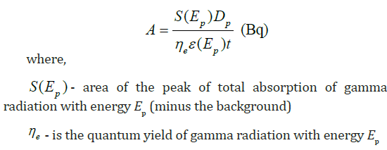
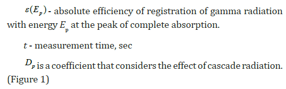
The content of natural radionuclides (226Ra, 228Th and 212Pb) in the bone and muscle tissue of some fish species of Lake Issyk-Kul has been investigated. 64 samples were prepared for gamma spectrometric analysis. The measurements were carried out on a GX4019 gamma spectrometer with Genie-2000 S 502, S501 RUS software. To obtain a stable equilibrium of radionuclides, the studied samples after their packing were stored from 25 to 180 days. The radionuclide activity in the sample on the day of spectrum measurement was calculated by the formula:

The Erica tool 1.3 application package is used for radioecological assessment of the state of terrestrial and aquatic ecosystems. Knowing the content of radioactive elements in soil or water, the program calculates the accumulation of radionuclides by living organisms, absorbed radiation doses, and an assessment of the radiation risk factor. The element of the radioecological assessment consists of three levels, at the first level the radiation risk factor is assessed, if its value is low, then level 1 can be limited (Figure 2). If the value of the radiation factor is increased, then the action is recommended to continue the assessment at level 2. To assess the statistical distribution of indicators, level 3 is used [13,14].

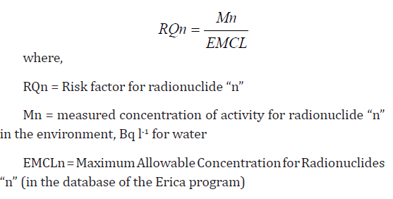
To calculate the radiation risk factor using the Erica tool 1.3 program, we entered the data on the specific activity of uranium in the water of Lake Issyk-Kul. If the calculated value of the risk coefficient is higher than 1, then there is a possibility of accumulation of radionuclides by living organisms. The risk factor (RQ) was determined using the following formula:
Results and Discussion
The species composition of the ichthyofauna of Lake Issyk- Kul is not rich and consists of 22 species of fish, including 12 aboriginal and 10 acclimatized [15,16]. We have investigated the distribution of natural radionuclides (226Ra, 228Th and 212Pb) in the bone and muscle tissue of some fish species of Lake Issyk-Kul. For the analyzes, fish species with wide distribution were selected, their habitat, type of food, commercial value were considered:
1. Pike perch (Lucioperca Lucioperca. 1958) is a predator, a widespread acclimatized species, the basis of its food is chironomids, mysids, amphipods, from about one year old it begins to switch to a predatory type of food. Forms large accumulations during the feeding period in shallow waters, and mainly in areas of the lake with a lower salinity of water.
2. Issyk-Kul trout (Salmo ischchan Issykogegarkuni Lushin. 1932) - an acclimatized species, distributed throughout the lake, with the formation of pre-spawning accumulations in the estuarine areas, near shallow waters with a spawning substrate. The diet of trout is predominantly benthic, but it can switch to a predatory type of diet.
3. Issyk-Kul Chebachok (Leuciscus bergi Kaschkarov. 1925) - an aboriginal and rare species, found in small quantities in the southwestern and southeastern parts of the lake.
4. The research results showed that natural radionuclides are predominantly found in bone tissue in comparison with muscles, especially 226Ra (Table 1). Concentrations of natural radionuclides vary within the background values, for example, the 226Ra content in the total mass of a two-year-old pike perch (Lucioperca Lucioperca) was 0.61 Bq/kg per wet weight, the absorbed dose level was 0.081 μGy/h, and the risk factor was 0.008. Lower concentrations are typical for 228Th and 212Pb.

With the average uranium content in the water of Lake Issyk- Kul - 0.809 Bq/l, the following results were obtained (Table 2). Among the reference organisms, uranium is able to accumulate in aquatic plants (190.09 Bq/kg). The absorbed dose level was 4.56 μGy/h, it can vary in the range of 0.47 - 17.9 μGy/h. According to the Erica tool 1.3 database, absorbed doses in the range (0-50 μGy/h) do not have statistically significant negative biological effects on aquatic plants. At low doses of radiation, a slightly stimulating effect on growth is possible (by 1.2 times). For Lake Issyk-Kul, the greatest species diversity falls on the group of blue-green (Cyanophyta), diatoms (Bacillariophyta) and green (Chlorophyta) algae. The average value of the hazard coefficient for aquatic plants is (0.45). If the calculated value of the risk coefficient is higher than 1, then there is a probability of accumulation of radionuclides by living organisms (Figure 3). The estimated average uranium content in pelagic fish is 7.11 Bq/kg wet weight. The absorbed dose level was 0.17 μGy/h, it can vary in the range of 0.06 - 0.42 μGy/h (Figure 4).
According to the database of the Erica tool 1.3 software, no statistically significant negative biological effects on the fish organism are observed within these absorbed doses. It is known that uranium accumulates mainly in bone tissue, kidneys, gills, and then in the liver, and only a small part is retained in muscles. Since parts of fish organs: gills, liver and bones are usually not eaten by the population, the total daily dose for uranium established by the World Health Organization is no more than 50 Bq/kg [17,18].


Conclusion
Calculations using the Erica tool 1.3 software showed that the natural uranium content in the water of Lake Issyk-Kul (0.809 Bq/l) is safe for living organisms of the aquatic ecosystem, the level of absorbed dose, accumulation and risk factors vary within normal limits. According to the conclusions of the scientific of the UN Committee on the Effects of Atomic Radiation, the absorbed dose of 80 μGy/h is the threshold dose. The presented absorbed doses for reference organisms in Table 1 are much lower than this indicator. The content of uranium and other natural radionuclides (226Ra, 228Th and 212Pb) in certain fish species of Lake Issyk-Kul vary within the background values, mainly radionuclides are found in bone tissue as compared to muscles.
To Know more about Oceanography & Fisheries
Click here: https://juniperpublishers.com/index.php