Showing posts with label Inflammation. Show all posts

Monday, August 12, 2024

Inflammation and Cartilage Degradation in the Pathophysiology of Osteoarthritis: Potential for Targeted Therapies - Juniper Publishers


Orthopedics and Rheumatology Open Access Journal - Juniper Publishers

Abstract

Generally, osteoarthritis (OA) is considered to be a common degenerative joint disorder characterized by destruction of the articular cartilage, subchondral bone alterations and low-grade joint synovitis. Aging population and obesity are increasingly being associated with a concomitant increase in the incidence of OA and disability worldwide causing significant burden on the individual and society. The understanding of OA has evolved over years from a degenerative (wear and tear) non-inflammatory disease to a disease of joints that results from many predisposing factors including: aging, genetics, excessive exercise, obesity, genetic predisposition, inflammatory autoimmune disorders, poor nutrition, injury, metabolic disorders such diabetes mellitus, homocysteinaemia, joint trauma, hormonal imbalance, altered biomechanics, obesity and infection. All these factors contribute to an imbalance between catabolism and anabolism of joint cartilage leading to eventual joint damage and structural joint failure. This review focuses on the pathophysiological basis of OA highlighting the potential of specific targeted treatments.

Keywords: Osteoarthritis; Pathophysiology; OA treatment; Cytokines; Inflammation; Targeted therapy

Abbreviations: OA: Osteoarthritis; RMD: Rheumatic and Musculoskeletal Disease; DMOADs: Develop Disease Modifying Osteoarthritic Drugs; AGE: Advanced Glycation End Products; MMPs: Metalloproteinases; VIP: Vasoactive Intestinal Peptide; BMI: Body Mass Index; CGRP: Calcitonin Gene Related Peptide; IGF-1: Insulin-like Growth Factor 1; NO: Nitric Oxide; PGE2: Prostaglandin E2; ECM: Extra-Cellular Matrix; mPGES-1: microsomal PGE Synthetase-1; sPLA2: soluble Phospholipase A2; IFNϒ: Interferon Gamma; COX2: Cyclo-Oxygenase 2; BMLs: Bone Marrow Lesions

Introduction

Osteoarthritis (OA) is one of the most common non-inflammatory rheumatic and musculoskeletal disease {RMD}associated with progressive cartilage destruction which leads to structural and functional joint failure. Aging and other concomitant diseases such as diabetes mellitus are associated with significant increased risk of developing OA. The understanding of OA has evolved over years from that of a degenerative (wear and tear) non-inflammatory disorder to a disease of joints that results from many predisposing factors that include inter alia: aging, genetic predisposition, excessive exercise, obesity, genetic predisposition, hormonal imbalance, inflammatory autoimmune disorders, poor nutrition, injury, metabolic disorders such diabetes mellitus, homocysteinaemia, joint trauma, altered biomechanics, obesity and infection [1-3].

Despite OA being the most common joint disease, there is still is no definitive or preventive treatment other than symptomatic treatment. As the aging population increases globally, OA poses a significant financial and social burden. This highlights the need to develop disease modifying osteoarthritic drugs (DMOADs) [2]. A full understanding of the pathophysiologic and inflammatory pathways that initiate the onset and progression of OA will facilitate the development of targeted therapies [2,3]. Early diagnosis as well as understanding the risk factors and development of biomarkers of OA will aid early therapeutic intervention. The pathophysiologic mechanisms involved in OA are complex and multiple, and lead to the development of a state of chronic inflammation resulting in propagation and progression toward the phenotype of clinical OA [3]. Current studies have shown that inflammatory cytokines produced also by chondrocytes, induce chondrocytes to release metalloproteinases that degrade cartilage [4]. This review will focus on the pathophysiological basis of OA highlighting the potential of targeted treatments and need to continue to design such strategies.

Pathophysiology

Chondrocytes produce articular cartilage. Cartilage is comprised of extracellular collagen matrix and various proteoglycans. Cartilage is metabolically active and is constantly renewed throughout life [1-5]. The elasticity and compressibility of cartilage is due to the proteoglycan Aggrecan. Aggrecan is a central core protein comprising of numerous glycosaminoglycan chains of chondroitin sulphate and keratan sulphate moiety which are attached to hyaluronic acid [5]. The collagen functional and metabolic network is disrupted with ageing leading to fissuring and increased levels of advanced glycation end products (AGE). AGE binds specific advanced glycation end products (RAGE) receptors found on the surface of chondrocytes. This binding perturbates the catabolic activity in chondrocytes [6]. AGEs can also induce oxidative stress in cells triggering the secretion of many pro-inflammatory cytokines and chemokines.

A variety of cellular factors involved in the pathogenesis of OA are inter alia: mononuclear cells, chondrocytes, osteoblasts and osteocytes, and synovial lining cells [1-19]. A variety of soluble mediators such as: proinflammatory cytokines {tumour necrosis factor α (TNFα), interleukin 1β (IL-1 β), interleukin 6 (IL-6)}, transforming growth factor β (TGF β), metalloproteinases (MMPs), lipid mediators [Prostaglandin E2 (PGE2)], nitric oxide (NO), insulin-like growth factor1 (IGF-1), adipokines (leptin, adiponectin, resistin) and neuropeptides [neuropeptide Y, vasoactive intestinal peptide (VIP), substance P, and calcitonin gene related peptide (CGRP)] are also involved in the disease pathophysiology [2,7,8]. Neuropeptides induce pain in OA via afferent sensory nerve nociceptors which signal to the brain the via the spinal and spinothalamic tracts [9]. Nociceptors are found on several structures of the osteoarthritic joint such as the synovium, periosteum, subchondral bone and ligaments [7,8].

Chondrocytes express estrogen receptors which can mediate the production of growth factors [7]. The synthesis of these growth factors is decreased in menopausal women due to a reduction in estrogens [7]. One of the risk factors for OA in weight-bearing joints and also for non-weight-bearing joints such as hands is obesity. Adiposity contributes to a proinflammatory milieu since adipose tissue secretes many inflammatory mediators such as adipokines and leptin [10,13]. Leptin is also found in osteophytes and cartilage from patients with OA and its levels in synovial fluid correlate with body mass index (BMI). Leptin is pro-inflammatory and stimulates the synthesis of IGF-1 and TGFβ, and proteoglycans by chondrocytes [11,12]. Leptin enhances the capacity of pro-inflammatory cytokines to induce NO synthesis [12,24]. Visfatin is one of the adipokines which has been shown to enhance cartilage degradation [13]. Adiponectin levels are low in obese patients and are also negatively associated with hand OA progression [14].

Pro-inflammatory cytokines can activate chondrocytes, synovial cells and osteoblasts. OA Chondrocytes from OA patients can secrete a number of metalloproteinase enzymes such as MMP-1 (collagenase 1), MMP-3, MMP-9, MMP-13 (collagenase 3), aggrecanase, ADAMTS-4 and ADAMTS-5, that all degrade cartillage. Specific endogenous metalloproteinase tissue inhibitors (TIMPs) {TIMP1, TIMP2, TIMP3 and TIMP4} are also produced and inhibit MMPs activity. is determined by The MMPs and TIMPs balance ratio, determines the rate of cartilage matrix degradation in joints. Collagenase 1 and 3, and MMP-13 (collagenase 3) can cleave type II collagen [5]. Cartilage degradation can also be induced by Stromelysin-1 (MMP-3), Stromelysin-2 (MMP-10), Stromelysin-3 (MMP-11) [15]. Matrisylin (MMP-7) also plays a role in the degradation of proteoglycans [16]. IL-1β differential regulates MMPs and TIMP syntheses suggesting that the low the grade IL-1β production in joints may promote cartilage degradation by relative upregulation of MMPs versus TIMP, thereby creating an unfavorable balance between the level of the degrading enzymes and their inhibitors [17].

Abnormal mechanical forces and oxidative stress can stimulate chondrocytes, synovial cells and subchondral osteoblasts to produce proinflammatory cytokines {IL-1β, TNF-α} [18]. These cytokines can initiate a variety of catabolic and degradative processes in cartilage mediated by metalloproteinases that degrade cartilage extra-cellular matrix (ECM) [2,19]. The expression of ADAMTS-4 and ADAMTS-5 and enhanced production PGE2 can be induced by IL-1β and TNF-α via enhanced of the gene expression and/or activities of COX-2, microsomal PGE synthetase-1 (mPGES-1), and soluble phospholipase A2 (sPLA2). IL-1β and TNF-α can also stimulate nitric oxide synthetase (iNOS or NOS2) to upregulate NO production and stimulate other production of other proinflammatory cytokines such as IL-6, LIF (leucocyte inhibiting factor), IL-17 and IL-18, chemokines and IL-8. Serum levels of IL-6 and IL-8 are significantly higher OA patients when compared to healthy subjects. The genes associated with the differentiated chondrocyte phenotype, including aggrecan (AGAN) and type II collagen (COL2A1) are suppressed by IL-1β and TNF-α [5]. The inhibition of IL- 1β involves upregulation of: IL-1 Receptor antagonist (IL-1Ra), soluble form of IL-1R, and anti-inflammatory cytokines [2,20]. On the other hand, the anti-inflammatory cytokines IL1Ra, IL-4, IL-10, IL-13 and interferon gamma (IFNϒ) are also present in the OA joint. They inhibit the secretion of some MMPs and may differentially increase the synthesis of TIMPs [21]. Defective production of these anti-inflammatory factors could contribute to OA pathogenesis.

IL-1β and TNF-α, and lipopolysaccharides {produced by bacterial infections}, can upregulate iNOS gene expression and induces NO production. In chondrocytes. NO is involved in the degradation of cartilage by down-regulating the synthesis of IL-1Ra, aggrecan and collagen, enhancing MMPs activity. NO may contribute to chondrocyte apoptotic cell death by downregulating cell survival signals from the ECM [22,23]. In experimental animal models of OA, inhibitors of NO synthesis retard the development of histological changes and clinical features of experimental OA [24]. The eicosanoid pathway is also involved in chondrocyte activation [25,26]. Pro-inflammatory cytokines induce Prostaglandin E2 (PGE2), via the PLA2 pathway, cyclo-oxygenase 2 (COX2) and mPGES1. The activity of synovial cells, macrophages, chondrocytes is modulated by PGE2, which also induces bone resorption [27]. PGE2 induces several MMPs and can potentiate the action of other inflammatory mediators.

A loss of chondrocyte maturational arrest which pushes chondrocytes towards a more differentiated, hypertrophic-like state, has been observed in cartilage from OA patients. This chondrocyte hypertrophy increases the expression of type X collagen, upregulation of matrix metalloproteinases-13 and induces the synthesis of type II collagen and aggrecan, (MMP-13), synthesis of shorter proteoglycans and initiation of pathological calcification [27]. Chondrocytes can also interestingly, acquire a de-differentiated phenotype associated with increased types I and III collagen synthesis whilst type II collagen synthesis is inhibited [27]. Chondrocytes also acquire an activated phenotype corresponding to a pro-degradative state producing several proteinases induced prostaglandins), free radicals (NO, H2O2) and cytokines. These mediators perpetuate chondrocyte activation creating a vicious loop for cartilage degradation. NO induces NF-κB signaling that enhances pro-inflammatory cytokine production in the joint. NO also induces apoptotic chondrocyte cell death which further could decreases the synthesis of ECM components [21].

Cartilage breakdown products also enhance synovial inflammation which enhances catabolic and pro-inflammatory processes leading to excessive production of metalloproteinases that also break cartilage matrix establishing a positive feedback loop. The synovial tissue from OA joints is infiltrated with macrophages and T cells associated with enhanced inflammatory cytokine expression in both early and late OA. Another important feature of OA pathophysiology is also the presence of sclerosis, osteophytes, bone cysts and bone marrow lesions (BMLs) and subchondral bone remodeling [28]. BMLs can occur at the onset of the disease and are now considered to be a precursor for OA development as well [29,30]. Osteoarthritis subchondral osteoblasts can stimulate overproduction of IL-6, IL-8, C-terminal type I procollagen propeptide, alkaline phosphate, osteocalcin, TGF β1, IGF-1, urokinase, and osteopontin [28-31]. These osteoblasts in the affected joints, fail to respond to the bioactivity of parathyroid hormone, which might explain the dysregulated bone remodeling and osteophyte development [28].

Stimulation of trans-membrane G protein–coupled receptors by a variety of stimuli such as cytokines, hormones, neurotransmitters inter alia, initiates intracellular signaling by recruitment of by recruiting of the G protein-coupled receptor kinase 2 (GRK2). Perturbations of the GRK2 molecular pathway may also be involved in OA pathophysiology. GRK2 is involved in myocardial cell hypertrophy. Its expression has also been found to be increase in the chondrocytes from mouse models of OA and injured human cartilage and may also be involved in chondrocyte hypertrophy [31]. It has been observed that gene deletion of GRK2 in mice decreased experimental OA development [32]. This suggests a potential important role of GRK2 in OA pathophysiology. Finally, subchondral bone is involved in the pathogenesis of OA. Firstly, mechanical stress induce osteoblasts to produce the degradative MMP-1, MMP-13, PGE2 and IL-6. Secondly, enhanced expression of IGF-1 and TGF β in subchondral bone initiates new bone formation and development of osteophytes and subchondral sclerosis.

In conclusion, OA) is now considered to be a slow progressive inflammatory disease characterized by low grade inflammation, degradation of articular cartilage and joint function failure and pain. The balance between catabolic and anabolic mediators and their regulators is dysregulated. In addition, cartilage, the pathophysiologic processes also involve the entire joint, with active participation by subchondral bone, ligaments, capsule, synovial membrane and periarticular muscles. The understanding of the molecular and cellular pathophysiology of the disease is opening up novel therapeutic options using existing treatment modalities and potential to design and develop new ones.

Novel Therapeutic Approaches

The classical mode for the management of OA has been control of symptoms with a variety of symptom modifying OA drugs (SMOAD). The understanding of the pathophysiology of OA has given hope to the potential of disease modifying OA drugs (DMOAD) that are expected to modify the underlying OA pathophysiology, thereby inhibiting structural damage to prevent and/or reduce the development of mechanical joint failure and associated disability, and provide symptomatic relief of pain as well as mitigate the need for surgery. A number of targets have been identified for target therapy approaches:

Targeting Inflammatory sites

a. IL-1 inhibition

i. Anakinra

ii. Canakinumab

iii. Gevokizumab

iv. Lutikizumab

Targeting Bone Marrow Lesions

a. Bisphosphonates

b. Strontiun ranelate

c. Teripatide

d. Vid D3

e. Calcitonin

Targeting Cartilage Metabolic pathway

a. Growth factors

b. Wnt signaling pathway

c. Cathepsin-K

d. MMP/ADAMTS

e. AMPK pathway

Pain Mechanisms

a. NGF

b. Intra-articular corticosteroid injections

A number of trials have been helping with existing drugs with mixed results and outcomes [33-44]. A number of challenges are there. The existing drugs have an unfavorable safety profile given the nature of OA. There is therefore an unmet medical need for DMOADs which would be expected to bring significant clinical effects to OA management in the future. However, since catabolic and proinflammatory mediators (intracellular signaling, cytokines, GRP2, nitric oxide, neuropeptides prostaglandins) are involved in perturbating balance between cartilage matrix production, degradation and repair as well as OA synovitis, there is a potential for targeted therapeutic management.


To Know more about our Juniper Publishers Juniper Publishers
Click here: https://juniperpublishers.com/index.php


Monday, August 8, 2022

 JOJ Ophthalmology - Juniper Publishers


Abstract

Age-related macular degeneration (ARMD) is the leading cause of blindness in the developed world. No effective therapy exists for the dry form of ARMD, which makes up about 90% of cases. Major modifiable risk factors have been identified such as cigarette smoking, obesity, nutritional factors, and alcoholism. The risk of dry ARMD is increased by several modifiable etiologic factors in particular – lack of blue light filtration, oxidative stress, and inflammation. Two components of plant foods, lutein and zeaxanthin, screen out blue light, resulting in greater macular pigment optical density (MPOD), and act as antioxidants in the macular. A plant-based diet also reduces inflammation and lowers hsCRP. These factors combine to give vegetarians a lower risk of dry ARMD. Epidemiological studies have shown that consuming meat raises the risk of dry ARMD while consuming plant foods lowers the risk. Studies have shown significant correlations between high lutein concentration in ocular tissues or in serum and reduced risk of ARMD. Vegetarians have been found to have a greater MPOD. A broad range of antioxidants from plant foods, sulforaphane, can reduce oxidative damage, and may decrease the risk of dry ARMD. Elevated levels of hsCRP, a marker of inflammation, predict greater future risk of ARMD. On a vegan diet hsCRP was reduced 32%, even more than on the American Heart Association diet.

Keywords: Antioxidants; ARMD; Blindness; Inflammation; Lifestyle; Lutein; Macular pigment optical density; Oxidative stress; Vegan; Zeaxanthin

Abbreviations: ARMD: Age-Related Macular Degeneration, HSCRP: High-Sensitivity C-Reactive Protein, MPOD: Macular Pigment Optical Density; PUFA: Poly-Unsaturated Fatty Acids; ROS: Reactive Oxygen Species; SFN: Sulforaphane

Introduction

Age-related macular degeneration (ARMD) is the leading cause of blindness in the developed world [1,2]. It is becoming similarly important in the developing world in association with increasing longevity and Westernization of diet and lifestyle [3]. ARMD may be classified as dry or wet. While geographic atrophy may occur in both the wet and dry forms of ARMD, it is better characterized in the dry form [4]. It is characterized by sharply demarcated areas of hypopigmentation in which choroidal blood vessels are more visible than in surrounding areas [5,6]. Choroidal neovascularization (CNV) is more commonly seen in the wet form of ARMD. The dry form of ARMD is also known as non-exudative, non-neovascular, or atrophic ARMD. This is the more common form of ARMD, seen in about 90% of cases [7]. At present, no effective therapy exists for dry ARMD [8]. Dry ARMD is a multifactorial disease, having both genetic, age and modifiable risk factors [9]. The strongest risk factor is age: late ARMD can be found primarily in those aged 70 years and older [10,11]. The risk of ARMD is greater in persons with a family history of the disease than in those without [12] Observational studies have identified major modifiable risk factors such as cigarette smoking, obesity, nutritional factors, and alcoholism [9]. The risk of dry ARMD is increased by three modifiable etiologic factors in particular – lack of blue light filtration, oxidative stress, and inflammation. Two components of plant foods, lutein and zeaxanthin, screen out blue light and act as antioxidants in the macular. A plant-based diet also reduces inflammation. Therefore, we would expect a plant-based diet to contribute to a lower incidence of dry ARMD.

Epidemiology

Epidemiological studies have demonstrated that consuming meat increases the risk of dry ARMD while consuming plant foods decreases the risk. However, it should be noted that most epidemiological studies on ARMD do not distinguish between wet and dry ARMD [13]. Since most cases are the dry form of the disease, the results of research can be said to be broadly applicable. One study showed that a high consumption of red meat (fresh or processed) increased the risk of developing ARMD. That risk was over two times greater with processed meat, such as salami or sausage [14]. The Mediterranean diet pattern is characterized by high consumption of plant-based foods and fish and low consumption of meat and dairy products. In one study, adherence to the diet was associated with a 41% reduced risk of incident advanced ARMD. These findings support the role of a diet rich in healthful nutrient-rich foods such as fruits, vegetables, legumes, and fish in the prevention of ARMD [15]. People who follow a vegetarian diet have a greater macular pigment optical density (MPOD), affording them greater protection from blue light and greater levels of antioxidants, [16,17] resulting in a lower risk of dry ARMD. One study showed that their MPOD was about 18% higher in vegetarian subjects compared to non-vegetarian subjects [18].

Pathophysiology

. Lutein and zeaxanthin

Lutein and zeaxanthin are structural isomers that belong to a class of molecules called carotenoids. Carotenoids, which are primarily plant-derived lipophilic pigments, are essential factors in human health and development. Specifically, they function in a wide range of biological processes, including reproduction, embryonic development, immunity, and vision. In the eyes, they help provide coloration and absorption of light energy. Carotenoids may be divided into two general classes: carotenes and xanthophylls. Carotenes are non-polar molecules, which contain only carbon and hydrogen atoms, whereas xanthophylls are polar carotenoids, containing at least one oxygen atom [19]. Lutein and zeaxanthin are xanthophylls biochemically distinct from other carotenoids due to the presence of hydroxyl groups located at each end of these molecules. This functionality allows xanthophylls to be oriented in lipid membranes exposed to aqueous environments in a special and possibly protective way [20]. The xanthophylls (lutein and zeaxanthin) account for 20–30% of total carotenoids in human serum and 80–90% of total carotenoids in the human retina [21] the highest concentrations anywhere in the human body, suggesting an important functional role for these molecules in the retina [22]. Chemical structure of Zeaxanthin, Meso-Zeaxanthin and Lutein [23].

Lutein and zeaxanthin confer macular protection via antioxidant and light-screening properties [24]. Meso-zeaxanthin is a relatively recently discovered structural isomer of zeaxanthin and appears to be a metabolite of lutein or zeaxanthin. It also has both light filtering and antioxidant properties [25]. It has been shown that lutein and zeaxanthin are entirely of dietary origin, humans cannot synthesize lutein and zeaxanthin de novo. [16,17] This is evidenced by the fact that lutein and zeaxanthin levels in the diet, serum, and retina correlate. [26]. We have adapted a capacity for efficient lutein and zeaxanthin uptake [27,28], transport [29,30], retention [31-34] and protection [35] in the retina. The efficient operation of these processes may be indicated by the physiologic significance of lutein and zeaxanthin in retinal health and disease. The levels of macular pigments, usually measured in terms of the macular pigment optical density (MPOD), can reflect retinal health status [36-40]. An inverse association between macular pigment density and ARMD has long been recognized [41-42]. Studies have shown significant correlations between high lutein concentration in ocular tissues or in serum and reduced risk of ARMD [43-45].

Oxidative stress

Oxidative damage may play an important role in the pathogenesis of dry ARMD. Oxidative stress, which refers to cellular damage caused by reactive oxygen species (ROS), has been implicated in many disease processes, especially age-related disorders. ROSs include free radicals, hydrogen peroxide, and singlet oxygen, and they are often the byproducts of oxygen metabolism [46]. Oxidative stress is a major risk factor for the pathological development of retinal diseases and vision impairment. With age, the retinal pigment epithelium (RPE) and the space between the RPE and Bruch’s membrane gradually accumulate lipofuscin, a heterogeneous fluorescent mixture rich in lipid-protein complexes, which is also composed of by-products of vitamin A metabolism and lipid peroxidation [47-49]. Lipofuscin is undegradable and also acts as a plausible photosensitizer, generating ROS [50]. The retina is particularly susceptible to oxidative stress because of its high oxygen consumption, its exposure to light (photo-oxidation), and its exposure to high levels of ROS acting on PUFA content, including docosahexaenoic acid (DHA) [51-52]. Complement proteins have been found in histological specimens of eyes with dry ARMD. Altered levels of both intrinsic complement proteins and activated products have been found in the circulation of patients with dry ARMD. Complement activation may be triggered by oxidative stress, resulting from retinal exposure to incoming light [53].

Unfortunately, as we age, oxidative damage increases and antioxidant capacity decreases, as does the efficiency of reparative systems [54]. It appears that these age-related oxidative changes are characteristic indicators of early dry ARMD, which, in combination with hereditary susceptibility and other retinal modifiers, can progress to the pathology and visual morbidity associated with advanced dry ARMD. A broad range of antioxidants, known to have chemical properties that can reduce oxidative damage, have been proposed to decrease the risk of dry ARMD, [55,56] sulforaphane. Sulforaphane (SFN) is an isothiocyanate molecule present in cruciferous vegetables, broccoli being the most relevant example of a natural SFN source. (57) Two studies demonstrated the antioxidant potential of SFN on human RPE cells exposed to oxidant agents, including different chemical oxidative stressors and light exposition [58,59]. The mechanism by which sulforaphane accomplishes this has been determined [60]. SFN induces the expression of the phase 2 genes, through activation of the Nrf2. These genes encode for different antioxidant enzymes, including glutathione transferases or NAD(P)H dehydrogenase quinone [60]. The mechanism of action of SFN is based on the dissociation of the transcription factor Nrf2 of Keap1, a cytosolic repressor, promoting its translocation to the nucleus and inducing an antioxidant response. The binding of Nrf2 to the DNA promoter region antioxidant-responsive element (ARE), triggers the nuclear antioxidant response, through an increased expression and activity of reductive systems [61,62]. A plant-based diet protects against chronic oxidative-stress-related diseases. Dietary plants contain variable chemical families and amounts of antioxidants. Plant antioxidants may contribute to the beneficial health effects of dietary plant foods [63]. On average plant foods provide 11.57 mmol/100gm antioxidant content, while animal foods provide only on average 0.18 mmol/100gm [63]. It is therefore to be expected that a plant-based diet could decrease the risk and progression of dry ARMD through reducing oxidative stress.

Inflammation

Inflammation also plays a significant role in the incidence and progression of dry ARMD [64-66]. Drusen, subretinal deposits indicative of the onset of ARMD, have been shown to contain fibrinogen, vitronectin, complement components, and high-sensitivity C-reactive protein (hsCRP), proteins associated with generalized inflammation [67-69]. Inflammatory cell debris has also been isolated from the outer surface of the Bruch’s membrane in eyes with dry ARMD [70]. Pooled findings from 5 prospective cohorts show that elevated levels of hsCRP predict greater future risk of ARMD [71]. Lower levels of hsCRP were found in those following a vegetarian diet for more than 2 years [72-73]. An interventional study found that after 8 weeks on a vegan diet hsCRP was reduced 32%, even more than on the American Heart Association diet [74].

Clinical considerations

To help prevent dry ARMD, patients should be encouraged to eat plenty of foods containing lutein and zeaxanthin along with other plant foods containing antioxidants, as a part of a healthy plant-based diet. Lutein can be found in several vegetables such as kale, spinach, romaine lettuce, broccoli, and some nuts such as pistachio nuts. Zeaxanthin can be found in foods such as corn, orange peppers, mango, and orange juice. [75-76]. Supplements are also available, and some studies indicate that they may slow the progression of ARMD [77]. Bilberry and lingonberry extracts may also have a protective effect against blue light photooxidation through their antioxidant properties [78-81]. Despite initial enthusiasm, a meta study found that fish oil supplements (omega 3 LCPUFA) supplementation in people with ARMD for periods up to five years does not reduce the risk of progression to advanced ARMD or the development of moderate to severe visual loss [82]. A plant-based diet can be very efficacious in the prevention and treatment of comorbidities such as hypercholesterolemia and type 2 diabetes. For instance, the plant-based diet is as efficacious as Lovastatin in treating hypercholesterolemia and is more efficacious than Metformin in treating type 2 diabetes [83,84]. It also reduces the risk of several other pathologies such as coronary artery disease, [83] stroke, [85], osteoarthritis, [86] prostate and colon cancer, [87,88] diverticular disease, [89] ulcerative colitis, [90] Crohn’s disease, [91] Grave’s disease, Hashimoto’s thyroiditis [92] and rheumatoid arthritis [93] just to name a few. When treating comorbidities, it’s important to titrate relevant medications as the effect of the plant-based diet become evident. Lab work should also be done before starting treatment with a plant-based diet and then 6 to 8 weeks afterwards, since the therapeutic effects of a plant-based diet often take several weeks to become evident.

Discussion

There is currently no treatment for non-neovascular ARMD. Therefore, prevention, always of prime importance, becomes even more important. As longevity has increased in the developed world, the prevention of chronic diseases is needed even more than before. Oxidative stress seems to be one of the etiologic factors in dry ARMD. The plant-based diet, rich with antioxidants, can help reduce oxidation and therefore help prevent dry ARMD. Vegans have lower levels of hsCRP which indicates less inflammation, which reduces the risk of dry ARMD. The increased consumption of lutein and zeaxanthin and the greater MPOD that vegetarians have can also help prevent dry ARMD.

It’s notable that plant foods supply both zeaxanthin and lutein along with fiber but with little saturated fats. The plant-based diet has no contraindications or adverse effects. It can help treat several common comorbidities. One weakness of this research review is that much of the research fails to report the type of ARMD studied. Since 90% of the cases of ARMD are dry ARMD, this is unlikely to change the conclusion.

To Know more about JOJ Ophthalmology 

Click here: https://juniperpublishers.com/index.php  

Tuesday, November 16, 2021

Evaluation of Biofield Energy Healing Treatment Based Proprietary Test Formulation on Gut Health Potential in Colon Cancer Cell Line (HT-29) - Juniper Publishers

 Pharmacology & Clinical Research - Juniper Publishers

Abstract

The present study was aimed to evaluate the anti-inflammatory potential of Biofield Energy Healing (the Trivedi Effect®-Consciousness Energy Healing) on the test formulation in colon cancer cell line (HT-29). Each ingredient of the test formulation was divided into two parts, one part was denoted as the untreated test formulation and the other part was demarcated as the Biofield Energy Treated test formulation, which received Biofield Energy Healing Treatment by a renowned Biofield Energy Healer, Mr. Mahendra Kumar Trivedi. MTT assay showed that the test formulation was found safe and non-toxic upto 122 μg/mL in HT-29 cells with more than 78% cell viability. The level of interleukin-6 (IL-6) expression was significantly reduced by 34.09% and 59.41% (p≤0.001) at 3 and 15 μM, respectively compared to the vehicle control (VC) group under the stimulation of tumor necrosis factor - alpha (TNF-α). Moreover, IL-8 level was significantly suppressed in the Biofield Energy Treated test formulation by 57.09% and 42.88% at 0.1 and 3 μM, respectively compared with the VC group. However, the Biofield Energy Treated test formulation further substantial altered the level of interferon gamma compared to the VC group. The Trivedi Effect®-Consciousness Energy Healing significantly regulate the inflammatory condition after treatment with the test formulation in colon cancer cell line (HT-29). This experimental data suggested that the Biofield Treated test formulation can be utilized for many inflammatory disease conditions such as rheumatoid arthritis, multiple sclerosis, psoriasis, inflammatory bowel diseases, scleroderma, and type 1 diabetes mellitus.

Keywords: Biofield Energy Healing; The Trivedi Effect®; Inflammation; Colon cancer cell line (HT-29); Pro-inflammatory cytokines

Abbreviations: FBS: Fetal bovine serum; MTT: 3-(4,5-Dimethylthiazol-2-yl)-2,5-Diphenyltetrazolium Bromide; ELISA: Enzyme-linked immunosorbent assay; NCCAM: National Center for Complementary and Alternative Medicine; CAM: Complementary and Alternative Medicine

Introduction

The relation among of the gut health, micro biota, and cytokines have been well studies and reported in past two decades against various inflammatory bowel diseases (IBD) and associated mucosal inflammations. The cytokines play an important role in the mechanism in inflammation especially in Crohn’s disease and ulcerative colitis. The pathogenesis of IBD is not completely understood, but the role of cytokines in the intestinal immune system has significant impact in disruption of normal state of controlled gut inflammations [1,2]. Innate immune response is the major response in gut inflammation and its related diseases. Most of the cytokines are secreted by activated dendritic cells and the macrophages, which regulates the inflammatory response in gut inflammatory diseases. Once, these cytokines are secreted by antigen presenting cells, they triggers and differentiate various T cells by activating adaptive immune response. Gut inflammation dysregulates the T-cells, and manage the over-reactive and auto-reactive cells. T-cell regulation or its overproduction leads to the development of gut inflammatory diseases [3]. These cells along with various types of cytokines play a complex role in inflammatory gut diseases [4, 5]. Thus, there is the need of some novel formulation which alters the level of cytokines to improve the gut health. The present study was aimed to test the impact of the Biofield Energy Treated test formulation comprised of zinc chloride, ferrous sulphate, copper chloride (II-cupric), vitamin B6 (pyridoxine HCl), vitamin B12 (cyanocobalamin), magnesium (II) gluconate, and cholecalciferol (vit. D3) against the colon cytokines. The novel test formulation was treated with Biofield Energy Healing Treatment, as one of the best CAM approach with significant therapeutic outcomes. Biofield Energy Healing is one of the emerging frontiers aspect to CAM and various clinical approach has been used with significant results [6-9]. CAM therapies have been recommended by The National Center for Complementary/Alternative Medicine (NCCAM) and there therapies exist in various forms such as external qigong, Johrei, Reiki, therapeutic touch, yoga, Qi Gong, polarity therapy, Tai Chi, pranic healing, deep breathing, chiropractic/osteopathic manipulation, guided imagery, meditation, massage, homeopathy, hypnotherapy, progressive relaxation, acupressure, acupuncture, special diets, relaxation techniques, Rolfing structural integration, healing touch, movement therapy, pilates, mindfulness, Ayurvedic medicine, traditional Chinese herbs and medicines in biological systems both in vitro and in vivo. Human Biofield Energy has subtle energy that has the capacity to work in an effective manner [10] with its various clinical benefits [11]. This energy can be harness and transmit it into living and non-living things by the process of Biofield Energy Healing Treatment. Biofield Energy Treatment (the Trivedi Effect®- Consciousness Energy Healing Treatment) has been extensively studied with significant outcomes in the field of pharmaceuticals [12-14], nutraceuticals [15,16], metals and ceramics [17-19], microbiology [20-22], microbial genetics [23, 24], cancer research [25,26], livestock, agriculture science [27-29], improved bioavailability of many compounds [30-32], improved skin health [33, 34], improved properties of nutraceuticals [35, 36], improved overall bone health [37-39], human health and wellness. Thus, the study was planned on colon cytokines estimation that could significantly helped to improve the prevalence of gut inflammatory diseases using novel test formulation consisting of minerals such as Mg, Zn, Fe, Cu and vitamins including B6, B12, D3 in colon cancer cell line (HT-29).

Materials and Methods

Chemicals and Reagents

Antibiotics solution (Penicillin-Streptomycin) was purchased from HiMedia, India. 3-(4, 5-dimethyl-2-thiazolyl) 2, 5 diphenyl-2 H-tetrazolium) (MTT), Dulbecco’s Modified Eagle’s Medium (DMEM), NaHCO3, and EDTA were purchased from Sigma Chemical Corp. (St. Louis, MO), a subsidiary of Sigma-Aldrich Corporation. ELISA (enzyme-link immunosorbent assay) assay kits for all cytokines tumor necrosis factor alpha (TNF-α), macrophage inflammatory protein-1α (MIP-1α), and interleukin-1 beta (IL-1β) were purchased from R&D Systems, USA. Fetal bovine serum (FBS) was purchased from GIBCO, USA. Iron sulfate, copper chloride, and cholecalciferol (vitamin D3) were obtained from Sigma Chemical Co. (St. Louis, MO). Zinc chloride and magnesium (II) gluconate hydrate were obtained from TCI, Japan. Pyridoxine- HCL (vit-B6), cyanocobalamin (vit-B12) were procured from Alfa Aesar, USA. All other chemicals used in this study were analytical grade available in India.

Test Formulation and Reference Standard

The test formulation contained a combination of vitamins with minerals viz. iron sulfate, copper chloride, zinc chloride and magnesium (II) gluconate hydrate, cholecalciferol (vitamin D3), pyridoxine-HCL (Vit-B6), and cyanocobalamin (Vit-B12). Tumor necrosis factor alpha (TNF-α) was used as an inflammatory stimulant, while epigallocatechin-3-gallate (EGCG) was used as a reference standard (positive control) for immunomodulatory action in colon cancer cell line (HT-29).

Biofield Energy Healing Strategies

One part of each ingredient of the test formulation did not receive any sort of treatment and was defined as the untreated test formulation group, while another part received Biofield Energy Treatment known as Biofield Treated Test formulation by Mr. Mahendra Kumar Trivedi, a renowned Biofield Energy Healer under standard laboratory conditions for ~3 minutes. This treatment was provided through the Biofield Energy Healer unique Energy Transmission process (the Trivedi Effect®) to the test formulation. Further, the untreated test formulation was treated with a “sham” healer for comparison purposes. The “sham” healer did not have any knowledge about the Biofield Energy Treatment. After that, the Biofield Energy treated and untreated test formulations were kept in similar sealed conditions and used for the in vitro study on colon cancer cell line (HT-29) for cytokines estimation.

Experimental Design

The colon cancer cell line (HT-29) was divided into four different groups. Group 1 comprised of the HT-29 cells vehicle was denoted as the vehicle control, group 2 included cells with epigallocatechin-3-gallate (EGCG) as positive control at various concentrations. Group 3 and 4 included the cells with the untreated and Biofield Energy Treated test formulation, respectively at concentration range 0.1 to 15 μg/mL in presence of tumor necrosis factor - alpha (TNF-α).

Cytotoxicity by MTT Assay

The effect of the Biofield Energy Treated and untreated test formulations at a wide range concentration were tested for cell viability using 3-(4,5-dimethylthiazol-2-yl)-2,5-diphenyl tetrazolium bromide (MTT) assay. The number of viable cells were determined by the ability of mitochondria to convert MTT to formazan dye. The details procedure was followed as per Plikerd et al. 2017 [40]. The effect of the test formulation on cell viability of HT-29 cells was determined as:

%Cell viability=100-%cytotoxicity (1)

Where; % cytotoxicity = [(O.D. of control cells – O.D. of cells treated with the test formulation)/O.D. of control cells]*100.

The results of the concentrations that showed >75% viability were selected subsequently for cytokine estimation.

Determination of Cytokine levels by ELISA

The HT-29 cell suspension in DMEM medium containing 10% FBS was plated at a density of 0.5 X 10⁶ in 12-well cell culture plates. Cells were incubated at 37 °C for 24 hours. Cells were sera starved by replacing medium with DMEM containing 0% FBS and again incubated at 37 °C for another 24 hours. Cells were treated with proprietary test formulation at selected noncytotoxic concentrations and stimulated with Hu-TNF-α. Cells treated with TNF-α + EGCG were included as the positive control. After treatment, cells were incubated in a 5% CO2 incubator for 72 hours. After incubation, culture supernatants were collected from each well and stored at -20 °C until analysis. The level of cytokines (IL-6, IL-8, and IFN-γ) in culture supernatants of HT-29 cells were determined using ELISA as per manufacturer’s instructions.

Statistical Analysis

All the data were expressed as mean of three replicates ± SEM and were subjected to one-way analysis of variance (ANOVA) followed by Dunnett’s test and Student’s t-test for two groups comparison. Statistical significance was considered at p≤0.05.

Results & Discussion

MTT Assay

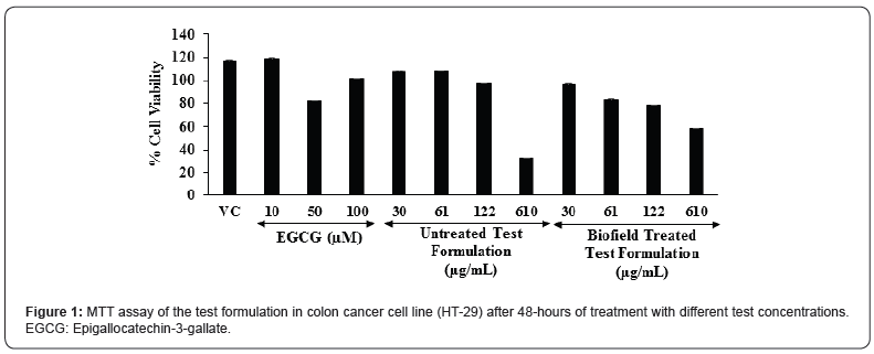
The cell viability results are summarized in the Figure 1. The percent cell viability in the vehicle control (VC) group was found as 117.5%. Moreover, the positive control, epigallocatechin-3- gallate (EGCG) showed 119.2%, 83%, and 101.2% cell viability at the concentration of 10, 50, and 100 μM, respectively. Further, the untreated test formulation showed more than 97% cell viability upto 122 μM; while the Biofield Treated test formulation showed more than 78% cell viability upto 122 μM. Based on the MTT cell viability assay the test formulation was found as safe and nontoxic upto the concentration of 122 μM. MTT assay is widely used for the cell toxicity against any test formulations. In addition, this assay was found as more rapid, less costly, less time consuming, and non-radioactive method as compared with the other assays. This assay display cell proliferation results on the basis of the cell growth and metabolic activity [41]. MTT assay suggest that the concentrations of the test formulation were found safe up to 122 μg/mL with respect to the viability in the colon cancer cell line (HT-29).

Estimation of Interleukin-6 (IL-6) Expression

The level of interleukin-6 (IL-6) expression in colon cancer (HT-29) cells is represented in the Figure 2. The positive control, epigallocatechin-3-gallate (EGCG) was significantly reduced the level of IL-6 by 38.71%, 92.43%, 91.74%, and 98.20% (p≤0.001) at the concentrations of 1, 10, 50, and 100 μM, respectively as compared to the vehicle control (VC) group. Moreover, the untreated test formulation showed significant reduction of IL-6 by 11.96%, 35.94%, and 52.3% (p≤0.001) at 0.1, 3, and 15 μM, respectively as compared to the VC group. Further, the Biofield Energy Treated test formulation group showed 14.19%, 34.09%, and 59.41% (p≤0.001) reduction of IL-6 at 0.1, 3, and 15 μM, respectively as compared to the VC group under the stimulation of TNF-α stimulation. Besides, the Biofield Treated test formulation also significantly reduced the level of IL-6 by 14.87% as compared to the untreated test formulation group. Overall, the minerals and vitamin-based Biofield Energy Treated test formulation showed an anti-inflammatory activity by reducing the level of IL-6 under the stimulation of TNF-α as compared with the vehicle control as well as untreated test formulation groups. Hence, the Biofield Energy Treated test formulation could be used a major role in immune-related disorders and also defined as controlling factor for many diseases [42]. Thus, it can be suggested that the Biofield Energy Treated test formulation can be used in many inflammatory disorders.

Estimation of IL-8 Expression

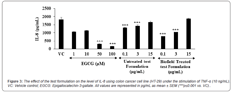
The level of interleukin-8 (IL-8) expression in colon cancer (HT-29) cells is represented in the Figure 3. The positive control, epigallocatechin-3-gallate (EGCG) was significantly reduced the level of IL-8 by 41.49%, 36.77%, 82.89% (p≤0.001), and 91.21% (p≤0.001) at the concentrations of 1,10,50, and 100 μM, respectively as compared to the vehicle control (VC) group. Moreover, the untreated test formulation showed significant reduction of IL-6 by 27.34% (p≤0.001), 21.03% ( ≤0.001), and 9.08% at 0.1, 3, and 15 μM, respectively as compared to the VC group. Further, the Biofield Energy Treated test formulation group showed significant (p≤0.001) reduction of IL-8 by 57.09% and 42.88% at 0.1 and 3 μM, respectively as compared to the VC group under TNF-α stimulation. Besides, the Biofield Treated test formulation also significantly reduced the level of IL-8 by 40.94% and 38.25% at 0.1 and 3 μg/mL, respectively as compared to the untreated test formulation group. Overall, the minerals and vitamin-based Biofield Energy Treated test formulation showed an anti-inflammatory activity by reducing the level of IL-8 under the stimulation of TNF-α as compared with the vehicle control as well as untreated test formulation group. Chronic inflammatory conditions leads to the massive production of proinflammatory factors such as chemokines. IL-8 is one of the chemokine in chronic inflammation and it is initially act as a neutrophil chemotactic and activating factor [43,44]. Overall, the experimental data suggested that Biofield Energy Healing Treatment has the significant capacity to reduce the level of IL-8 with respect to vehicle control and untreated test formulation.

Estimation of IFN-γ Expression

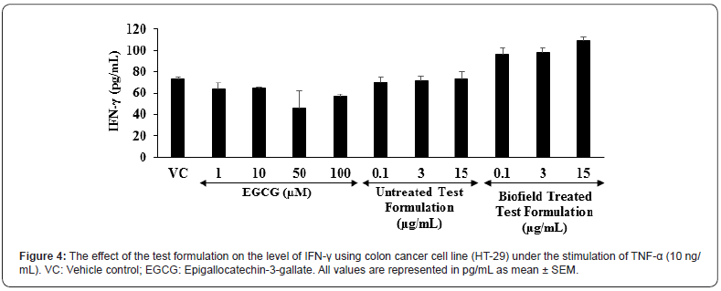
The level of interferon gamma (IFN-γ) expression in colon cancer (HT-29) cells is shown in the Figure 4. The positive control, epigallocatechin-3-gallate (EGCG) was significantly reduced the level of IFN-γ by 13.64%, 12.64%, 37.64%, and 22.99% at the concentrations of 1, 10, 50, and 100 μM, respectively as compared to the vehicle control (VC) group. Moreover, the untreated test formulation showed significant reduction of IFN-γ by 4.55% and 2.78% at 0.1 and 3 μM, respectively as compared to the VC group. Further, the Biofield Energy Treated test formulation significantly altered the level of IFN-γ as compared to the VC group under TNF-α stimulation. This suggests that the Biofield Energy Treated test formulation has significant immunomodulatory activity. Scientific reports suggest that various immunological and inflammatory functions of chemokines play significant role in controlling the immune response during infections. Overall, the immunomodulatory effect might be the result of specific modulation of NF-κB, a transcription factor involved in the activation of many inflammatory mediator genes [45,46].

Worldwide scope of alternative medicine and its outcomes have been increased significantly. However, an important phytoconstituents along with minerals and vitamins are reported to have beneficial role against many diseases such as diabetes, indigestion, inflammation of intestine, osteomalacia, blood disorders, infertility, potent revitalizer, etc. [47]. Due to high safety profile with the wide therapeutic action of alternative medicines, the scope has been increased worldwide [48]. Besides, the individual constituents of the novel proprietary test formulation has been reported to have substantial immunomodulatory action, and Biofield Energy Healing Treatment significantly alters the action of cytokines. Overall, the Biofield Energy Healing Treatment on the test formulation can be a novel approach in supports of the use of Biofield Treated test formulation for various types of autoimmune disorders in colon cancer cell line (HT-29).

Conclusion

On the basis of current study findings, it is concluded that the novel proprietary test formulation showed significant antiinflammatory action on the tested cytokines (IL-6, IL-8, and IFN-γ) in colon cancer cell line (HT-29) after administration of the Biofield Energy Treated formulation. MTT assay in the Biofield Energy Treated colon cancer cell line (HT-29) suggest that the test formulation showed more than 78% cell viability and found as safe and non-toxic. In addition, the levels of cytokine, interleukin-6 (IL-6) was significantly reduced by 34.09% and 59.41% (p≤0.001) at 3 and 15 μM, respectively as compared to the vehicle control (VC) group. Moreover, IL-8 level was reported to be significantly suppressed in the Biofield Energy Treated test formulation by 57.09% and 42.88% at 0.1 and 3 μM, respectively as compared with the VC group. On the basis of experimental results of various tested cytokines and their expression, significant anti-inflammatory activity in colon cancer cells was reported in the new test formulation after treated with the Trivedi Effect®- Biofield Energy Healing. Biofield Energy Treated test formulation can be used as a Complementary and Alternative Medicine (CAM) to prevent the immune-mediated diseases such as Irritable Bowel Syndrome, Rheumatoid arthritis, Ulcerative colitis and Crohn’s disease, Stress, Asthma, and many more with safe therapeutic index. Besides, it can also be utilized in organ transplants (for example kidney transplants, liver transplants and heart transplants), various autoimmune disorders such as Lupus, Addison Disease, Celiac Disease (gluten-sensitive enteropathy), Dermatomyositis, Graves’ Disease, Hashimoto Thyroiditis, Multiple Sclerosis, Myasthenia Gravis, Pernicious Anemia, Aplastic Anemia, Sjogren Syndrome, Systemic Lupus Erythematosus, Diabetes, Alopecia Areata, Fibromyalgia, Vitiligo, Psoriasis, Scleroderma, Chronic Fatigue Syndrome and Vasculitis, Type 1 to improve the overall health and quality of life.

Click here: https://juniperpublishers.com/index.php 
Friday, August 27, 2021

Evaluation of Biofield Energy Healing Treatment Based Proprietary Test Formulation on Gut Health Potential in Colon Cancer Cell Line (HT-29) - Juniper Publishers

Pharmacology & Clinical Research - Juniper Publishers


Abstract

The present study was aimed to evaluate the anti-inflammatory potential of Biofield Energy Healing (the Trivedi Effect®-Consciousness Energy Healing) on the test formulation in colon cancer cell line (HT-29). Each ingredient of the test formulation was divided into two parts, one part was denoted as the untreated test formulation and the other part was demarcated as the Biofield Energy Treated test formulation, which received Biofield Energy Healing Treatment by a renowned Biofield Energy Healer, Mr. Mahendra Kumar Trivedi. MTT assay showed that the test formulation was found safe and non-toxic upto 122 μg/mL in HT-29 cells with more than 78% cell viability. The level of interleukin-6 (IL-6) expression was significantly reduced by 34.09% and 59.41% (p≤0.001) at 3 and 15 μM, respectively compared to the vehicle control (VC) group under the stimulation of tumor necrosis factor - alpha (TNF-α). Moreover, IL-8 level was significantly suppressed in the Biofield Energy Treated test formulation by 57.09% and 42.88% at 0.1 and 3 μM, respectively compared with the VC group. However, the Biofield Energy Treated test formulation further substantial altered the level of interferon gamma compared to the VC group. The Trivedi Effect®-Consciousness Energy Healing significantly regulate the inflammatory condition after treatment with the test formulation in colon cancer cell line (HT-29). This experimental data suggested that the Biofield Treated test formulation can be utilized for many inflammatory disease conditions such as rheumatoid arthritis, multiple sclerosis, psoriasis, inflammatory bowel diseases, scleroderma, and type 1 diabetes mellitus.

Keywords: Biofield Energy Healing; The Trivedi Effect®; Inflammation; Colon cancer cell line (HT-29); Pro-inflammatory cytokines

Abbreviations: FBS: Fetal bovine serum; MTT: 3-(4,5-Dimethylthiazol-2-yl)-2,5-Diphenyltetrazolium Bromide; ELISA: Enzyme-linked immunosorbent assay; NCCAM: National Center for Complementary and Alternative Medicine; CAM: Complementary and Alternative Medicine

Introduction

The relation among of the gut health, micro biota, and cytokines have been well studies and reported in past two decades against various inflammatory bowel diseases (IBD) and associated mucosal inflammations. The cytokines play an important role in the mechanism in inflammation especially in Crohn’s disease and ulcerative colitis. The pathogenesis of IBD is not completely understood, but the role of cytokines in the intestinal immune system has significant impact in disruption of normal state of controlled gut inflammations [1,2]. Innate immune response is the major response in gut inflammation and its related diseases. Most of the cytokines are secreted by activated dendritic cells and the macrophages, which regulates the inflammatory response in gut inflammatory diseases. Once, these cytokines are secreted by antigen presenting cells, they triggers and differentiate various T cells by activating adaptive immune response. Gut inflammation dysregulates the T-cells, and manage the over-reactive and auto-reactive cells. T-cell regulation or its overproduction leads to the development of gut inflammatory diseases [3]. These cells along with various types of cytokines play a complex role in inflammatory gut diseases [4, 5]. Thus, there is the need of some novel formulation which alters the level of cytokines to improve the gut health. The present study was aimed to test the impact of the Biofield Energy Treated test formulation comprised of zinc chloride, ferrous sulphate, copper chloride (II-cupric), vitamin B6 (pyridoxine HCl), vitamin B12 (cyanocobalamin), magnesium (II) gluconate, and cholecalciferol (vit. D3) against the colon cytokines. The novel test formulation was treated with Biofield Energy Healing Treatment, as one of the best CAM approach with significant therapeutic outcomes. Biofield Energy Healing is one of the emerging frontiers aspect to CAM and various clinical approach has been used with significant results [6-9]. CAM therapies have been recommended by The National Center for Complementary/Alternative Medicine (NCCAM) and there therapies exist in various forms such as external qigong, Johrei, Reiki, therapeutic touch, yoga, Qi Gong, polarity therapy, Tai Chi, pranic healing, deep breathing, chiropractic/osteopathic manipulation, guided imagery, meditation, massage, homeopathy, hypnotherapy, progressive relaxation, acupressure, acupuncture, special diets, relaxation techniques, Rolfing structural integration, healing touch, movement therapy, pilates, mindfulness, Ayurvedic medicine, traditional Chinese herbs and medicines in biological systems both in vitro and in vivo. Human Biofield Energy has subtle energy that has the capacity to work in an effective manner [10] with its various clinical benefits [11]. This energy can be harness and transmit it into living and non-living things by the process of Biofield Energy Healing Treatment. Biofield Energy Treatment (the Trivedi Effect®- Consciousness Energy Healing Treatment) has been extensively studied with significant outcomes in the field of pharmaceuticals [12-14], nutraceuticals [15,16], metals and ceramics [17-19], microbiology [20-22], microbial genetics [23, 24], cancer research [25,26], livestock, agriculture science [27-29], improved bioavailability of many compounds [30-32], improved skin health [33, 34], improved properties of nutraceuticals [35, 36], improved overall bone health [37-39], human health and wellness. Thus, the study was planned on colon cytokines estimation that could significantly helped to improve the prevalence of gut inflammatory diseases using novel test formulation consisting of minerals such as Mg, Zn, Fe, Cu and vitamins including B6, B12, D3 in colon cancer cell line (HT-29).

Materials and Methods

Chemicals and Reagents

Antibiotics solution (Penicillin-Streptomycin) was purchased from HiMedia, India. 3-(4, 5-dimethyl-2-thiazolyl) 2, 5 diphenyl-2 H-tetrazolium) (MTT), Dulbecco’s Modified Eagle’s Medium (DMEM), NaHCO3, and EDTA were purchased from Sigma Chemical Corp. (St. Louis, MO), a subsidiary of Sigma-Aldrich Corporation. ELISA (enzyme-link immunosorbent assay) assay kits for all cytokines tumor necrosis factor alpha (TNF-α), macrophage inflammatory protein-1α (MIP-1α), and interleukin-1 beta (IL-1β) were purchased from R&D Systems, USA. Fetal bovine serum (FBS) was purchased from GIBCO, USA. Iron sulfate, copper chloride, and cholecalciferol (vitamin D3) were obtained from Sigma Chemical Co. (St. Louis, MO). Zinc chloride and magnesium (II) gluconate hydrate were obtained from TCI, Japan. Pyridoxine- HCL (vit-B6), cyanocobalamin (vit-B12) were procured from Alfa Aesar, USA. All other chemicals used in this study were analytical grade available in India.

Test Formulation and Reference Standard

The test formulation contained a combination of vitamins with minerals viz. iron sulfate, copper chloride, zinc chloride and magnesium (II) gluconate hydrate, cholecalciferol (vitamin D3), pyridoxine-HCL (Vit-B6), and cyanocobalamin (Vit-B12). Tumor necrosis factor alpha (TNF-α) was used as an inflammatory stimulant, while epigallocatechin-3-gallate (EGCG) was used as a reference standard (positive control) for immunomodulatory action in colon cancer cell line (HT-29).

Biofield Energy Healing Strategies

One part of each ingredient of the test formulation did not receive any sort of treatment and was defined as the untreated test formulation group, while another part received Biofield Energy Treatment known as Biofield Treated Test formulation by Mr. Mahendra Kumar Trivedi, a renowned Biofield Energy Healer under standard laboratory conditions for ~3 minutes. This treatment was provided through the Biofield Energy Healer unique Energy Transmission process (the Trivedi Effect®) to the test formulation. Further, the untreated test formulation was treated with a “sham” healer for comparison purposes. The “sham” healer did not have any knowledge about the Biofield Energy Treatment. After that, the Biofield Energy treated and untreated test formulations were kept in similar sealed conditions and used for the in vitro study on colon cancer cell line (HT-29) for cytokines estimation.

Experimental Design

The colon cancer cell line (HT-29) was divided into four different groups. Group 1 comprised of the HT-29 cells vehicle was denoted as the vehicle control, group 2 included cells with epigallocatechin-3-gallate (EGCG) as positive control at various concentrations. Group 3 and 4 included the cells with the untreated and Biofield Energy Treated test formulation, respectively at concentration range 0.1 to 15 μg/mL in presence of tumor necrosis factor - alpha (TNF-α).

Cytotoxicity by MTT Assay

The effect of the Biofield Energy Treated and untreated test formulations at a wide range concentration were tested for cell viability using 3-(4,5-dimethylthiazol-2-yl)-2,5-diphenyl tetrazolium bromide (MTT) assay. The number of viable cells were determined by the ability of mitochondria to convert MTT to formazan dye. The details procedure was followed as per Plikerd et al. 2017 [40]. The effect of the test formulation on cell viability of HT-29 cells was determined as:

%Cell viability=100-%cytotoxicity (1)

Where; % cytotoxicity = [(O.D. of control cells – O.D. of cells treated with the test formulation)/O.D. of control cells]*100.

The results of the concentrations that showed >75% viability were selected subsequently for cytokine estimation.

Determination of Cytokine levels by ELISA

The HT-29 cell suspension in DMEM medium containing 10% FBS was plated at a density of 0.5 X 10⁶ in 12-well cell culture plates. Cells were incubated at 37 °C for 24 hours. Cells were sera starved by replacing medium with DMEM containing 0% FBS and again incubated at 37 °C for another 24 hours. Cells were treated with proprietary test formulation at selected noncytotoxic concentrations and stimulated with Hu-TNF-α. Cells treated with TNF-α + EGCG were included as the positive control. After treatment, cells were incubated in a 5% CO2 incubator for 72 hours. After incubation, culture supernatants were collected from each well and stored at -20 °C until analysis. The level of cytokines (IL-6, IL-8, and IFN-γ) in culture supernatants of HT-29 cells were determined using ELISA as per manufacturer’s instructions.

Statistical Analysis

All the data were expressed as mean of three replicates ± SEM and were subjected to one-way analysis of variance (ANOVA) followed by Dunnett’s test and Student’s t-test for two groups comparison. Statistical significance was considered at p≤0.05.

Results & Discussion

MTT Assay

The cell viability results are summarized in the Figure 1. The percent cell viability in the vehicle control (VC) group was found as 117.5%. Moreover, the positive control, epigallocatechin-3- gallate (EGCG) showed 119.2%, 83%, and 101.2% cell viability at the concentration of 10, 50, and 100 μM, respectively. Further, the untreated test formulation showed more than 97% cell viability upto 122 μM; while the Biofield Treated test formulation showed more than 78% cell viability upto 122 μM. Based on the MTT cell viability assay the test formulation was found as safe and nontoxic upto the concentration of 122 μM. MTT assay is widely used for the cell toxicity against any test formulations. In addition, this assay was found as more rapid, less costly, less time consuming, and non-radioactive method as compared with the other assays. This assay display cell proliferation results on the basis of the cell growth and metabolic activity [41]. MTT assay suggest that the concentrations of the test formulation were found safe up to 122 μg/mL with respect to the viability in the colon cancer cell line (HT-29).

Estimation of Interleukin-6 (IL-6) Expression

The level of interleukin-6 (IL-6) expression in colon cancer (HT-29) cells is represented in the Figure 2. The positive control, epigallocatechin-3-gallate (EGCG) was significantly reduced the level of IL-6 by 38.71%, 92.43%, 91.74%, and 98.20% (p≤0.001) at the concentrations of 1, 10, 50, and 100 μM, respectively as compared to the vehicle control (VC) group. Moreover, the untreated test formulation showed significant reduction of IL-6 by 11.96%, 35.94%, and 52.3% (p≤0.001) at 0.1, 3, and 15 μM, respectively as compared to the VC group. Further, the Biofield Energy Treated test formulation group showed 14.19%, 34.09%, and 59.41% (p≤0.001) reduction of IL-6 at 0.1, 3, and 15 μM, respectively as compared to the VC group under the stimulation of TNF-α stimulation. Besides, the Biofield Treated test formulation also significantly reduced the level of IL-6 by 14.87% as compared to the untreated test formulation group. Overall, the minerals and vitamin-based Biofield Energy Treated test formulation showed an anti-inflammatory activity by reducing the level of IL-6 under the stimulation of TNF-α as compared with the vehicle control as well as untreated test formulation groups. Hence, the Biofield Energy Treated test formulation could be used a major role in immune-related disorders and also defined as controlling factor for many diseases [42]. Thus, it can be suggested that the Biofield Energy Treated test formulation can be used in many inflammatory disorders.

Estimation of IL-8 Expression

The level of interleukin-8 (IL-8) expression in colon cancer (HT-29) cells is represented in the Figure 3. The positive control, epigallocatechin-3-gallate (EGCG) was significantly reduced the level of IL-8 by 41.49%, 36.77%, 82.89% (p≤0.001), and 91.21% (p≤0.001) at the concentrations of 1,10,50, and 100 μM, respectively as compared to the vehicle control (VC) group. Moreover, the untreated test formulation showed significant reduction of IL-6 by 27.34% (p≤0.001), 21.03% ( ≤0.001), and 9.08% at 0.1, 3, and 15 μM, respectively as compared to the VC group. Further, the Biofield Energy Treated test formulation group showed significant (p≤0.001) reduction of IL-8 by 57.09% and 42.88% at 0.1 and 3 μM, respectively as compared to the VC group under TNF-α stimulation. Besides, the Biofield Treated test formulation also significantly reduced the level of IL-8 by 40.94% and 38.25% at 0.1 and 3 μg/mL, respectively as compared to the untreated test formulation group. Overall, the minerals and vitamin-based Biofield Energy Treated test formulation showed an anti-inflammatory activity by reducing the level of IL-8 under the stimulation of TNF-α as compared with the vehicle control as well as untreated test formulation group. Chronic inflammatory conditions leads to the massive production of proinflammatory factors such as chemokines. IL-8 is one of the chemokine in chronic inflammation and it is initially act as a neutrophil chemotactic and activating factor [43,44]. Overall, the experimental data suggested that Biofield Energy Healing Treatment has the significant capacity to reduce the level of IL-8 with respect to vehicle control and untreated test formulation.

Estimation of IFN-γ Expression

The level of interferon gamma (IFN-γ) expression in colon cancer (HT-29) cells is shown in the Figure 4. The positive control, epigallocatechin-3-gallate (EGCG) was significantly reduced the level of IFN-γ by 13.64%, 12.64%, 37.64%, and 22.99% at the concentrations of 1, 10, 50, and 100 μM, respectively as compared to the vehicle control (VC) group. Moreover, the untreated test formulation showed significant reduction of IFN-γ by 4.55% and 2.78% at 0.1 and 3 μM, respectively as compared to the VC group. Further, the Biofield Energy Treated test formulation significantly altered the level of IFN-γ as compared to the VC group under TNF-α stimulation. This suggests that the Biofield Energy Treated test formulation has significant immunomodulatory activity. Scientific reports suggest that various immunological and inflammatory functions of chemokines play significant role in controlling the immune response during infections. Overall, the immunomodulatory effect might be the result of specific modulation of NF-κB, a transcription factor involved in the activation of many inflammatory mediator genes [45,46].

Worldwide scope of alternative medicine and its outcomes have been increased significantly. However, an important phytoconstituents along with minerals and vitamins are reported to have beneficial role against many diseases such as diabetes, indigestion, inflammation of intestine, osteomalacia, blood disorders, infertility, potent revitalizer, etc. [47]. Due to high safety profile with the wide therapeutic action of alternative medicines, the scope has been increased worldwide [48]. Besides, the individual constituents of the novel proprietary test formulation has been reported to have substantial immunomodulatory action, and Biofield Energy Healing Treatment significantly alters the action of cytokines. Overall, the Biofield Energy Healing Treatment on the test formulation can be a novel approach in supports of the use of Biofield Treated test formulation for various types of autoimmune disorders in colon cancer cell line (HT-29).

Conclusion

On the basis of current study findings, it is concluded that the novel proprietary test formulation showed significant antiinflammatory action on the tested cytokines (IL-6, IL-8, and IFN-γ) in colon cancer cell line (HT-29) after administration of the Biofield Energy Treated formulation. MTT assay in the Biofield Energy Treated colon cancer cell line (HT-29) suggest that the test formulation showed more than 78% cell viability and found as safe and non-toxic. In addition, the levels of cytokine, interleukin-6 (IL-6) was significantly reduced by 34.09% and 59.41% (p≤0.001) at 3 and 15 μM, respectively as compared to the vehicle control (VC) group. Moreover, IL-8 level was reported to be significantly suppressed in the Biofield Energy Treated test formulation by 57.09% and 42.88% at 0.1 and 3 μM, respectively as compared with the VC group. On the basis of experimental results of various tested cytokines and their expression, significant anti-inflammatory activity in colon cancer cells was reported in the new test formulation after treated with the Trivedi Effect®- Biofield Energy Healing. Biofield Energy Treated test formulation can be used as a Complementary and Alternative Medicine (CAM) to prevent the immune-mediated diseases such as Irritable Bowel Syndrome, Rheumatoid arthritis, Ulcerative colitis and Crohn’s disease, Stress, Asthma, and many more with safe therapeutic index. Besides, it can also be utilized in organ transplants (for example kidney transplants, liver transplants and heart transplants), various autoimmune disorders such as Lupus, Addison Disease, Celiac Disease (gluten-sensitive enteropathy), Dermatomyositis, Graves’ Disease, Hashimoto Thyroiditis, Multiple Sclerosis, Myasthenia Gravis, Pernicious Anemia, Aplastic Anemia, Sjogren Syndrome, Systemic Lupus Erythematosus, Diabetes, Alopecia Areata, Fibromyalgia, Vitiligo, Psoriasis, Scleroderma, Chronic Fatigue Syndrome and Vasculitis, Type 1 to improve the overall health and quality of life.

To Know more about Pharmacology & Clinical Research


To Know more about our Juniper Publishers

Click here: https://juniperpublishers.com/index.php   
Congenital lens dysfunction as a new, undiagnosed cause of decreased visual acuity based on observation over a period of 3 years- Juniper Publishers

  Ophthalmology- Juniper Publishers Abstract Two groups of patients with diagnosed myopia and astigmatism underwent surgery to have their na...