Friday, April 24, 2020

Is Reinforcement Learning a Panacea for Solving All Contingencies in UAVs? - Juniper Publishers

Robotics & Automation Engineering - Juniper Publishers


Abstract

We would like to point out that reinforcement learning (RL) needs to be used with caution in contingency planning for UAVs. A more practical approach is proposed in this opinion.

Keywords: Contingency planning; UAVs; Reinforcement learning


Introduction

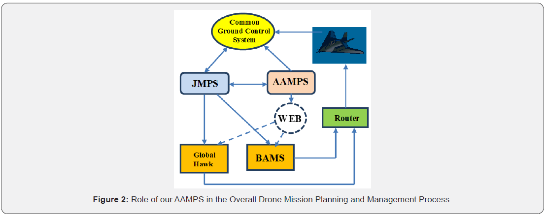
Reinforcement learning (RL) has two important elements: “critic” and “reward” based on performance. As shown in Figure 1, the “reward” is generated based on a performance evaluator for a system and the “critic” element generates proper actions to a system. In recent years, RL has gained a lot of attention because of its success in games such as the Alphago, which beat the human world champion in straight sets [1]. However, there are several recent blogs [2-4] by researchers in artificial intelligence (AI), who heavily criticized the capability of RL in numerous applications. Some notable criticisms include the requirement of huge amount of training data, lack of mechanism to incorporate metadata (rules) into the learning process, the requirement of starting from scratch in the learning process, etc. In short, those researchers think that RL is not a mature technology yet and has found success in only a few applications such as games, collision avoidance, etc.

Drones, also known as Unmanned Air Vehicles (UAVs), have much higher failure rates than manned aircraft [5]. In 2019, there is a Small Business Innovative Research (SBIR) topic [6] seeking ambitious ideas by using RL to handle quite a few contingencies in drones, including 1) preset lost-link procedures; 2) contingency plans in case of failure to reacquire lost links; 3) abort in case of unexecutable commands or unavoidable obstacles; 4) terminal guidance. After carefully analyzing the requirements in this topic, we believe that a practical and minimal risk approach is to adopt a hybrid approach, which incorporates both conventional and RL methods. Some of the requirements in this topic can be easily handled by conventional algorithms developed by our team. For instance, we have developed preset lost-link procedures to deal with lost links for NASA. Our procedures can satisfy FAA and air traffic control (ATC) rules and regulations. We also have systematic procedures to deal with contingency in case of failure to re-acquire lost links. All these procedures can be generated using rules and do not require any RL methods. We believe that RL should be best used in collision avoidance in dynamic environments be cause there is mature development in using RL to tackle clutter and dynamic environments for mobile robots.


Practical Approach

In the past seven years, our team has been working on contingency planning for UAVs to deal with lost links for NASA and contingency planning for engine failures, change of mission objectives, missed approach, etc. for the US Navy. We also have a patent on lost link contingency planning [7]. To tackle the contingencies in drones, a practical approach has several components. First, we propose to apply our previous developed system [7-10] to deal with lost-link and other contingencies. Our system requires some pre-generated databases containing FAA/ATC rules, locations of communication towers, emergency landing places, etc. for a given theater. Based on those databases, we can generate preset plans to handle many of the contingencies related to lost-link, engine failures, mission objective changes, etc. Our pre-generated contingency plans can also handle terminal guidance. We plan to devise different plans to handle different scenarios in the terminal guidance process. For example, if the onboard camera sees a wave-off signal, the UAV can immediately activate a contingency plan to guide the UAV to an alternative landing place. Second, we propose to apply RL to handle some unexpected situations such as dynamic obstacles in the path. RL has been successfully used in robot navigation in clutter and dynamic environments and hence is most appropriate for collision avoidance in UAVs.

Figure 1 shows the relationship of our Advanced Automated Mission Planning System (AAMPS), Joint Mission Planning System (JMPS), and Common Control System (CCS). Our AAMPS first uses JMPS to generate a primary flight plan for a given mission. JMPS has the advantage of containing airport and constraint zone information in its database. Second, we applied Common AAMPS to generate contingency plans for the primary flight path. The Ground Control System contingency plans can deal with major situations such as engine out, lost communications, retasking, missed approaches, etc. To avoid damage to ground structure, we propose to apply an automatic landing place selection tool, which selects appropriate landing places for UAVs so that UAVs can use these landing sites during WEB Route normal flight or emergencies.
JOJ Urology & Nephrology
In our approach, RL is used for path planning to avoid dynamic threats and obstacles. Recent works showed that RL could be used for autonomous crowd-aware robot navigation in crowded environments [11-12]. However, the performance of these techniques degrade as the crowd size increases since these techniques are based on a one-way human-robot interaction problem [13]. In a recent paper [13], the authors introduces an interesting work which uses RL for robot navigation in crowded environments. The authors of [13] name their method Self- Attention Reinforcement Learning (SARL). They also use the name local map SARL (LMSARL) for the extended version of SARL. The authors approach the crowd-aware navigation problem different than other techniques and the human-human interactions which affects robot’s anticipation capability for navigation are also considered in their method, SARL [13]. SARL can anticipate crowd dynamics resulting in time-efficient navigation paths and outperforms three state-ofthe- art robot navigation in crowded scenes methods which are Collision Avoidance with Deep Reinforcement Learning (CADRL) [11], Long Short-Term Memory-RL (LSTM-RL) [12], and Optimal Reciprocal Collision Avoidance (ORCA) [14]. We find similarities between the crowd-aware robot navigation application and the autonomous collision-free UAV navigation in crowded air traffic and SARL can be used as a promising technique along this line. We believe that we can customize SARL for autonomous collision-free UAV navigation in contingency situations such as when the link between operator and UAV is lost and the UAV needs to make a forced emergency landing.
JOJ Urology & Nephrology


Conclusion

It was argued that RL may not be able to solve contingency planning for UAVs. Instead, we advocate a practical approach to solving this problem.

 To know more about Robotics & Automation Engineering
Click here: https://juniperpublishers.com/raej/index.php
To know more about Juniper Publishers

Thursday, April 23, 2020

Rare Species Milk as a New Source for Potential LABs- Juniper Publishers

Nutrition & Food Science - Juniper Publishers 


Abstract

Present exploration depicts rare species milk as the new source in search of novel starter cultures i.e. lactic acid bacteria (LABs). Camel milk itself is very nutritional and rich source of initial feed to infants. The term probiotics is very popular from the last few decades so as the result search of new sources for the hunt of novel probiotic strains increased. Efficient probiotics fulfill the current market demand for the development of new industrial products. This study focuses on the significance of rare species milk i.e. camel milk and the essential probiotic attributes concerned for starters might be used in industrial applications.
Keywords: Camel milk; Probiotics; Micro flora; Lactic acid bacteria;


Introduction

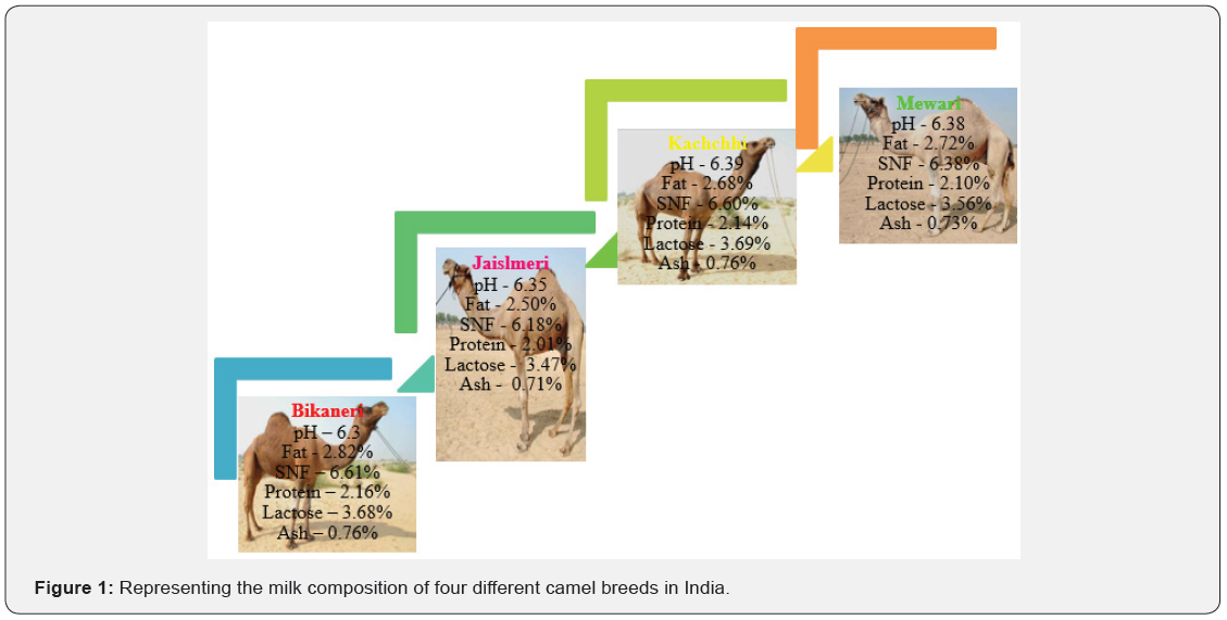
In today’s life every single (human being) is consuming the functional foods in the form of dairy or non-dairy products. Functional foods comprised of probiotics i.e. live bacteria. Etymologically probiotics are pro (for) and bios (life). According to FAO probiotics are defined as “live microorganisms which when administered in adequate amounts confer a health benefit on the host” [1]. Lactic acid bacteria genus considered to be safe and exhibit the properties to be called as probiotics. In current market scenario as the demand of probiotics is increasing so as the demand of new starter cultures for the product development. Therefore, to find the new sources is very important for the isolation of novel starter cultures. Among the dairy sources many rare species Milk might be used as a source, and one of them is camel milk. In India vast diversity of mammalian species are present but our society totally depends on cow and buffalo milk for the initial nutrition only because no one can access the milk from other thousands of species. And it is due to the fact of lack of knowledge and awareness regarding the benefits of rare species milk. The worlds camel population is 23.9 million out of which 0.45 million is contributed by India [2]. Different breeds of camel are present in India (Bikaneri, Mewari, Kachhi and Jaisalmeri) [3]. Being a ship of the desert camel does tolerate harsh climatic conditions and even in the scarcity of water they produce more milk with longer lactation period other than any species. Camel milk being rare species milk is very rich in nutrients like proteins, minerals and vitamins. Milk composition values of different breeds are shown in (Figure 1). It specifically contains lot of protective proteins and immunoglobulin’s which helps in improving the immune system. It lacks the allergic proteins which are present in cow’s milk. This is the solution towards the cow milk allergies, this might act as a substitute as a weaning feed for babies. Even for adults this rare milk is very valuable and beneficial for health because it is good in many disorders like allergies, autism and even in cancer.

With all these good prospects of camel milk its indigenous micro flora is also rich in LABs (lactic acid bacteria) which are termed as a safe species group of bacteria or GRAS (generally recognized as safe) [4,5]. LABs are known for their probiotic potential and they might use as starter cultures for dairy and non-dairy product development. The examples of LABs with probiotic application are Lactobacillus plantarum, Lactobacillus brevis, Lactococcus lactis, Bifidobacterium and many more. LABs are gram positive and catalase negative species which are able to produce lactic acid as an end product from fermentation. Further these strains might act as a probiotic feed for weaning babies and come up as the solution of wholesome food for the nutrition in growing stage. It is predominant to comment that probiotic potential of bacteria is very much strain specific. It is very important to recognize and identify the bacterial species so that it might be apt for industrial applications.


Probiotic Attributes and Associated Health Benefits

It is mandate for potential probiotic; bacterial species must exhibit some probiotic attributes within and exert beneficial effects on the host. Major traits to be called as probiotics are determined by in vitro tests:
a. Acid and bile salt tolerance is important criteria for strains;
b. Bile salt hydrolase activity;
c. Cell surface hydrophobicity;
d. In vitro cell adhesion to mucosal epithelial surfaces;
e. Antimicrobial activity against pathogenic bacteria;
f. Antibiotic resistance [1].
These in vitro parameters are the prerequisites for the probiotic strains and shown in Figure 2. As far as dose of probiotics is concerned, the lowest concentration 106 CFU/mL is consumed daily for the visible good probiotic effect. Different probiotic mechanisms are associated with the human health which may include the production of antimicrobial substances like bacteriocins, acidic pH of gut, and competitive adherence to mucosal epithelial surface, providing the gut barrier functions as well as enhancing the immune system [6]. There are clinically proven evidences that actually prove the associated health benefits of the probiotics. According to Russo et al. [7] and Orlando et al. [8] probiotic Lactobacillus rhamnosus strain GG (LGG) and Bifidobacterium adolescentis SPM0212 showed a significant anti proliferative role and inhibit human gastric cancer cells and three colonic cancer cells lines including HT- 29, SW 480, and Caco-2 [7,8]. The probiotic mechanism for decreasing the proliferation of cells and treatment still needs to be understood and more research is required. Probiotics are also helpful in allergies by moderating the allergic response. Allergic reactions occur when an immune system reacts with an allergen. Numbers of bacterial cultures were studied are very limited for their ability in the treatment and prevention of allergies in infants. Studies showed that L. rhamnosus GG has been successful in preventing the occurrence of atopic eczema in infants, when delivered to mothers who had already firstdegree atopic eczema, allergic rhinitis or asthma [9]. Health benefits of probiotics are not limited there are other; they contribute in reduction of cholesterol levels and eventually leads to reduction in coronary heart diseases, autism and bacterial vaginosis in women’s. The mechanism of probiotics behind the reduction of cholesterol level in serum is due to the presence of BSH activity which helps in absorbing the cholesterol from the gut. These properties are strain specific in nature and vary with strain to strain. There is more need of valuable research regarding the clinical evidences of health benefits of probiotics.


Conclusion

It can be concluded from the present study that, rare species milk are the good source for the isolation of novel LABs. Utilization of rare species milk needs to be considered by creating the awareness among the society. As far as their probiotic activity is concerned, remains to be validated in future studies.


Click here: https://juniperpublishers.com/nfsij/index.php

To know more about Juniper Publishers

Tuesday, April 21, 2020

Effect of Labor Augmentation Using Oxytocin on Neonatal Outcome, A Case Control Study - Juniper Publishers

Gynecology and Women’s Health - Juniper Publishers


Abstract

Aims: To re-evaluate whether augmentation of labor with oxytocin in dysfunctional labor, has a hazardous effect on neonatal Apgar scores, umbilical cord pH or blood gases
Materials and Methods: Prospective study where one hundred and fifty-three (153) patients in spontaneous labor were involved in the analysis. Seventy-nine patients (Group I) with hypo-contractile uterine activity were managed by low-dose oxytocin protocol for labor augmentation, while 74 patients (Group II) were not managed by augmentation of labor using oxytocin. Primary outcomes were Apgar score at 5th minutes of birth and umbilical artery pH, while secondary outcomes were Apgar score at 1st minute, arterial pO2, pCO2 and base deficit of all neonates.
Result: In this study, there was no significant difference between cases and controls regarding; Apgar score at 1st minute, Apgar score at 5th minute, mean umbilical artery pCO2 and pO2 while mean umbilical artery pH was significantly lower and umbilical artery base deficit was significantly higher in the case group compared to the control group. Moreover, umbilical artery pH was negatively correlated to duration of oxytocin administration.
Conclusion: Oxytocin use to augment labor, although affects umbilical cord blood gases, doesn’t significantly affect neonatal outcome if used in a proper dose to produce efficient uterine contractions

Keywords: Apgar score Augmentation Cord gases Oxytocin


Introduction

Approximately 20 percent of labors involve either protraction or arrest disorders [1]. The active management of labor was introduced during the 1960’s at the National Maternity Hospital in Dublin, Ireland to augment labor in nulliparous women. Proponents of this approach have suggested that it also safely reduces the number of Cesarean deliveries [2]. Oxytocin increases frequency of contractions, causes initial transient increase in the base tone (incomplete relaxation) and increases the amplitude and duration of phasic contractions [3]. During parturition, prolonged tonic contractions might be harmful for fetuses because an increase of the intrauterine pressure would disturb the feto-maternal circulation and may occlude blood vessels within the myometrium [4]. Utero-placental hypo-perfusion is the major cause of both respiratory and metabolic acidemia, with progression from the former to the latter over time if decreased utero-placental blood flow is not corrected [5], hence various studies were designed to study the safety of using oxytocin infusion during the course of labor on neonatal clinical and acid-base condition at delivery and the likelihood of causing perinatal asphyxia.


Materials and Methods

The study was conducted in Ain Shams University Maternity Hospital and El-Sahel Teaching Hospital in Cairo, Egypt, which are two big tertiary hospitals, from January till October 2010, where 180 patients in spontaneous labor were initially enrolled in this case-control study after informed consent and approval of the study protocol by the institute ethics committee. Ninety patients who fulfilled the selection criteria, were diagnosed as primary failure of progression in labor due to hypo-contractile uterine activity and managed by augmentation of labor using low-dose oxytocin (Syntocinon® 10 IU/ml concentrate for solution for infusion) (Group I). Another group of ninety patients also fulfilled the selection criteria but were not managed by augmentation of labor using oxytocin (Group II). Participants were all aged between 18 and 45 years old, free of medical disorders, planned for vaginal delivery having singleton pregnancy at full term admitted at 3-5cm cervical dilatation with vertex presentation of estimated fetal weight between 2500 to 4000 grams with average amniotic fluid volume and morphologically normal fetus, with no evidence of fetal distress or placental insufficiency and with uncomplicated pregnancy as detected by antenatal course.
All neonates were assessed using Apgar score at 1st and 5th minutes by same observer and umbilical cord blood samples were obtained from all neonates where a segment of 10-20cm of umbilical cord was doubly clamped and excised immediately after delivery then 1-2ml blood withdrawn in a heparinized syringe; one from the artery and the other from the vein then kept in ice and transported to the laboratory within 60 minutes and analyzed using blood analyzer (i-STAT300F, Abbott Point of Care Inc., USA).

Prior to start of the study, all participants were assessed to confirm eligibility then were consistently managed according to the local institutional intra-partum protocol and received proper monitoring and supportive care. Normal FHR pattern and assessment of uterine activity were confirmed using Cardiotocography (CTG) for 30 minutes before starting interventions. Intermittent auscultation was used in low-risk women while continuous electronic fetal monitoring was used for the high-risk cases. Vaginal examination was performed two hourlies, unless clinically indicated, to monitor progress of labor. Artificial rupture of fetal membranes (ROM) was considered, in women with intact membranes, if there was poor progress of labor (cervical dilatation <1cm/hour) with inefficient uterine contractions 2hours after ROM, the woman was assigned to Group I, receiving low-dose oxytocin protocol using oxytocin dilution of 10mU/mL, initial dose of 2mU/min (12mL/hr), and incrementalincrease of 2mU (12 mL) every 45 minutes until adequate contractions with the maximum dose being 16mU/min (96mL/ hr) [6]. Interpretation of intrapartum fetal heart rate pattern and uterine contractions was done according to ACOG guidelines [7,8]. Cases of uterine hyper-stimulation, antepartum hemorrhage, cases needed instrumental delivery or emergency Cesarean section, as well as neonates with apparent congenital anomalies at birth were all excluded (11 from group I and 16 from group II) and not involved in the analysis. The primary outcomes considered were Apgar score at 5th minute of birth and umbilical artery pH, while secondary outcomes were Apgar score at 1st minute of birth, umbilical artery pO2, pCO2 and base excess of all neonates.


Statistical Analysis

Statistical analysis was done on a personal computer using IBM® SPSS® Statistics version 19 (IBM® Corporation, Armonk, NY, USA). Kolmogorov-Smirnov test of normality was applied to all measured variables where quantitative variables were described as mean and standard deviation (SD), number and percentage, or median and inter-quartile range (IQR) as appropriate. The independent sample t-test was used to compare the two groups as regards quantitative variables in parametric data while Mann Whitney U test was used for non-parametric data and Chi square test was used for qualitative data. Relative risk and Odds ratio were used to calculate likelihood of negative effect with a confidence interval of 95%. Pearson’s correlation was used for measuring correlation between different variables. Significance level was always set at 0.05.


Result

Statistical analysis included 153 patients; 79 in Group I and 74 in Group II. Table 1 shows no significant difference between cases and controls regarding mean maternal age, BMI, gestational age, parity and mean cervical dilatation at enrollment (P>0.05).
Journal of Gynecology and Women’s Health
BMI body mass index [calculated as weight (kg) divided by squared height (m2)] Kg, Kilograms; m2, square meters; cm, centimeters. Analysis using independent student’s t-test or Mann Whitney U test as appropriate Data are presented as mean ± standard deviation or median (interquartile range, IQR) as appropriate.
Journal of Gynecology and Women’s Health
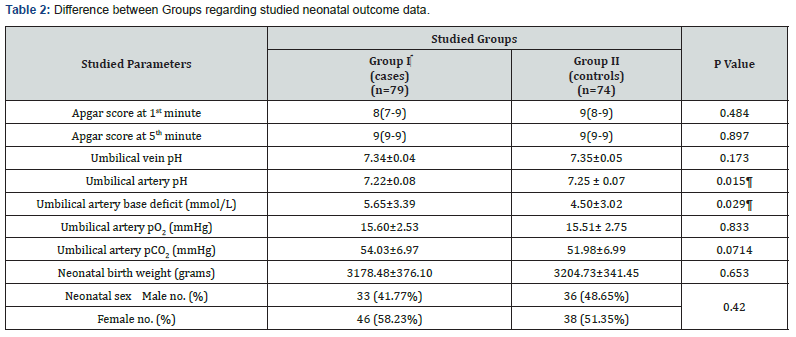
Analysis using independent student’s t-test, Chi square or Mann Whitney U test as appropriate Data are presented as median (interquartile range, IQR), number (%) or mean ± standard deviation as appropriate. ¶ Indicates statistical significance.

Table 2 illustrates studied neonatal outcomes showing no significant difference regarding neonatal weight, neonatal sex distribution, Apgar score at 1st and 5th minutes, as well as mean umbilical artery pCO2, pO2 and mean umbilical vein pH (P>0.05). However, umbilical artery base deficit was significantly more and pH was significantly less in case group (P=0.015; P=0.029; respectively).
Journal of Gynecology and Women’s Health
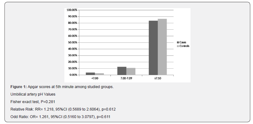
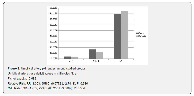
Interestingly, Figure 1 illustrates no significant difference between cases and controls regarding number of neonates with arterial pH less than 7.1 (13 (16.5%) versus 10 (13.5%); respectively) (RR=1.218, P=0.612; OR=1.261, P=0.611; 95%CI), while Figure 2 shows that number of neonates with significant impairment of arterial base deficit (>8mmol/L) didn’t show significant difference between cases and controls (16(20.3%) versus 11(14.9%); respectively) (RR=1.363, P=0.386; OR=1.455, P=0.384; 95%CI).
Journal of Gynecology and Women’s Health
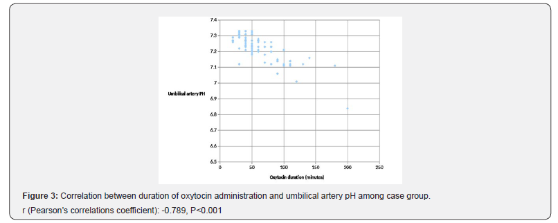
In this study, Apgar scores at 1st and 5th minute were not correlated to any of maternal age, parity, gestational age, neonatal sex or neonatal weight. Also, umbilical artery pH and base excess have non-significant correlations to these variables (P>0.05; Table 3). Figure 3 illustrates Pearson’s correlation test between duration of oxytocin administration and umbilical artery pH among group I, showing a significant negative correlation (P<0.001).


Discussion

Although labor augmentation using oxytocin has been investigated in few trials, most of these trials have focused on various outcomes mainly duration of labor, rate of Cesarean sections or instrumental deliveries and maternal experience with few of them [9-11], have measured neonatal outcomes as a primary outcome. The current study reported that low-dose oxytocin use is associated with reduction of neonatal umbilical artery pH and increase in base deficit without compromising the neonatal clinical condition or affecting Apgar scores

Oxytocin transiently compromises fetal circulation by increasing duration, frequency and strength of uterine contractions where this effect was studied by Olofsson et al. [12], through ultrasound and Doppler assessment where uterine and umbilical artery flow resistance increased significantly during uterine contractions reflecting rapid and exaggerated increase of vascular resistance in both arteries.
The biochemical disturbance must be significant before Apgar score is affected [13]. Moreover, a number of factors may influence Apgar score, including trauma, drugs, infections, congenital anomalies, hypovolemia, hypoxia and preterm birth. Accordingly, it is inappropriate to use an Apgar score alone to establish the diagnosis of asphyxia [14].

In the year 2008, oxytocin was added to the list of medications designated as high alert by the Institute for Safe Medication, USA [15]; such drugs are defined as those bearing a heightened risk of harm when used in error. Not following the guidelines regarding use of oxytocin is probably one reason for adverse neonatal outcomes. The use of oxytocin is on the increase and it is estimated that about 50% of nulliparous and 20% of parous women receive oxytocin during delivery [16]. A recent Cochrane review by Kenyon et al. involved 4 randomized trials and found no significant difference for Apgar scores, umbilical cord blood gases, admission to special care baby unit, or neonatal mortality between cases managed by high-dose and those managed by lowdose oxytocin for labor augmentation [17].

The current study found no statistical difference neither in Apgar scores at 1st and 5th minutes nor in distribution of Apgar score values between both groups, where 5 neonates (6.3%) of the case group and 4 neonates (5.4%) of the control group had a score less than 7 at 1st minute while one neonate of each group had ascore less than 7 at 5th minute. No neonatal deaths were reported during the study. Three Neonates were diagnosed with TTN and no special care admissions occurred for any of the neonates involved.

These results are comparable to those of Chan and To, in their prospective study using low-dose oxytocin, who found that 6% of the neonates of the study group had Apgar scores less than 7 at one minute, while 100% of the neonates of their study group had Apgar scores more than 7 at 5 minutes [18]. Also, our results agree with Merrill and Zaltnik (3.9% of the studied neonates had Apgar score < 7 at 5 minutes) [19]. Melin et al. [20] have classified neonatal acidemia according to umbilical artery pH at birth as mild or pre-acidemia at 7.10 to 7.19, moderate acidemia at 7.00 to 7.09, and severe acidemia at pH less than 7.00. In a similar context, and for the purpose of prediction of the likelihood of neonatal encephalopathy, Low et al. [21] have classified neonatal acidosis, as regards umbilical arterial base deficits, as mild; 4–8 mmol/L, moderate; 8-12mmol/L, and severe acidosis; greater than 12mmol/L. Accordingly, in the current study, numbers of neonates with pH<7.1 and those with base deficits>8 mmol/ Litre, were comparable between both groups with no significant difference (OR=1.261 and 1.455; respectively), while, surprisingly, the case group showed a significantly lower arterial pH and a significantly higher arterial base deficit as compared to controls, which emphasizes the undeniable effect of oxytocin on the arterial acid-base status but this effect was not enough to increase the risk of developing neonatal academia, which agrees with Merrill and Zaltnik in their randomized double masked trial [19]. Also, Thorp et al. [10] conducted a study on 1423 primigravid women where 43% of them received high-dose oxytocin augmentation and no significant difference was noticed in mean arterial pH between oxytocin and no oxytocin groups. Contradictorily, Oscarsson et al. [16] in their massive retrospective study on 106,755 deliveries, concluded that oxytocin use increased the risk of low Apgar score at 5th minute and neonatal special care admission but their study remains limited by being retrospective and hence subject to recall bias as well as heterogeneity of population recruited from 10 different hospitals and not considering factors like uterine hyperstimulation.

In the current study, fifteen umbilical artery pH values (20.3%) of the control group were less than 7.2 which agrees with those of Akoury et al. [9] (20%) but they have used base deficit of 7mmol/L as a cut-off to diagnose neonatal acidosis and hence, 29% of cases and 27% of controls in their study were acidotic. Different modes of delivery might be another reason for different results.
The fact of occurrence of acidemia in non-augmented labors was explained by Yoon and Kim who concluded a normal significant fall in umbilical artery pH and bicarbonate with the presence of labor and increased duration of second stage of labor in healthy term neonates [22]. The current study hasn’t considered neonates with mild acidemia/acidosis as it is thought that mild acidosis occurs in almost all normal labors [23].

The current study uniquely reported the significant negative correlation between duration of oxytocin administration among the case group and umbilical artery pH which signifies the compromising effect of oxytocin on neonatal circulation studied by Olofsson et al. [12] and correlates with the duration of use not only the dose which has been studied in few trials. As oxytocin impairs the utero-placental and umbilical circulations, there might be a gradual accumulation of lactates or gradual affection of the fetal ability to compensate and wash lactates out of the circulation even in absence of uterine hyper-stimulation.

The limitation of this study is that it hasn’t studied the incidence of meconium staining at birth and risk of meconium aspiration as a neonatal outcome. Also, the progress of labor hasn’t been correlated with the results. Moreover, only low-dose oxytocin was studied and further studies are needed to evaluate effect of highdose oxytocin compared to controls and emphasize the effect of duration of oxytocin use on neonatal outcome with larger number of patients and correlate it to other neonatal parameters.  

 To know more about Gynecology and Women’s Health 
To know more about Juniper Publishers

Monday, April 20, 2020

Therapeutic Alternatives for The Treatment and Prevention of Dilated Cardiomyopathy (DCM): The Era of Biological Agents-Juniper Publishers

Pharmacology & Clinical Research-Juniper Publishers


Abstract

Dilated Cardiomyopathy is a rare condition that can be caused by a variety of common morbidities. It is an immune mediated disease, characterized by chronic stimulation of inflammatory responses. The so far available treatment consists of agents that ameliorate only the functional burden off the circulatory system. Biological therapies have been successfully applied in several diseases derived from hyper activation of the immune system. In this mini review we investigate whether abatacept, a biological agent targeting co-stimulation, could be beneficial in inhibiting pathophysiological progression of DCM.

Dilated Cardiomyopathy–Introduction

Dilated cardiomyopathy is the most common cardiomyopathy. It reflects a functional state where the anatomic structures of the heart cannot support a proper blood circulation. Dilated cardiomyopathy is not a common disease. Nevertheless, its progression is crucial for the survival of the patient, since it can lead even rapidly to the need of transplantation. The a etiopathogenesis of the disease includes some very common morbidities of the general population, such as hypertension, coronary heart disease and heart attack. Metabolic diseases such as diabetes mellitus, and thyroid disease have been associated with DCM. Also, infections predominately viral can lead to DCM. All these conditions can affect the heart muscle, which in genetically predisposed patients can favor the development of DCM [1].


Pathophysiology of DCM: The Role of The Immune System

The role of the immune system has been long ago documented in the aetiopathogenesis and evolution of the disease. A proinflammatory microenvironment favors the development of Th1 and Th17 responses of the acquired immunity, This polarization of the adaptive immunity leads, thru a type IV hypersensitivity reaction (delayed, cell mediated type of tissue damage), to chronic inflammation, resulting in functional deregulation of the heart muscle. On this regard, insufficient mobilization of the homeostatic anti-inflammatory Treg response confers to further propagation of tissue damage and disease progression [2].


Therapy: Current Available Agents

Therapeutic interventions consist of cardiovascular agents that regulate the equilibrium of systemic circulation, thus compensating the functional impairment of the heart. The so far therapeutic approach only retards the progression to a non-compensative heart failure and does not intervene in the pathophysiological evolution of the disease. It has been already pointed out that DCM is an immune mediated condition. As a result, several immunosuppressive drugs have been appointed as potential therapeutic regimes for achieving a certain degree of disease repression. Nevertheless, the subclinical progression of the disease is a crucial factor for which the onset of clinical symptoms is often associated with substantial damage, annulling practically any benefits of a classical immunosuppressive intervention, which either way, per se, needs a certain degree of time in order to produce clinical amelioration.

Biological Therapy

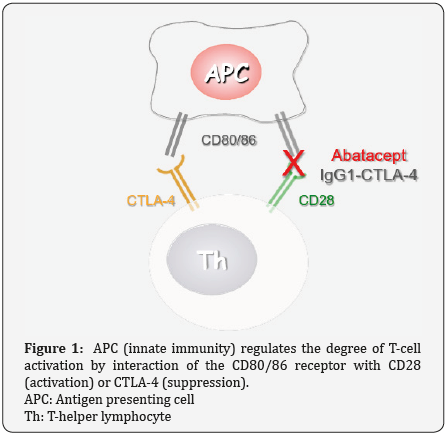
Beyond classical immunosuppression, the era of biological agents permitted pharmacological manipulation of several aspects of the immune response, in order to favor the rearrangement of an immune effective environment that will retard disease progression in several diseases. Central role in the establishment of a chronic inflammatory microenvironment holds the co- stimulatory pathway. This pathway consists of receptors and ligands that regulate the level of activation of the adaptive immune response from cells of the innate immune compartment. The main receptor of this pathway on antigen presenting cells is CD80/86 that can bind to either CTLA-4 (suppressive) or CD28 (activating) receptors on the surface of T-cells. The fact that the same receptor on APCs can either activate of suppress T-cell function, is a sophisticated homeostatic evolution of the immune system acting as a dimmer that can properly regulate the level of activation of T-cells (Figure 1).


Abatacept for DCM

In autoimmune diseases, suppression of T-cell activation, by inhibiting CD80/86-CD28 interaction has been proven clinically beneficial in conditions such as Rheumatoid Arthritis. Could the application of such a therapeutic strategy have beneficial effects of DCM? First of all, it has been documented that DCM is associated with an enhanced overexpression of constitutive molecules (CD25) on behalf of T-cells presupposing CD28 overexpression, both propagating continuous activation [1]. Furthermore, other reports discuss that the alternative CD80/86-CTLA-pathway is associated with down regulation of the inflammatory Th17 response [2], a helper T-cell response that contributes to the development of DCM. Therefore, the authors suggest that potential use of the monoclonal antibody abatacept in DCM, could indeed down regulate T-cell activation in patients manifesting premature clinical symptoms of DCM (Figure 1). Blockade of the CD80/86-CD28 pathway with abatacept will also leave unaffected the suppressive part of the dimmer complex (CD80/86-CTLA4) further favoring suppression of inflammatory Th responses (Th17).

Conclusion

Manipulation of the immune system has been proven so far effective for the treatment of autoimmune diseases and certain types of malignancies. Sophisticated application of current biological therapies may still provide therapeutic solutions in other immune mediated diseases such as DCM.


To know more about Juniper Publishers

Wednesday, April 15, 2020

Hybrid Nanoparticles Advance in siRNA Therapeutics - Juniper Publishers

Global Journal of Nanomedicine-Juniper Publishers

Abstract

Engineering organic-inorganic nanoparticles play the greatest advantages in regulating efficacy and safety when targeted delivery of the small interference RNA (siRNA). This opinion summarizes the current achievements and looks to the future in this area.

Keywords: siRNA therapeutics; Hybrid nanoparticles; Delivery; Efficacy; Safety

Introduction

Comparing with the traditional treatment modalities, small interference RNA (siRNA) has immense potential for the treatment of various human diseases including cancer, since Fire and co-workers discovered the siRNA in 1998 [1]. Despite the great versatility of RNA interference (RNAi) technology which could down-regulate any protein in targeted cells and tissues, many physiological and biological obstacles still stand in the way of successful, safety and efficient delivery in application for clinical. The major challenge indeed for RNAi-based therapy is the development of delivery system [2]. Up to now, a great number of systems were reported for delivery siRNA including the non-virus-based platforms [3]. According resurgence in clinical trials using RNAi occurred in 2012, more than 20 RNAi-based therapeutic cases are currently in clinical trials, and several of these are Phase III trials [4].

These encourage us to indicate the advantages of such platforms, leading to pave the way for the next generation of the siRNA therapeutics. Particularly, an excited news released from the U.S. Food and Drug Administration (FDA) has approved the first siRNA therapeutics for the treatment of peripheral never disease (polyneuropathy) in the 20th anniversary of siRNA Discovery. This great breakthrough encourages the scientist and industries would be more enthusiastic to develop and design functional vectors for siRNA therapies to fight the human diseases. Here, we summarize the advantages of hybrid nanoparticles including metal/non-metal cores further modification with the organic shells. Additionally, some typical examples are briefly illustrated here to provide translational strategies for the siRNA therapeutics in the near furfure.


Hybrid Nanoparticles in siRNA Therapeutics

Non-viral siRNA delivery hybrid nanoparticles have shown promise and have travelled to clinical trial applications. According with the understanding of characterization for the cell types and targeted tissues to rationally design the efficient vectors for siRNA therapeutics. Importantly, these functional systems have the advantages, such as low/non-toxicity, easy fabrication, biodegradation, biocompatibility, non-immunogenicity, low-price and high efficacy comparing with the commercial agents [5]. Gold and Quantum dots are the typically metal-based nanoparticles for the siRNA delivery [6-7]. Nanoparticles with an organic shell embedding a gold core are used for example in highly efficient, stable, organic light emitting diodes and organic photovoltaics [8]. For this application the dual purpose of the nanoparticle is used: gold core offers a plasmatic effect while a mono-dispersed polystyrene shell gives stability and solvent resistance [9].

An example of photothermal therapy is given by proteinases i.e. biological nanoparticles with a gold core and an organic shell engineered to present specified peptides or proteins [10]. Since biological nanoparticles are synthesized inside living cells, they present a much lower toxicity compared to synthetic nanoparticles when use these gold nanoparticles as siRNA delivery systems. Quantum dots possess particular photochemical properties. For instance, it is possible to use selective fluorescent to tag proteins as is traditionally done in classical immunocytochemistry. In addition, quantum dots present minimal photobleaching and a much higher signal to noise ratio compared to traditional methods [11]. They possess a broad absorption spectrum while maintaining a very narrow emission spectra, allowing multiplexing of many quantum dots of different colors in the same sample. This is unique and cannot be achieved using traditional fluorophores. These nanoparticles could be used in theragnostic siRNA platforms.

The other serial of the hybrid Nano systems is non-metalbased core with further functional organic shells. Two classical materials like silica/silicon or carbon-based nanoparticles widely used for siRNA delivery in vitro and in vivo [12,13]. Precise nanopore formation and ease of surface modifications are the main factors that brought interest silicon and silica nanoparticles to light. Silica nanoparticles have intrinsic biocompatibility, biodegradability and are efficiently bio-eliminated in vivo. However, metabolic changes and increased toxicity was observed especially in vivo due to active silanol groups used for surface modifications [14]. Some of the modifications applied to silica nanoparticles involve the synthesis of a cationic polymer layer or addition of quaternized dendrimers to the surface to permit electrostatic loading of siRNA, PEG (a protective polymer) addition to protect nanoparticles and to improve cellular uptake and delivery of MSNPs-siRNA conjugates [15,16].

Carbon nanotubes possess interesting physical and chemical properties that allow to easily cross the plasma membrane and translocate into the cytoplasm of cells. This property is due to their particular needle shape that exploits the cell’s endocytosisindependent mechanism without inducing cell death [17]. It has been shown successful delivery of siRNA using Carbon Nanotubes by functionalizing the nanotubes with functional groups. Using this functionalization siRNA was expelled from the sidewalls of the nanotube to silence telomerase reverse transcriptase expression inducing tumor growth suppression [18]. It has also been shown that amino- functionalized multiwalled carbon nanotubes (f-MWNT) can effectively deliver in vivo an siRNA sequence while triggering cell apoptosis resulting in human lung xenograft eradication and prolonged survival [19]. These nanotube-based siRNA transfer vectors have shown minimal cytotoxicity and effective delivery and gene-silencing capabilities.

Conclusion and Outlook

A central message of this opinion is that inorganic-organic hybrid functional nanoparticles for the siRNA delivery. Under well understand the properties of such nanoparticles when they serve as siRNA delivery systems, which successively addressing each of these barriers, innovative design features can be rationally incorporated that will create a new generation of nanotherapeutics, realizing a paradigmatic shift in nanoparticlebased siRNA delivery.

To know more about Global Journal of Nanomedicine
Click here: https://juniperpublishers.com/gjn/index.php

To know more about Juniper Publishers

Unveiling the Intersection of Age and Gender in the Emotional Well-being of Older Students: A Pilot Study at the Permanent University for Adults in Alicante (Spain) - Juniper Publishers

 Gerontology & Geriatric Medicine - Juniper Publishers Abstract This study assesses the emotional impact and well-being of older stud...