Thursday, September 30, 2021

Antiangiogenic Targeting of Early Developing Glioblastoma Behind a Weakened Blood Brain Barrier - Juniper Publishers

 Tumor Medicine & Prevention - Juniper Publishers

Abstract

Glioblastoma is the most devastating and most common brain tumor in adults. According to the (US) National Cancer Institute (NCI), there were an estimated 23,380 new cases and 14,320 deaths from brain and other nervous system cancers in the US in 2014. I will emphasize here therapy using Antiangiogenic agents, particularly in the case of tumors in their early development, taking advantage of the associated weakening of the blood brain barrier. As a prelude to this new development, I review below the current lines of treatment for Glioblastoma, making a distinction between primary and secondary tumors, and recurrent tumors. I will also review the brain protective barriers, chiefly the blood brain barrier which, as its name implies, is the primary impediment for the delivery of therapeutic drugs. I will also analyze the drug targeting difficulties and the mechanisms for drug targeting.

Keywords: Bevacizumab; Bradykinin; Cediranib (an antiangiogenics); Cilengitide; Mannitol; Temozolomide

Abbrevations: BBB: Blood Brain Barrier; BPB: Brain Protective Barriers; DFS: Disease Free Survival; CNS: Central Nervous System; CSF: Cerebro Spinal Fluid; EGFR: Epidermal Growth Factor Receptor; EVGF: Endothelial Vascular Growth Factor; GB: Glioblastoma; GBM: Glioblastoma Multiform; GY: Grey; HIFU: High Intensity Focused Ultrasound; MGMT: Methylated Promoter Gene (O6-alkylguanine DNA alkyltransferase); NCI: (US) National Cancer Institute; OS: Overall Survival; PFS: Progression-free survival; P-gp: P-glycoprotein; TMZ: Temozolomide

Introduction

Glioblastoma (GB), also known as Glioblastoma Multiform (GBM), is the most common primary brain tumor in adults. It remains an unmet need in oncology. The survival rate is ~1 year, and only 5% of the people affected live for 5 years. Figure 1 is an illustration of the appearance of GBM while Figure 2 contrasts an actual GBM with the corresponding contrastenhanced CT images. In a recent review article [1], the various treatment modalities devised so far for primary and recurring GBMs after treatment and their metastases, have been described in detail. These included surgery, conformal radiotherapy, boron neutron therapy, intensity modulated proton beam therapy, Antiangiogenic therapy, alternating electric field therapy, micro RNA, immunotherapy, adjuvant therapy, gene therapy, stem cell therapy and intranasal drug delivery without neglecting palliative therapies. Unfortunately, to date, the trials have not shown a benefit in overall survival (OS) either in the case of Antiangiogenic agents alone or in combination with chemo radiotherapy.

As is known, GBMs are highly vascular tumors and overexpress endothelial vascular growth factor (EVGF), a proantigenic cytokine. Premised on earlier studies that had shown promising radiographic response rates, delayed tumor progression, and a relatively safe profile for anti-EVGF agents, several randomized phase III trials were conducted. It was thus preliminarily concluded that Antiangiogenic agents might not be beneficial in unselected populations of patients with GBM [2].

Even for selected patients, biomarker identification has lagged behind the process of drug development and, apparently, no validated biomarker for patient stratification exists. Nonetheless, phase II trials have revealed an association between increased perfusion and oxygenation (which are consequences of vascular normalization) and survival. This suggests that earlyimaging biomarkers could help to identify that subset of patients who will most likely benefit from anti-EVGF agents.

will now report on potentially more promising results in which advantage is taken of the weakening of the blood brain barrier (BBB) by GBMs. This other approach may lead to new ways to kill brain tumors that use the BBB weakness to get targeted drugs into the brain during the early stages of the cancer. But first, let us briefly review the current lines of treatment for GBMs.

Current Lines of Treatment for Glioblastoma

First-line treatments have been clearly defined since 2005; however, no standard second-line treatment has yet been determined. No prevention strategy is known although several possible risk factors have been discussed. Most treatments cannot eradicate all tumor cells, thus, surgery is often insufficient given the diffuse nature of the disease, chemotherapy has major limitations because most drugs cannot cross the BBB, and penetration into brain cells is limited. In addition, the cells in brain tumors are greatly heterogeneous, which limits the treatment efficacy and explains the high rate of progression of the disease. The standard treatment consists of:

a) Surgery (maximal resection).

b) Radio chemotherapy (6 weeks of radiotherapy at a dose of 60 Grey [GY] together with concomitant chemotherapy with temozolomide (TMZ) at a rate of 75mg/ml daily); and once chemo radiotherapy is completed.

c) Adjuvant treatment (a minimum of 6 months with TMZ is started at a dose of 150-200mg/ml for 5 days every 28 days) [3-11].

Prognosis and response rates with TMZ are known to be better in patients presenting with a Methylated MGMT promoter gene (O6-alkylguanine DNA alkyltransferase). Survival of patients with Methylated MGMT is 21.7 months compared with 15.3 months for patients with a non-Methylated gene. Recent clinical trials in elderly patients (more than 65 years of age) diagnosed with GBM showed that TMZ is not inferior to radiotherapy. Patients with MGMT promoter methylation experienced the best results, facilitating decision-making in this fragile elderly population.

Two recent phase III clinical trials investigated the addition of bevacizumab to standard treatment with TMZ. They showed no increase in overall survival (OS) but disease-free survival (DFS) increased. In addition, several clinical studies have looked at:

a) The use of various drugs (e.g. integrand inhibitors such as cilengitide and other Antiangiogenic such as cediranib).

b) Vaccines against the epidermal growth factor receptor (EGFR), specifically the EGFR variant 3 (EGFRV3), which is detected in 30% of the patients.

Another innovative strategy is the application of alternating intermediate-frequency electrical fields (100–300kHz) as an adjunct to standard treatment. Immunotherapy has not yet demonstrated any conclusive results to date. There are no cures at present [12].

The use of cytotoxic drugs (chemotherapy) is essentially an educated trial and error approach with one or a combination of approved drugs. It does not stem from the deep understanding of tumor biology nor does it consider the braiding of both normal and cancerous cells in our genome [13-17]. As a result, chemotherapy has historically provided little durable benefit as the tumors recur within several months even in the case of the more accessible tumors that are located outside the brain. For brain tumors, the access is even more challenging because of the presence of the brain protective barriers (BPBs), chiefly the BBB, compounding the difficulties [18,19].

Distinction between primary and secondary tumors

It is important to distinguish primary from secondary GBMs. The former tumors occur spontaneously and the latter have progressed from a lower-grade glioma. Primary GBMs have a worse prognosis, different tumor biology, and may have a different response to therapy, resulting in a critical evaluation to determine patient prognosis and therapy. Over 80% of secondary GBMs carry a mutation in IDHI, whereas this mutation is rare in primary GBMs (5-10%). Thus, IDHI mutations are a useful tool to distinguish primary and secondary GBMs since histopathologically they are very similar and the distinction without molecular biomarkers is unreliable.

Targeted biomarkers and molecular targets that are specific to each person’s genomics can also be judiciously used to personalize the treatment to the selected patient. Biomarkers and molecular targets contain information that can be gathered from a person’s cancer cells and then used to tell what chemotherapeutic agents may work best for that specific patient. These biomarkers can also help locate natural phototherapeutics, which aid in personalizing a treatment program.

Case of recurrent glioblastoma

Disease recurs in almost all patients, and most patients will not be candidates for new surgery or a new course of irradiation so that therapeutic options are limited. Progression-free survival (PFS) after recurrence or progression is ~10 weeks, and OS is ~30 weeks. If the patient’s condition allows, the second-line treatment consists of anti-cancer drugs. Other approaches have therefore also been studied, including: Antiangiogenic, EGFR inhibitors, Nitrosoureas (alkylation agents characterized by high lipophilicity, allowing them to cross the BBB) and retreatment with TMZ. However, a standard second-line therapy has not yet been established.

It must further be emphasized that certain patients will be candidates only for symptomatic treatment because of their poor general condition or co-morbidities. Ensuring appropriate management and support for the complications that typically occur during the course of the disease (convulsions, thrombosis, and cognitive deteriorations) are essential.

The Brain Protective Barriers

Description

Being the most delicate organ of the body, the brain is protected against potentially toxic substances by barriers that restrict the entry of most pharmaceuticals into the brain. The collective term “blood-brain barrier” (BBB) actually describes four main interfaces between the central nervous system (CNS) and the periphery:

a) The BBB proper formed by complex tight junctions between the endothelial cells of the cerebral vasculature. Its primary manifestation is the impermeability of the capillary wall due to the presence of the junctions and a low endocytic activity.

b) The blood-cerebra spinal fluid (CSF) barrier formed by tight junctions between epithelial cells of the choroid plexus. Both (a) and (b) extend down the spinal cord.

c) The outer CSF-brain barrier formed by tight junctions between endothelial cells of the arachnoids vessels (pia arachnoids).

d) The inner CSF-brain barrier formed by strap junctions between the neuro-ependymal cells lining the ventricular surfaces. It is present only in early development and absent in the adult [18,19].

The above barriers and the blood retinal barrier (not studied here) are parts of a whole realm of barriers. The following considerations are limited to the BBB proper (Figure 3).

Function

The BBB behaves as a continuous lipid bilayer surrounding the brain. It is the major obstacle preventing the passage of polar and lipid-insoluble substances and drugs that are potentially useful for combating diseases affecting the CNS. Unfortunately, in its neuro protective role the BBB functions to hinder the delivery of many potentially important diagnostic and therapeutic agents to the brain. Therapeutic molecules and antibodies that might otherwise be effective in diagnosis and therapy do not cross the BBB in adequate amounts. Essential nutrients are delivered to the brain by selective transport mechanisms (glucose transporter, amino acid transporters, etc). Although most drugs can enter the brain by passive diffusion through the endothelial cells depending on their lipophilicity, degree of ionization, molecular weight, relative brain tissue and plasma bindings some others can use specific endogenous transporters. In such cases, bindi competition on the transporter with endogenous products or nutrients can occur and limits drug transfer. Extensive efforts are therefore being made to come up with drug delivery strategies that would enable the passage of therapeutic molecules safely and effectively. Such strategies involve modifying the drug itself or coupling it to a vector for receptor-mediated or adsorptionmediated transcytosis [2,3]. In other cases such as, for example, in Antiangiogenic treatment discussed here, advantage is taken of modifications (weakening, disruption, modification, etc.) of the BBB for drugs to pass through.

Drug targeting difficulties

The BBB can be a major impediment and the main limitation for the treatment of neurological diseases caused by inflammation, tumors or neurodegenerative disorders. The close intercellular contact between cerebral endothelial cells due to tight junctions also prevents the passive diffusion of hydrophilic components from the bloodstream into the brain. Thus, many drugs are unable to reach this organ at therapeutic concentrations. Various attempts have been made to overcome the limited access of drugs to the brain e.g. chemical modification, development of more hydrophobic analogs, or linking an active compound to a specific carrier. Transient opening of the BBB in humans has been achieved by intracarotid infusion of hypertonic Mannitol solutions or of Bradykinin analogs. Another way to increase or decrease brain delivery of drugs is to modulate the P-glycoprotein (P-gp) whose substrates are actively pumped out of the cell and into the capillary lumen. Many P-gp inhibitors or inducers are available to enhance the therapeutic effects of centrally acting drugs or to decrease central adverse effects of peripherally active drugs [4]. Nonetheless, overcoming the difficulty of delivering therapeutic agents to specific regions of the brain presents a major challenge to the treatment of most brain disorders.

Mechanisms for drug targeting

Mechanisms for drug targeting in the brain involve going either “through” or “behind” the BBB: In the former case, this entails: a) Disrupting

a) Disrupting the BBB by osmotic means. This can be accomplished biochemically by the use of vasoactive substances such as Bradykinin or even by localized exposure to high-intensity focused ultrasound (HIFU).

b) Modulating the expression and/or the activity of efflux transporters. Several specific transport systems (via transporters expressed on cerebral endothelial cells) are implicated in the delivery of nutrients, ions and vitamins to the brain; other transporters expressed on cerebral endothelial cells extrude endogenous substances or xenobiotics (which have crossed the cerebral endothelium) out of the brain and into the bloodstream.

c) Using the physiological receptor-dependent BBB transport. Other methods used to get through the BBB by increasing the permeability of the BBB entail the use of endogenous transport systems, including carrier-mediated transporters such as glucose and amino acid carriers.

Receptor-mediated trancytosis for insulin or transferring; the blocking of active efflux transporters such as the transferring receptor of active efflux transporters such as P-gp. However, vectors targeting BBB transporters, such as the transferring receptor have been found to remain entrapped in brain endothelial cells of capillaries, instead of being ferried across the BBB into the cerebral parenchyma. Still, other methods create new viral or chemical vectors to cross the BBB [5].

Drug delivery methods “behind” the BBB by-pass the BBB by central drug administration. They include intracerebral implantation (such as with needles) and convection-enhanced distribution. Mannitol can be used to bypass the BBB.

Antiangiogenic Therapy in Presence of a Weakened Blood Brain Barrier

Recent experiments with mice [20] have shown that cancer cells hijack and feed off blood vessels. In the brain, these results in a weakened BBB and this may provide one reason for the rapid spread of GBMs. This observation may lead to new ways to kill brain tumors using the BBB weakness to get targeted drugs into the brain during the early stages of the cancer. In these studies, the researchers used fluorescent dyes and a variety of imaging techniques in mouse models of GBMs to see how tumor cells travel to the brain and how they relate to other cells and blood vessels. They focused on interactions among GBM cells, astrocytes and blood vessels.

Outside the main tumor mass, these researchers found that nearly all GBM cells gather in the space between the astrocytic end feet and the outer surface of blood vessels. It appeared that the cancer cells were using the network of small blood vessels as a scaffold to guide their migration along the blood vessels as they extracted nutrients from the blood inside. It also appeared that the GBM cells hijacked control over blood flow in the BBB away from the astrocytes, loosening the tight junctions, and resulting in a breakdown in the barrier. Very small groups of GBM cellseven individual cells - could weaken the BBB in the early stages of the disease. At an early stage of the disease, as they invade the blood vessels, it looks like the tumor cells are not completely protected by the BBB, thus making them more vulnerable to targeted drugs delivered via the bloodstream to the brain.

If the above findings hold true in humans, treatment with anti-invasive agents might be beneficial in newly diagnosed GBM patients. A better understanding of how tumor cells and the BBB interact may lead to more successful ways to treat GBM patients. This includes a better understanding of how the BBB is regulated and exactly how tumor cells hijack control over blood vessels to grow and spread.

Summary and Conclusion

Glioblastoma (or Glioblastoma multiform) is the most common primary brain tumor in adults. It remains an unmet need in oncology. I have reviewed the difficulties encountered in devising treatment lines and the correspondingly associated risks, making a distinction between a primary and the often recurring tumor. I have also briefly analyzed the various treatment options whether singly or in combination. Chemotherapy (or treatment with cytotoxic drugs) has historically been not beneficial in general, and a fortiori not in the brain which is generally surrounded by protective barriers that represent serious impediments. Over recent years, multiple drugs have been assessed both in immunotherapy or combination therapy. Multiple trials have been conducted; unfortunately, most of these trials do not have a control arm, have used different assessment tools, and may have been influenced by many events including repeat surgery. As a consequence, the trial conclusions are rather controversial.

In addition to chemotherapy, I have again briefly reviewed the other treatment strategies that are used either singly or in combination with chemotherapy, including: surgery; conformal radiotherapy; boron neutron therapy; intensity modulated proton beam therapy; Antiangiogenic therapy; alternating electric fields; and even vaccines and palliative therapies and lifestyle changes [21-25].

In present-day practice, it is recommended that treatment of recurrent Glioblastoma be individualized according to several factors: performance status, age, tumor histology, biomarkers, possibility for repeat surgery, time to recurrence and response to prior treatment. It is also recommended that patients be enrolled into well-designed clinical trials to maximize their clinical benefits. Research into these and other treatment options is ongoing including but not limited to: micro RNA, immunotherapy, adjuvant therapy, gene therapy, stem cell therapy and intranasal drug delivery. It is too early to report definitely on their corresponding benefits and associated risks.

Recent experiments with mice have shown that cancer cells hijack and feed off blood vessels. In the brain, these results in a weakened BBB and this may provide one reason for the rapid spread of GBMs. This observation may lead to new ways to kill brain tumors using the BBB weakness to get targeted drugs into the brain during the early stages of the cancer. Outside the main tumor mass, nearly all GBM cells gather in the space between the astrocytic end feet and the outer surface of blood vessels as if the cancer cells were using the network of small blood vessels as a scaffold to guide their migration along the blood vessels as they extracted nutrients from the blood inside. It also appeared that the GBM cells hijacked control over blood flow in the BBB away from the astrocytes, loosening the tight junctions, and resulting in a breakdown in the barrier. Very small groups of GBM cellseven individual cells-could weaken the BBB in the early stages of the disease. At an early stage of the disease, as they invade the blood vessels, it looks like the tumor cells are not completely protected by the BBB, thus making them more vulnerable to targeted drugs delivered via the bloodstream to the brain. If the above findings hold true in humans, treatment with anti-invasive agents might be beneficial in newly diagnosed GBM patients. A better understanding of how tumor cells and the BBB interact may lead to more successful ways to treat GBM patients. This includes a better understanding of how the BBB is regulated and exactly how tumor cells hijack control over blood vessels to grow and spread.

Click here: https://juniperpublishers.com/index.php  

Wednesday, September 29, 2021

Modified Cellulose-Based Edible Polymer Coating: An Investigation of Castor Oil Concentration for Application in Apple Preservation - Juniper Publishers

Academic Journal of Polymer Science - Juniper Publishers

Abstract

The purpose of this study was to evaluate the effect of adding various concentrations of castor oil (2% and 4% v/v) on the properties of edible films based on carboxymethyl cellulose (CMC). Moisture content, water solubility, tensile strength, elongation at break, elastic modulus, water vapor permeability, optical and thermal properties and antioxidant activity of the films were examined. The results demonstrated that the presence of castor oil led to a decrease in moisture content, water solubility and mechanical strength of the films. The film containing 4% of oil showed the highest water vapor permeability. The optical properties measurement represented that all of the samples were transparent. By the addition of castor oil, the antioxidant activity of the films improved largely. Thermal properties of the samples had also been investigated and it was determined that the effect of castor oil on the melting point was negligible. Finally, the performance of the coatings for protecting fresh apples was studied with measuring the pH value of apples and performing some field test.

Introduction

Every year a large portion of foods, especially fruits and vegetables, from the time they are harvested until they reach to consumers, go through different kinds of qualitative and quantitative deterioration and loss [1]. Various factors are involved in food deterioration such as microorganisms, mechanical damages, time and light. Therefore, if we fail to protect the food, it can be damaged within hours or days [2]. Currently, there are numerous technologies used for the purpose of fruits and food preservation. Some include controlled atmosphere storage and modified atmosphere packaging, innovative osmotic dehydration technologies, electro-osmotic dewatering , thermal pasteurization, and gaseous ozone and ozonated water treatments [3,4]. One way to prevent or delay the food deterioration is the use of edible coatings. During the past decade, edible coating has been widely used for this purpose [5-9]. Some advantages of edible coatings include extending shelf-life of fresh fruits, reducing amount of waste produced from packaging processes, improving appearance and fulfilling environmental safety, as well as enhancing nutritional properties due to containing lots of beneficial biopolymers and biological compounds including polysaccharides, proteins, and lipids. Moreover, they decrease water loss and retard ripening of fruits due to barrier mechanism, and are also able to enhance antioxidant properties of fruits and vegetables [10-13].

Any material used to cover foods in order to enhance the shelf life, which may be eaten with the food, is an edible coating [2]. Various studies have been conducted on the properties and applications of edible films and coatings [14-18]. The initial studies on edible coatings can be traced back to 1967, where Hardenburg studied the application of wax used by Chinese on citrus fruits [19]. Since 1986 there were various studies on edible coating including wax emulsions, oils, cellulose, chitin and chitosan, and their impact on diverse properties of fruits and food like shelf-life [20-26]. However, more research is needed because there are no edible films that can be used for all purposes. As other technologies, edible coatings can also bring some challenges like undesirable tastes caused by presence of wax materials, hydrophilic nature of edible coatings which makes them moisture-sensitive and limits their moisture-barrier performance, poor adhesion and coverage, and insufficient mechanical properties of biopolymers comparing to the synthetic ones [27,28]. In general, an ideal edible film must have characteristics such as being nontoxic, allergenic or indigestible materials, protecting foods from mechanical damages, good adhesion, maintaining the appearance of the product and having an easy and economical production [2]. Proteins, carbohydrates and lipids are the main film forming agents in edible coatings [29]. Generally, lipids will reduce water transmission in edible coatings, polysaccharides are good for controlling the transmission of gases and protein-based films have proper mechanical strength [29]. These materials can be used either individually or as a blend (composite films) in edible coatings [29].

Carboxymethyl cellulose (CMC) is a conventional polysaccharide, which is one of the common derivatives of cellulose. CMC is an anionic polysaccharide, which unlike cellulose is soluble in water [30]. CMC has wide variety of uses in different industries such as foods and coatings [31] and therefore is an excellent choice for edible coatings because it has no toxic or allergic effects. It is one of the most desirable polysaccharide polymers due to its excellent properties including perfect film forming ability, availability, low price and high viscosity, which can be used to produce both edible and degradable films and coatings [31]. In most cases, a plasticizer must be added to the film forming solution in order to reduce the brittleness of CMC films. However, using plasticizers will affect the mechanical and permeability properties of the films [32]. The most common food grade plasticizers are some polyols including glycerol, mannitol, sorbitol, and sucrose [33]. Glycerol is a clear, colorless and odorless liquid, which is soluble in water due to its hydrogen bonds. This component reduces the film's fragility by being located between the CMC chains (enhancing chain mobility) and also by absorbing water [34]. The beneficial properties of some lipids, including their good compatibility with other film constituents and good barrier properties against water vapor and other gases, make them an ideal choice for edible coatings and films [35,36]. Lipid compounds that are commonly used to make films and edible coatings include edible oils, fatty acids and waxes [37]. The efficiency of lipid used in edible films and coatings depends on the nature of the lipid, particularly its structure, hydrophobicity and its interaction with other components in the system [2]. Castor oil is a viscous and non-volatile liquid with a pale yellow color. High amount of both resinoleic acid (RA) and double bonds results in oxidative stability of the castor oil as well as long shelf life [37]. The antioxidant activity of castor oil also makes it an excellent choice for use in edible coatings in order to prevent food degradation.

In this study, the effect of castor oil on mechanical, optical, physical and thermal properties of CMC-based edible films along with their antioxidant activity have been investigated. The results showed potential use for castor oil in edible coatings.

Materials and Methods

Materials

Carboxymethyl Cellulose (CMC) with purity of 99.6% and viscosity of 2787 cps ; Castor oil with density of 0.959 g/mL [38] and Polysorbate 80 (Tween 80) were purchased from Pasargad Novin chemical Co. Glycerol (USP grade) was purchased from PALMAC. Analytical grade of Ascorbic acid was obtained from Merck (Darmstadt, Germany). CMC is a white, granule-shaped powder used for increasing the viscosity. Castor oil consists of various fatty acids mostly containing ricinoleic acid (12-hydroxyoctadecenoic acid) [39]. Tween 80 which is a highly viscous liquid, is used as a surfactant [40,41].

Preparation of films

Film forming solution was prepared by adding 1 g of CMC to 100 ml of distilled water (1% w/v). The system was under continuous agitation by magnet stirrer and the temperature was set on 75℃. After 40 minutes, a clear solution was achieved. Then 1 ml of Glycerol (as plasticizer) was added to the film forming solution and stirring went on for another 15 minutes. At this point, the film forming solution was ready in order to prepare the control films (films containing no castor oil). To do so, 25 g of the solution was poured in the middle of plastic circular plates (with 10 cm diameter and 1 cm height) and dried at room temperature in about 48 hours. Other film samples were prepared as described below:

a. CO-2: 0.2 ml of Tween 80 (proportional to castor oil) was added to the film forming solution under continuous stirring. After 15 minutes, 2 ml of castor oil was also added to the solution and the stirring continued for another 30 minutes.

b. CO-4: 0.4 ml of Tween 80 was added to the film forming solution under continuous stirring. After 15 minutes, 4 ml of castor oil was also added to the solution and the stirring continued for another 30 minutes.

c. AA1: 1 g of ascorbic acid was added to the film forming solution and the system was under stirring for 10 minutes.

d. AA2: 0.4 ml of Tween 80 was added to the film forming solution under continuous stirring. After 15 minutes, 4 ml of castor oil was also added to the solution and the stirring continued for another 30 minutes. Following that, 1 g of ascorbic acid was added to the solution and stirring continued for 10 minutes.

The procedure of preparing film from all of the mentioned solutions was similar to the control films. Also, to do some field tests and measuring pH the sample films were applied on apple by silicon brush. All the dried films were peeled of the plates and kept in a desiccator at 25℃ and 50% relative humidity (RH) for 72 hours until further evaluations.

Thickness

A hand-held micrometer was used to measure the film thickness. For each film, the thickness was measured in several areas (at least at five locations) and the final thickness was reported as the average value.

Optical properties

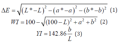
The color of each film was monitored using a calorimeter in the CIE Lab system. The films were placed on a standard white background (L*= 89.7584, a* = -0.5083 and b* = -2.0585) and their “Lab” values were determined. The color difference (ΔE) between the films and the standard background, whiteness index (WI) and yellowness index (YI) were calculated using the following equations:

Where “L” value represents the lightness of the sample (varies from 0 (black) to 100 (white)), “a” represents the redness-greenness balance of the sample (from negative (green) to positive (red) values) and “b” indicates the yellowness-blueness balance of the film (between negative (blue) and positive (yellow) values) [37].

Moisture content (MC)

The moisture content of each film was determined based on weight loss. In this method, the film was first weighed and then placed inside the oven at 110°C. The weight of the film was measured every hour until it reached a constant value. Finally, the amount of moisture in the film was obtained from the difference between the initial and the final weight. The percentage of water in the film was calculated by the following equation:

Where Wi is the weight of the film before being placed in the oven and Wf is the weight of the film after that. The test was repeated three times for each sample to secure a more accurate result.

Water solubility (WS)

The solubility of the film in water was evaluated based on the method described by Gontard et al. with some modifications [42]. The initial weight of the film was measured after drying the sample at 110 °C. Then the dry samples were immersed in 100 ml water for 5 hours at 25°C under continuous stirring. After filtration, to determine the final dry weight, the portion of the film, which was undissolved in water, was placed in an oven at 110°C to reach a constant weight. The water solubility percentage of the film was calculated by the following equation:

Where Wi is the initial dry weight and Wf is the final dry weight of the films (after immersing in water).

In order to obtain a consistent result, water solubility of each film was evaluated three times and the average value was reported.

Water vapor permeability (WVP)

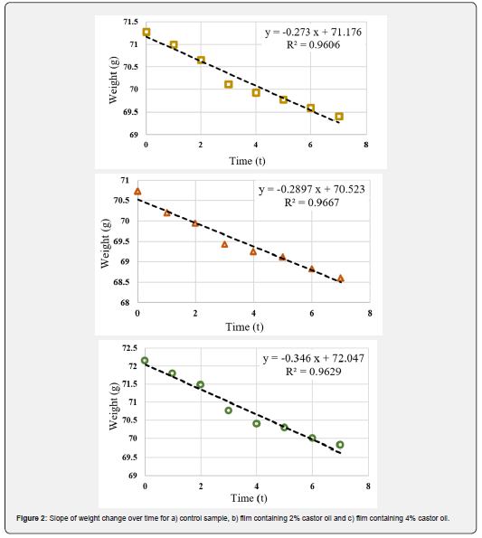
The water vapor permeability of each film was determined based on the instructions provided by ASTM E96 [43]. Cups with an average diameter of 3.5 cm and a depth of 1 cm were used to determine WVP. The films were cut in circle shapes with slightly larger diameter than of the cup. After placing 3 g of water (RH = 100%) in each cup, they were covered with sample films. Each cup was placed in a desiccator containing silica gel (RH = 0%) and the temperature was maintained at 25°C. The cups were weighed every 24 hours (With a precision of 0.0001 g). The weight change of the cup was plotted as a function of time and the slope of this curve (weight change versus time) was calculated by a linear regression. Water vapor transmission rate (WVTR) was obtained by dividing this slope by the transmission area (m2). Finally, the WVP was evaluated from the following equation:

Where P is the saturated vapor pressure of water at test temperature (25℃), R1 is the RH inside the desiccator, R2 is the RH inside the cup and X is the film thickness (in meter).

Antioxidant activity

The antioxidant activity of the film samples was investigated through DPPH radical scavenging activity based on instruction of Brand-Williams et al. [44] with some modification. This test is based on the ability of the samples to donate hydrogen or electron and is evaluated by measuring the amount of color reduction of 2,2-diphenyl-1-picrylhydrazyl (DPPH) solution in methanol (a solution with purple hue). DPPH radical is absorbed at 517 nm but the amount of absorbance decreases when it is exposed to an antioxidant. To evaluate this property, 25 mg of the sample film was added to 5 ml of distilled water and the mixture was stirred continuously by a magnet Stirrer. Then 0.1 ml of the resulted solution was added to 3.9 ml of DPPH solution (0.1 mM methanol solution) and the sample was incubated in darkness for one hour at room temperature. The sample’s absorbance was then measured at 517 nm and the scavenging activity was calculated by the following equation:

Where A is the absorbance at 517 nm.

Mechanical properties

Tensile strength, elongation at break and elastic modules of the films were measured by the universal testing machine. The test was conducted based on the ASTM D882-91 standard [45]. For this purpose, the films were cut into rectangle shapes (about 2 cm x 10 cm) and placed between the grips. The initial space between the grips and the crosshead speed were set at 50 mm and 10 mm/min, respectively (with 1 kg load cell). Mechanical properties were evaluated for three replications of each sample.

Thermal properties

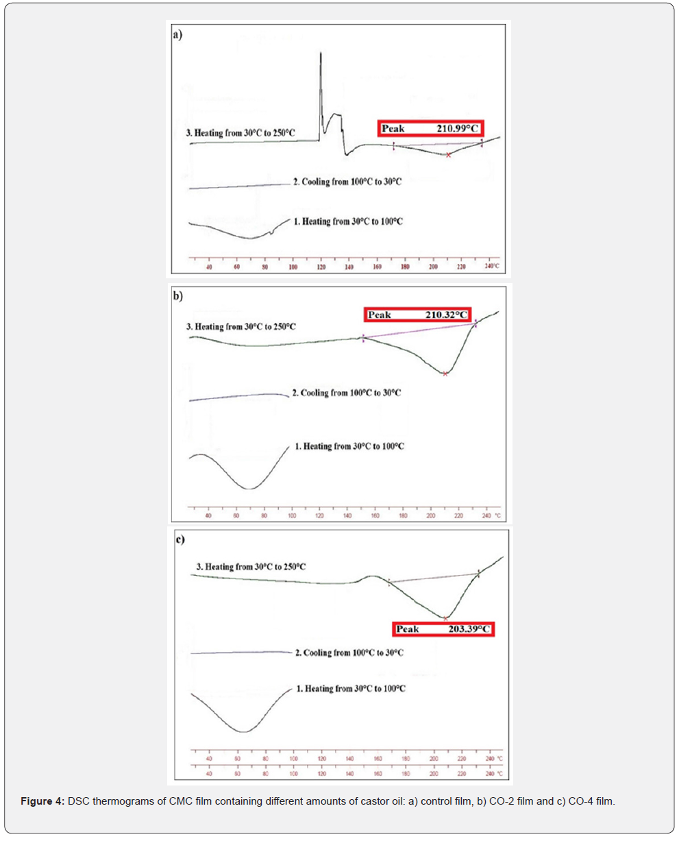
The thermal properties of the films were investigated by DSC test, which is described below. Approximately 5 mg of the film was placed in an aluminum pan. The reference was also an empty aluminum pan. Thermal behavior of different films were measured at a thermal scanning rate of 5℃/min in three steps. First heating from 30℃ to 100°C, then cooling from 100℃ to 30°C and last heating again from 30℃ to 250°C. The melting point (Tm) of the films was determined in this process.

pH value

To evaluate the pH of apples, 10 g of each sample (with its coating) was added to 100 ml of distilled water and then stirred for 30 minutes. After filtering the mixture, its pH was measured and reported by a digital pH-Meter. In order to compare the results, the same process was performed on control apples (apples with no coating). The pH measurement was started immediately after applying coating on the apples and was repeated every 4 days. During the test process, apples were kept at room temperature.

Field test

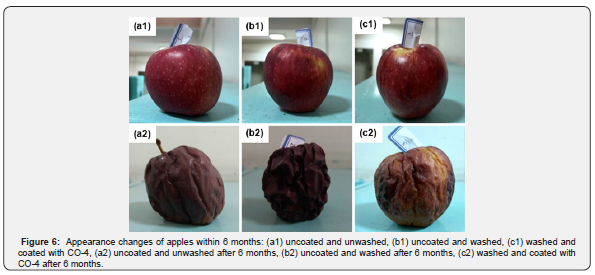
To evaluate the performance of the coating, three apples were picked from a store and processed as described below:

a. The first apple was kept unwashed.

b. The second one was washed.

c. The last one was washed and then the CO-4 film forming solution was applied on it with a silicon brush.

All three samples were then kept at a dark place at room temperature for six months.

Statistical analysis

All the Data were analyzed using MATLAB and Statistics Toolbox Release R2018b, The MathWorks, Inc., Natick, Massachusetts, United States. The results were reported as mean ± standard deviation.

Results and Discussion

Thickness

Thickness is an important parameter that affects some properties of the films such as transparency, permeability and mechanical strength [46]. The thickness of the films varied from 45 to 95 microns. As shown in Table 1, the thickness of the control film (without castor oil) was 45 microns and with increasing the oil content in the samples, their thickness increased until it reached 95 microns for the film containing 4% castor oil. The reason is that by adding oil to the film-forming solution, the solid content of the final coating increases, because castor oil enters the CMC matrix, which results in an increase in thickness. Same result have been reported by Shojaee-Aliabadi et al. [47].

Optical properties

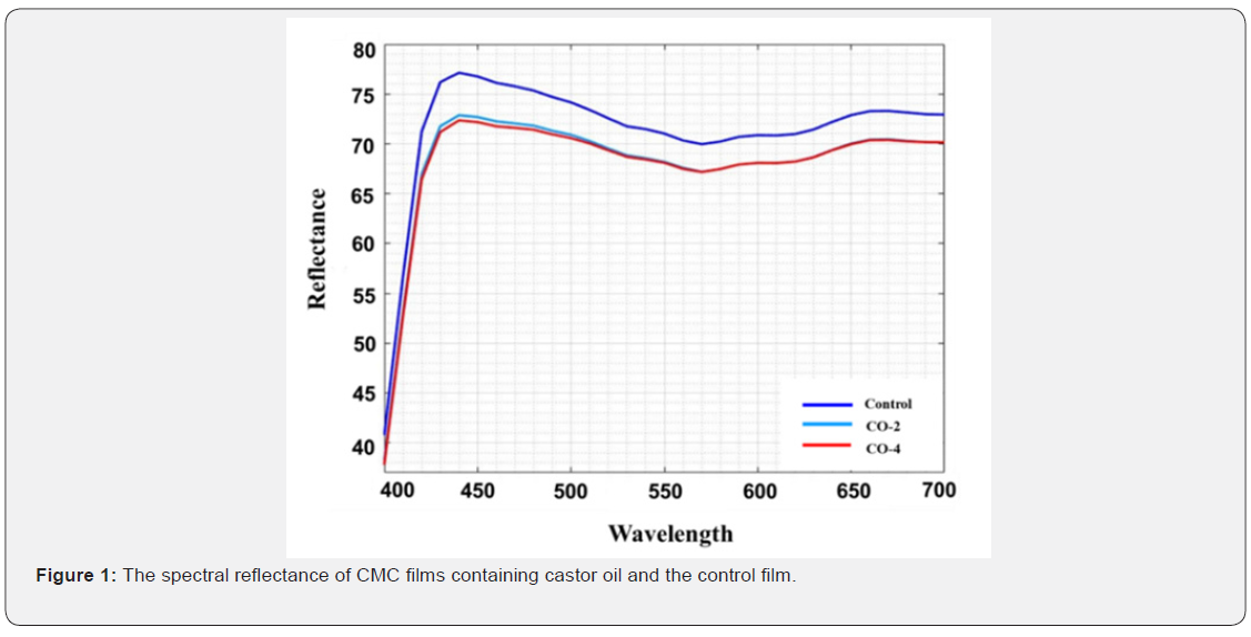
Optical properties or appearance is an important factor of edible coatings and films as they affect the acceptancy of a product by the consumers. The least expectation from a coating is to maintain the appearance of the product if cannot improve it. The CIE Lab, color difference (ΔE), whiteness index (WI) and yellowness index (YI) values used to describe the optical properties are given in Table 2. The spectral reflectance curves of the samples are also shown in Figure 1. As illustrated, the control film had the highest area under the curve and therefore the highest brightness. The L value (brightness) of the samples decreased slightly with increasing oil content. In addition, the yellowness index of the specimens increased marginally with increasing oil content in the film structure, which could be due to the absorption of light by castor oil at low wavelengths. For the same reason, the color difference of the samples increased moderately with the addition of oil to the film (the color difference between the CO-2 and CO-4 samples having different oil content was not significant). Finally, the whiteness index of the samples decreased slightly with the addition of oil to the system. The presence of castor oil and increasing the amount of it reduced the transparency of the film to some negligible degree. The reason is that by adding oil to the film forming solution, which is a water-insoluble fraction, the system becomes an emulsion, resulting in a decrease in the transparency of the film relative to the oil-free sample [48]. Overall, the addition of castor oil to CMC films had no major negative effects on their appearance.

MC

Moisture content of films is an important factor in determining coatings quality. High moisture content can provide an ideal environment for the growth of microorganisms and cause deterioration during long storage periods. The MC also helps edible films to melt in mouth when eaten [2]. The MC of each specimen is reported in Table 1. As can be seen, samples containing castor oil had a lower moisture content than the control film, and the MC decreases with increasing oil content from 2% to 4%. The reason for this decrease can be mainly attributed to the increased hydrophobicity of the films due to the increased oil content. In addition to increasing hydrophobicity, the interaction between castor oil and the hydroxyl groups present in the CMC impedes water absorption by these groups, thereby reducing the amount of water in the film. Similar results were found by previous studies [47,49].

WS

Moisture content of a film can also affect its solubility in water. The higher the moisture content of the film, the greater the solubility in water. The water solubility of each film sample is reported in Table 1. As shown in the diagram, the CMC film containing no castor oil (control sample) is completely soluble in water. The solubility in water decreased with increasing castor oil concentration in samples due to the high amount of hydrophobicity in the film matrix and preventing CMC hydroxyl groups from absorbing water. The lowest water solubility was observed in CO-4 sample containing 4% castor oil in its structure.

WVP

WVP is an important factor to investigate the performance of edible films and coatings and can be influenced by characteristics such as film integrity, hydrophobic ratio and film thickness [50]. Knowing WVP of the film is very helpful for preventing the mass transfer from food to the surrounding environment and therefore extending the shelf life of the product. The values of G/t, which is the slope of the weight change curve over time of the samples, are shown in Figure 2. Using these slopes, the WVP values were calculated for the samples (Table 1). It is observed that with increasing oil content, the slope of the curve increased, resulting in a higher WVP. The film containing 4% oil shows the highest amount of WVP. While the common expectation is that increasing oil content would decrease the permeability of the samples due to the increased hydrophobicity of the films, the exact opposite result has been achieved. This could be due to the fact that the increase in castor oil concentration in CMC films, besides increasing the hydrophobicity ratio, had a negative effect on the cohesion of the film matrix. The weakening of the cohesion forces in the matrix accelerates the transfer phenomenon and ultimately increased the water vapor permeability. Furthermore, castor oil also has a softening effect similar to glycerol. This makes the movement of polymer chains in the film easier, which increases permeability. Similar result was observed in the work of Dashipour et al. [49].

Antioxidant activity

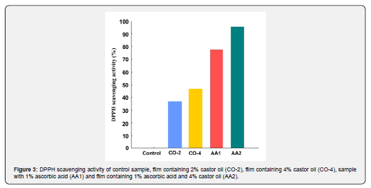
The ability of an edible coating to prevent foods from oxidation is an important factor in determining the coating's performance to extend the shelf life of the product. The higher the antioxidant activity of the coating, the better its function in protecting the food. In this study, the antioxidant activity of the samples was measured by DPPH scavenging activity method, and the results are shown in Figure 3. As expected, the control film showed no antioxidant activity, as there is no compound in its structure capable of radical absorption. The amount of antioxidant activity in oil-containing films is significantly higher than that of control sample. By increasing the oil concentration from 2% to 4%, the scavenging activity of the film also increased and reached 46.3%, which is desirable. The antioxidant activity of films containing just castor oil was lower than that of ascorbic acid containing samples. The highest scavenging activity was for AA2 sample, which had both castor oil and ascorbic acid (95.4%). The radical scavenging activity of the oil-containing specimens results from the unsaturated double bands present in castor oil structure. These double bands become saturated by absorbing free radicals; therefore, prevent the product from oxidizing.

Mechanical properties

Characteristics of edible films such as tensile strength (TS), elastic modulus (EM) and elongation at break (EB) are very important in coatings and packaging and can be helpful to predict coatings performance [51]. Table 1 shows the TS, EM and EB values of samples. As illustrated, the tensile strength of the control sample was 10.14 MPa, which decreased significantly with increasing oil content and reached 3.43 MPa in CO-4 film. Similarly, the elastic modulus of the films decreased with increasing castor oil from 76.91 MPa to 27.75 MPa. On the other hand, with increasing castor oil concentration, the flexibility of the films improved and therefore their elongation at break increased from 24.81% for control sample to 49.08% for CO-4 sample. This can be due to the fact that the addition of castor oil created a heterogeneous film, which results in decreasing mechanical strength and increasing flexibility [52]. In addition, as mentioned earlier, castor oil has some level of softening property. Therefore, its molecules can be placed between CMC chains and facilitate their movements and as a result increased the films’ flexibility, desirably.

Thermal properties

Determination of thermal properties, especially glass transition temperature, is very effective in evaluating the performance of edible films and coatings. If the glass transition temperature of the film is much higher than the ambient temperature, the film would be very brittle, but it would have low permeability. However, if the Tg is lower than the ambient temperature, the permeability would be very high and the film would be very soft and flexible, which will not provide the proper mechanical protection for the product. As a result, if the glass transition temperature of the films is slightly above ambient temperature (close to ambient temperature), it will be in a desirable range of permeability and mechanical strength [46]. Therefore, if castor oil could reduce the Tg and Tm of the specimens, it will be considered a beneficial effect. Thermal diagram of the samples obtained by performing DSC experiment and is shown in Figure 4. As shown in the diagrams, in the temperature range of 208℃ to 211°C there are large and endothermic peaks, which could be attributed to the melting temperature of the CMC matrix. The Tm of the control sample is 210.99°C, and with increasing castor oil content to 4% (CO-4), the Tm reached 208.39°C. As presented, no glass transition temperature was observed in the samples due to the low sensitivity of the test equipment.

pH value

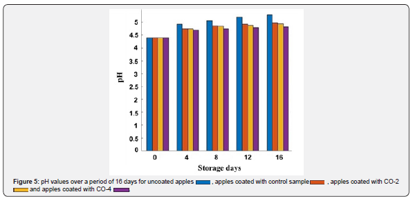
The microbial growth in the food produces nitrogen-containing compounds that increase the pH of the product [53]. The coating should prevent this increase in pH by restraining the microbial growth. The better the coating performs in this matter, the lower the pH increases and the longer the shelf life of the product will be. The pH values of control and coated apples were measured over 16 days, which are represented in Figure 5. As can be seen, the highest pH increase was for uncoated apples and the lowest increase was for apples with CO-4 coating. In general, coatings (especially the ones with castor oil) were successful in preventing pH increase and their pH was lower than non-coated apples after 16 days. This result shows that CMC-based coatings containing castor oil prevented pH value from increasing.

Field test

As mentioned earlier, one of the causes of food deterioration is time. Nowadays, the process of preparing foods until they are ready for consumers’ use takes a considerable amount of time. During this time-consuming process, foods could undergo water loss and deterioration. Therefore, the performance of edible coatings over time is very important. Figure 6 shows the changes of apples after being exposed to ambient temperature for 6 months. Although the coated apple (Figure 6, c2) was also suffered from high deterioration, it had less water loss and discoloration than the other two apples, which had no coating. The coated apple also had a much better physical state and was more firm and less wrinkled than the other two.

Conclusion

The presence of castor oil in the film forming solution increased the thickness of the resulting CMC films, but due to its hydrophobic nature, reduced the moisture content and water solubility of the films. The addition of castor oil in carboxymethyl cellulose films resulted in higher permeability due to the weakening of cohesion forces in CMC matrix and softening properties of the oil. The mechanical strength of the films was also weakened for the same reason. As the oil content increased, the color difference and yellowness index of the films increased, and the whiteness index decreased slightly. Due to the presence of unsaturated double bonds in the castor oil structure, this oil had the ability of radical absorption and therefore antioxidant activity. Addition of castor oil had no significant effect on the thermal properties of the carboxymethyl cellulose films and reduced the melting temperature of the samples by 3°C. The coatings prevented the early deterioration of apples and reduced the pH increasing rate by preventing the growth and activity of the microorganisms.

Click here: https://juniperpublishers.com/index.php  


Tuesday, September 28, 2021

Conservative Versus Surgical Management for Non-Traumatic Subarachnoid Hemorrhage: A Mini Review - Juniper Publishers

 Head Neck & Spine Surgery - Juniper Publishers 

Abstract

Subarachnoid hemorrhage diagnosis is always a challenge for patients and physicians because it has different presentations. There are two main causes of subarachnoid hemorrhage: traumatic and no traumatic subarachnoid hemorrhage. Both groups share clinical characteristics, but not treatment. Therefore, it is essential to recognize the signs, symptoms, and types of presentation for proper management. The objective of this article is to inform our audience about the significant difference between conservative and surgical treatment for non-traumatic subarachnoid hemorrhage because it carries a high risk of morbidity and mortality, requiring emergency management and well-trained physicians to evaluate patients suspicious for the diagnosis. This article is a compilation of several articles that have been selected from different databases, International Journal of Emergency Medicine, and Journal of Neurosurgery. Traumatic subarachnoid-related articles were excluded from our search. There is no consensus yet about the approach of patients with non-traumatic Subarachnoid Hemorrhage(ntSAH) among experts worldwide, so SAH diagnosis is often overlooked due to the clinical manifestations and inconsistencies in individual findings, especially atypical presentation arrives at the ER. The most significant limitations of timely and aggressive management of SAH are the lack of clinical suspicion and the delay from the CT scan order until the CT scan report is ready. The most repeated cause of subarachnoid hemorrhage is aneurysm rupture. A timely aneurysm repair is considered the most critical strategy to reduce the risk of aneurysm re-rupture. Therefore, detection of the cause of bleeding and prompt management can make the difference between life and death.

Keywords: Subarachnoid hemorrhage; Surgical Management of SAH; Conservative Management of SAH; Medical management of SAH; Nontraumatic Subarachnoid hemorrhage

Abbreviations: SAH: Subarachnoid Hemorrhage; NTSAH: Non-Traumatic Subarachnoid Hemorrhage; GCS: Glasgow Coma Scale; DCI: Delayed Cerebral Ischemia

Introduction

The most common cause of patients going to the emergency room is headache [1]. As physicians, we must be able to recognize headaches that could be life-threatening. The medical history and the physical findings will allow us to differentiate a simple headache from those that can be deadly. "The worst headache of my life," this is the way how medical students and physicians can start thinking about subarachnoid hemorrhage, but not all the patients can recognize the worst headache in their life; some of them die before they arrive at the emergency room. There are many tools and strategies to approach and treat patients with severe headaches, and we must understand the strengths and limitations of each strategy.

The clinical presentation should be considered before proceeding with the different diagnostic modalities for subarachnoid hemorrhage. Once a subarachnoid hemorrhage is suspected, a CT scan must be requested. Subarachnoid hemorrhage diagnosis is always a challenge for the physician. Every second and minute will determine a poor or great outcome in each patient. There are multiple causes of subarachnoid hemorrhage (SAH), but we can differentiate two big groups: Traumatic and no traumatic subarachnoid hemorrhage. Both groups share some similar clinical characteristics, but not the treatment.

There are several scales to categorize SAH. The systems used to predict the patient outcome are the Hunt and Hess score and World Federation of Neurological Surgeons grading, and the Fisher grade helps predict vasospasm. In terms of patient-centered results and prognosis, specific scores were not performed better than the Glasgow Coma Scale (GCS). As we search into the diagnosis of SAH, it is essential to note that some patients with SAH, for example, Hunt and Hess lower grades are more commonly failed to see because the clinical presentation is mild, and they may have smaller aneurysms with scant subarachnoid blood. These patients do not necessarily recover or have less morbidity with rupture or re-rupture [1].

Accurate data is not available about the management of subarachnoid hemorrhage, but some of the Egyptian, Greek, and Arabic literature report some clues of earliest management in 1800. Unfortunately, the management is still controversial, especially when the discussion is between surgical and medical management of SAH. That is why reviewing many studies worldwide will allow colleagues to understand how to face this particular situation.

The most crucial strategy to reduce the risk of aneurysm re-rupture is timely aneurysm repair is generally considered. However, evidence for the ideal timing of treatment is limited, and it is undefined if ultra-timely treatment (within 24 hours) is superior to timely aneurysm repair (within 72 hours) [2]. A recently published retrospective data analysis that compares ultra-early treatment with repair performed within 24-72 hours after hemorrhage suggests that aneurysm occlusion can be performed safely within 72 hours after aneurysm rupture [2]. The American Heart Association/American Stroke Association suggests as a Class IB guidance that surgical clipping or endovascular coiling of the ruptured aneurysm should be implemented as early as achievable in most patients to decrease the risk of re-bleeding after SAH [2]. The treatment modality option between surgical clipping and endovascular coiling is a complex endeavor that requires an interdisciplinary team's expertise, including neuro-intensivists, interventional neuroradiologists, and neurovascular surgeons. The endovascular approach is superior for aneurysms to be considered equally treatable by both modalities, associated with better long-term outcomes.

Retrospective data on clipping and coiling in poor-grade patients suggests that surgical clipping and endovascular are equally effective. An early and short course of an antifibrinolytic drug, including tranexamic acid, started as soon as the radiological diagnosis of SAH is made and stopped within 24-72 hours, has been associated with a decreased rate of ultra-early re-bleeding and a non-significant improvement in long-term functional outcome. This approach remains controversial, and short-term administration of tranexamic acid to prevent re-bleeding is being studied in a multicenter randomized trial (Dutch Trial Registry number NTR3272) [3]. The avoidance of extreme levels of blood pressure is another medical intervention applied to prevent aneurysm re-rupture. The American Heart Association/American Stroke Association and the Neurocritical Care guidelines advise keeping the mean arterial blood pressure below 110mm Hg or systolic blood pressure below 160mm Hg (or both) in the presence of a ruptured unsecured aneurysm.

Serum biomarkers to detect the risk of delayed cerebral ischemia (DCI) are showing promising results [3]. Changes in serum protein S100B levels interacted with DCI status (presence vs. absence): F= 3.84, p= 0.016. Patients with DCI had higher S100B concentration level on day 3 than those without DCI (3.54±0.50ng/ml vs. 0.58±0.43ng/ml, p= 0.001). S100B concentration on day 3 following a SAH predicted DCI (p= 0.006). The multivariate logistic regression analysis has shown that impaired cerebral autoregulation and elevated S100B concentration on day three increase the likelihood of DCI [3]. Subarachnoid hemorrhage (SAH) is a medical emergency that requires urgent management. Around Eighty-five percent of cases of atraumatic SAH result from a ruptured aneurysm. Other factors such as arteriovenous malformation, Ehlers-Danlos disease can also be the cause [4].

The diagnosis of SAH ought to be considered in any patient with a severe and sudden onset or rapidly escalating headache. With many such patients presenting to the ED with a chief complaint of headache, differentiating those with a benign cause from an emergent etiology such as SAH can be difficult. Establishing the diagnosis of SAH, the most critical time-sensitive goals include confirmation of airway security and stabilization of hemodynamics. In the setting of a low Glasgow Coma Scale Score or the lack of ability to protect the airway, intubation should be undertaken, but care should be taken to mitigate increases in mean arterial pressure during the intubation process [5]. These therapeutic modalities should be addressed with the admitting neuro-intensivist or neurosurgery team. In addition, continuous electroencephalogram monitoring may be started in the intensive care unit.

It is essential to determine adequate management in every case, as this can be the difference between life and death. According to preoperative neurologic function, location, size of the aneurysm, the timing of the operation, severe initial bleeding, re-bleeding (usually within two weeks), and delayed ischemia were the major preoperative problems; ten percent died, and 13 percent deteriorated before surgery. Operative mortality was 5 percent, ranging from 1.6 percent of patients with normal preoperative neurologic function to 35 percent of severely disabled patients. Intraoperative complications (5 percent of cases) related primarily to the size and location of the aneurysm, postoperative delayed ischemia (minor and reversible in 10 percent and severe in 5 percent) related to operation timing and occurred primarily in patients afflicted within the previous ten days [4]. The outcomes of surgical treatment, including preoperative deaths, were better than the natural history of the illness. The difference became apparent after one month of observation.

Once a bleeding aneurysm is identified, the ultimate therapeutic goal is to secure it surgically by coiling or clipping. While coiling is the preferred method since it is less invasive than open surgical clipping, data is indeterminate as to whether long-term outcomes are better with either procedure, but protocols propose that coiling should be performed if both are possible [6]. In some cases, tortuous vascular anatomy or other contraindications to coiling make open surgery necessary. Timely treatment and securing the aneurysm are associated with a lower risk of re-bleeding. If surgical treatment is delayed, antifibrinolytics such as aminocaproic acid may be used for a short time to mitigate the risk of re-rupture [6].

Nine articles have been selected from Pubmed, Google Scholar, International Journal of Emergency Medicine, Journal of Neurosurgery, International Journal of Emergency Medicine, and other Databases. The articles were published within the previous ten years and written in the English language. The studies reviewed include review articles, clinical articles, systematic reviews, single-center, retrospective studies, prospective, multicenter cohort studies, cross-sectional studies, observational studies, and clinical trials. Traumatic subarachnoid-related articles were excluded from our search. The objective of this article is to inform our audience about the significant difference between conservative and surgical treatment for non-traumatic subarachnoid hemorrhage.

Discussion

There is no consensus about treating patients with hemorrhage (no traumatic Subarachnoid Hemorrhage) among expert clinicians within the United States and worldwide. Many concerns arise from an attempt to establish a protocol for the individual patient. However, at least in some areas, the wide variety of management practice testifies to a lack of agreement in the medical community. Therefore, we sought to design a survey that would highlight areas of controversy in the modern management of ntSAH and identify specific areas of interest for further research. Additionally, we performed a comprehensive review of the existing literature on several of these controversial subtopics in the management of ntSAH [7].

Although the timing of surgical intervention after SAH is controversial, it should be based on the clinical-grade, site of the aneurysm, and patient's medical condition. There are many factors to consider when treating patients with SAH, such as patient neurological condition and aneurysm location (Ex. Basilar aneurysms) aneurysms, unusually large or irregular aneurysms [8]. Patients with a non-peri mesencephalic SAH have an increased risk of a worse neurological outcome. Therefore, these patients should be monitored attentively. When an aneurysm breaks down, patients require a calcium channel blocker to reduce vasospasm risk due to ischemia. For example, The Mayo Clinic experience of 1,947 patients who underwent surgical treatment because of aneurysmal SAH or aneurysmal repair for about 20 years shows the results after a follow-up that 1,445 had an excellent outcome, 231 had an acceptable outcome, 171 had a poor outcome, and 100 died. Aggressive management can benefit many patients with severe neurologic injury after SAH by preventing rupture of the aneurysm, attenuating the severity and sequelae of vasospasm, and decreasing the surgical complications [8].

Clinically, subarachnoid hemorrhage diagnosis is often missed due to the various clinical manifestations and inconsistencies in individual findings, especially when atypical presentation arrives at the ER. In addition, there are several etiologies of non-traumatic SAH, such as perimesencephalic SAH, intracranial arterial dissection, pituitary apoplexy, mycotic aneurysms, reversible cerebral vasoconstriction syndrome, cerebral venous sinus thrombosis, moyamoya, vasculitis, and even cocaine use [9]. When SAH is suspected, the best initial step would be a CT scan of the head or LP. Once the diagnosis of SAH hemorrhage has been made, it is essential to classify and grade the patient's risk to lead to the urgency of further management and prevent neurological consequences [9].

Subarachnoid hemorrhage carries a high risk of morbidity and mortality, requiring emergency medicine physicians to evaluate patients suspicious for the diagnosis cautiously. It is crucial to consider the restrictions of diagnostic modalities and early implementation of grading/scoring systems even in a nontraditional presentation. Giving the SAH complications, making a timely diagnosis, initiating management in the ED, and employing suitable consultations or admission for possible early intervention is crucial for care [9].

The two most significant limitations of timely and aggressive management of SAH are the lack of clinical suspicion from physicians and the delay from the CT scan order until the CT scan report is ready [8]. We suggest starting a SAH standardized protocol that includes the high priority of imaging studies (CT scan) to reduce the time from diagnosis and management. Performing a prospective cohort study using the protocol could lead us to better conclude aggressive and early management in non-traumatic SAH.

Limitation

This systematic review uses data collected in nine articles that included cohort studies, a cross-sectional study, and several observational studies and clinical trials. Given the nature of this investigation (secondary data review), the main limitation of this study is the lack of control over the desired study population, variables of interest, and the study design. Problems with secondary data could be that bias may have crept meanwhile obtaining the data; this bias will go unnoticed and may inadvertently affect the results.

Furthermore, the primary data may not include certain demographic information (e.g., respondent zip codes, race, ethnicity, and specific age) relevant to the study. For example, in the specific case of this investigation, age, availability of conditions to perform endovascular procedures, the severity of the SAH, and other variables could be ignored. In such cases, the data would create an aggregate pooled effect that may be misleading if there are important reasons to explain variable treatment effects across different types of patients.

In addition, secondary data analysis research cannot establish causality. This kind of investigation is limited to descriptive, exploratory, and correlational designs and nonparametric statistical tests. By their nature, they are retrospective, and the investigator cannot examine causal relationships (by a randomized, controlled design).

These significant limitations were addressed and minimized by:

1. Assuring that the correct type of studies was eligible for the review and guaranteeing that identifying all relevant information was comprehensive.

2. Considering publication bias.

3. Confirming that the methods used in each study were appraised and had an appropriate data abstraction.

Conclusion

Non-traumatic subarachnoid hemorrhage is a medical emergency. Early diagnosis and adequate management are crucial for a patient's survival. Therefore, conservative or surgical management should be promptly established. Intense headache is one of the most common alarm symptoms of non-traumatic subarachnoid hemorrhage that bring a patient to the emergency room; frequently described as “the worst headache of my life”. There are many tools and strategies to approach and treat our patients with severe headaches, and we must understand the strengths and limitations of each strategy.

One of the most frequent causes of subarachnoid hemorrhage is aneurysm rupture. This can be caused by certain conditions such as arteriovenous malformation, Ehlers-Danlos disease, collagen deficiencies, uncontrolled high blood pressure, uncontrolled Diabetes Mellitus. A timely aneurysm repair is considered the most vital strategy to reduce the risk of aneurysm re-rupture. However, evidence for optimum timing of management is insubstantial, and it is unclear whether ultra-early actions to resolve the subarachnoid hemorrhage (less than 24 hours) are superior to early aneurysm repair (within 72 hours) [2].

Retrospective data on clipping and coiling in low-grade patients suggests that surgical clipping and endovascular are equally effective. Early detection of the cause of bleeding and prompt determination of management can make the difference between life and death, as it requires prompt and adequate management.

To Know more about Head Neck & Spine Surgery 


To Know more about our Juniper Publishers

Click here: https://juniperpublishers.com/index.php  

Monday, September 27, 2021

Liponax® sol, A Patented Food Supplement in Liquid Dosage Form: Study of Pharmacokinetics Parameters of R-α-Lipoic Acid in Healthy Volunteers - Juniper Publishers

Open Access Journal of Toxicology - Juniper Publishers


Abstract

α-Lipoic acid (LA) is a strong antioxidant compound widely used in oxidative stress-related clinical conditions. LA exists in two enantiomeric forms, R-(+)-lipoic acid (R-LA) and S-(-)-lipoic acid (S-LA). Nowadays LA is synthetized mainly by a chemical processes that produce the racemic mixture, but only the R-LA is the biological active form. A lot of different formulation of LA are available on the market and the majority of them contains the racemic mixture, and few data are available on the impact of the inactive enantiomer on the pharmacokinetic profile. However, it is known that the S-LA is somewhat impacting the PK of the products. This study aims to evaluate the pharmacokinetic profile of a new patented oral liquid formulation (Liponax® sol) containing 300 mg of R-LA in 10 healthy volunteers. Blood samples were collected up to 180 minutes after the consumption of 300 mg R-LA in fasting subjects. Plasma concentrations of R-LA were determined by ultra-high performance liquid chromatographic coupled with mass spectrometer (UHPLC-MS). R-LA was rapidly absorbed showing a Cmax value higher than the commonly recognized lipoic acid therapeutic effect activation threshold and a high AUC if compared to other published data where 600 mg racemic LA tablets were administered.

Keywords: R- α-lipoic acid; UPLC-MS; Pharmacokinetics; Blood samples, Liponax sol

Abbreviations: LA: Lipoic Acid; UHPLC-MS: Ultra-High-Performance Liquid Chromatographic Coupled with Mass Spectrometer; LOD: Limits of Detection; LOQ: Limit of Quantification; SIR: Single Ion Recording; SD: Standard Deviation; AUC: Area Under the Concentration–Time Curve; NA: Naproxen

Introduction

α-lipoic acid (LA), also known as thioctic acid or 1,2-dithiolol-3-pentanoic acid, is a small amphiphilic organosulfur molecule produced by plants, animals, and humans [1]. Due to the chiral carbon in C6 position, LA exists as two enantiomeric forms: R-(+) lipoic acid (R-LA) and S-(-) lipoic acid (S-LA), of which R-LA is the naturally exsisting compound. In fact, R-LA is covalently bound to lysine residues as lypoyllysine and occurs in many vegetable (i.e. spinach, broccoli, peas and tomatoes) and animal foods (i.e. kidney, heart, and liver), in very low amounts, ranging from about 1 mg/g to 22 mg/g and 2 mg/g to13 mg/g, respectively [2]. R-LA is an essential cofactor for mitochondrial enzymes involved in energy production and cellular metabolism (i.e. pyruvate dehydrogenase, α-ketoglutarate dehydrogenase, and branched chain α-ketoacid dehydrogenase complex) [3]. In humans, due to the extensive tissue diffusion capacity, LA crosses the blood-brain barrier and is localized at cerebral cortex level [4]. The most important feature of this molecule is its powerful antioxidant activity. Along with its reduced form (dihydro-lipoic acid, DHLA), it is a potent redox couple with a redox potential of -320 mV [5] which makes it able to directly regenerate other natural antioxidants, such as oxidized form of glutathione (GSSG/GSH couple with a redox potential of -140 mV [6]) and vitamin C, and indirectly vitamin E. Moreover, DHLA can be recycled from LA [7,8] and for this reason the LA/DHLA couple has been called the “universal antioxidant”. R-LA can exert its antioxidant activity in both cellular membranes and cytosol, differently from other endogenous antioxidants, which exert their activity only in hydrophilic or hydrophobic environment. This molecule shows metal-chelating capacity [9,10] and the ability to scavenge hydroxyl radicals, hypochlorous acid, and single oxygen [11]. R-LA activates the insulin signaling pathway in insulin responsive tissues [12], stimulates glucose uptake by translocating and regulating the intrinsic activity of GLUT4 [13], participates in lipid metabolism [14,15], increases insulin sensitivity [16,17].

Although R-LA is the naturally occurring and most active form it is rarely used due to its intrinsic susceptibility to polymerize. Nowadays, LA is synthetized mainly by a chemical process that produce the racemic mixture (more stable) that is widely used as drug or food supplement ingredient [18,19].

A lot of different formulation of LA are available on the market and the majority of them contains the racemic mixture, and not much has been investigated on the impact of the inactive enantiomer on the pharmacokinetic profile. It is known that the S-LA is somewhat impacting the PK of the products due to its influence on the polymerization susceptibility of the R-LA few literature data underline the potential negative effects of the S-LA [19,20].

Administered intravenously, LA achieves the maximum plasma level leading to beneficial effects in the treatment of symptomatic diabetic polyneuropathy and other diabetic related conditions. The pioneering ALADIN study [21] shows that intravenous LA in diabetic patients with neuropathy causes a significant improvement in symptoms, such as burning, paraesthesia and pain, compared to placebo. On the other hand, LA is a food supplement available only for oral administration usually as tablets or capsules containing 600 mg racemic LA [22]. Unlike the intravenous administration, the bioavailability of LA tablets is lower through the oral route. In the past, the bioavailability of various solid formulation was investigated and it was established a low grade of LA absorption around 30% [23]. Indeed, phenomena such as reduced solubility in acidic environment and enzymatic degradation, which characterize its gastric and hepatic passage when administered orally, limit the potential of LA. Moreover, the oral administration involves various aspects that restrict the amount of LA absorbed, such as the disintegration of the solid formulations, the first pass effect in the liver, the inter-individual variability [10,23]. For this reason, various chemical interventions and formulations have been tested to achieve greater plasma bioavailability of LA even after oral administration and to ensure better therapeutic effects [18,24- 27]. To date, there is only one patented oral liquid formulation on the market containing 300 mg of the R-(+) enantiomer stabilized as Na salt in water and co-solvent [28]. This formulation has shown improved pharmacokinetic parameters in experimental animals when administered orally at 50mg/kg dose and in an experimental model of diabetes induced in Sprague Dawley rats it resulted to improve nerve conduction velocity, hyperglycaemia and hypertriglyceridemia [19]. Nowadays, an improved liquid formula, Liponax® sol, is available as food supplement and its biological activity, tolerability and safety were tested in different clinical studies.

In a group of 38 patients with peripheral neuropathy of various etiologies treated for 4 weeks with R-LA liquid formulation a significant reduction in the total value obtained using NPS (Italian Neuropathic Pain Scale) was registered [29]. Despite performed on a limited number of patients, this study confirms that the liquid solution of R-LA provides relief from the pain symptoms of peripheral neuropathy. Beside confirming the efficacy of LA in the relief of symptoms in neuropathic pain, the results obtained may be attributed also to the improved bioavailability of the new formulation. The aim of the present investigation is to confirm the high bioavailability of the liquid formulation Liponax® sol evaluating its pharmacokinetics parameters in human, considering that according to literature, beneficial effects of LA were also correlated to a threshold of activation reached with a maximum concentration (Cmax) of 4-5 μg/mL and an area under the curve (AUC) of 171 min × μg/mL (Hermann et al., 1996). For this purpose, Liponax® sol was administered in fasting healthy adult at 300 mg dose of R-LA.

Materials and Methods

Study design

To evaluate pharmacokinetic parameters (Tmax, Cmax, AUC and T1/2) a clinical study was performed by Comegen Medical Cooperative (Naples, Italy) on a healthy adult population. Blood samples were collected after the consumption of an oral dose of a food supplement (Liponax® sol) containing 300 mg of R-LA after an overnight fast of 12 h. The subjects received oral and written information concerning the study before they gave their written consent. Protocol, letter of intent of volunteers, and synoptic document about the study were submitted to the Scientific Ethics Committee of ASL NA 1, Naples, Italy. The study was approved by the Ethics Committee and the chairperson was Silvana Perna (Protocol N°122). The study was carried out in accordance with the Helsinki declaration of 1964 (as revised in 2000).

Materials

R-LA, naproxen (NA), acetic acid (analytical grade 99% pure), methanol (analytical grade 99% pure) and human heparinized plasma were purchased from Sigma Aldrich (Missuri, United States). Water and acetonitrile were LC/MS pure grade >99.9% and were purchased from Merck (Merck Company, Germany). Blood samples were collected in 10-mL EDTA coated tubes (Becton Dickinson, Plymouth, United Kingdom). Liponax® sol was supplied by I.B.N. SAVIO (via del Mare 36, Pomezia, 00040, Italy).

Sample preparation

R-LA stock solution (1mg/mL) was prepared in methanol and stored at 4°C. The calibration curve was obtained in a concentration range of 0.05 – 5.00 μg/mL with six concentration levels and performing triplicate analysis for each level. Naproxen (NA) was selected as internal standard at the concentration of 5μg/mL. All the solutions were stored at 4°C until the beginning of the analysis and there was no change in stability after 30 days (data not shown).

UHPLC-MS conditions

UHPLC-MS analyses were performed on an Acquity I class, equipped with a QDa single quadrupole mass detector (Waters, Milford (MA), U.S) system. The separation was performed on a Kinetex® Biphenyl 100 Å column with geometry (L × I.D) 10 cm × 2.1 mm, 2.6 μm (Phenomenex®, Bologna, Italy) employing as mobile phases: A) 0.1% CH3COOH in H2O and B) ACN, with the following gradient: 0 min, 45% B, isocratic for 2.50 min, 2.51 min, 99 % B, isocratic for 1 min. Returning to 45 % in 1.50 min. The flow rate was set to 0.4 mL/min. Column oven was set to 40°C, 5 μL of extract were injected.

The ESI was operated in negative mode. Source temperature was 600°C, Probe voltage -3.5 kV. Nitrogen was used as nebulizer gas (10 L/min). MS analysis were conducted in selected ion recording (SIR) mode, employing 205.0 m/z for R-LA and 229.0 m/z for NA.

Study population

Study participants were recruited by the Cooperative of physician, Comegen (Naples, Italy). Subjects of both sexes, aged 18-65 years, were considered eligible for enrolment if they are in healthy status with a BMI < 25 kg/m2. Pregnant women, women suspected of being pregnant, women who hoped to become pregnant, breastfeeding, subjects with R-LA allergy, smokers, subjects exposed to a high risk of cardiovascular events, subjects suffering from endocrine disorder (e.g. hypothyroidism, Cushing’s syndrome, polycystic ovary syndrome or PCOS), subjects taking corticosteroids, subjects who have taken food supplements in the two weeks before recruiting, subjects suffering from liver disease, and not self-sufficient were excluded from the study.

Blood samples (2.5 mL) for the determination of R-LA plasma concentration were collected at the time of 0, 2, 5, 10, 15, 20, 25, 30, 40, 50, 60, 90, 120, and 180 min after oral administration as reported in Figure 1.

Pharmacokinetic parameters

The pharmacokinetics parameters calculated for each subject included: maximum plasma concentration (Cmax) of R-LA after oral administration of Liponax® sol (10mL), time to reach maximum concentration (Tmax), area under the concentration–time curve (AUC) equivalent to the total amount of R-LA that reaches the systemic circulation unmodified, and elimination half-life (t1/2).

Method validation

Sample preparation: To an aliquot of 200 μL of plasma in a 1.5mL Eppendorf plastic tube, 30μL of the internal standard (100μg/mL) were added to achieve the concentration of 5μg/ mL. The solution was vortexed for 15 seconds. Then, 370μL of icecold acetonitrile were used for protein precipitation, vortexed for 30 seconds and centrifuged at 14.000rpm for 15 min. Thus, the supernatant was filtered (0.45μm pore size) and injected in the UPLC system.

Linearity: The linear regression was used to generate the calibration curve that correlates peak area versus analyte concentrations with values of correlation coefficient R2 ≥ 0.999. The peak areas were converted to the corresponding concentrations (μg/mL) and the amount of R-LA was expressed as μg of analyte per mL of plasma.

Precision, accuracy and absolute recovery: Precision was evaluated using the measurements of the repeatability (intraday) and intermediate precision (inter-day). Repeatability was established by five replicate injections of sample and solutions at low, medium, and high concentration levels of the calibration curve with the same chromatographic conditions and analyst at the same day and within three consecutive days. The results are expressed as the relative standard deviation percentage of the measurements (R.S.D. %). Accuracy of the method in term of recovery was measured by comparing the peak area of the spiked samples at three different concentration levels. Accuracy is given as a percentage of the recovered amounts, comparing experimental peaks with those obtained from the calibration curve. Limits of detection (LODs) and quantification (LOQs) were calculated by the ratio between the standard deviation (SD) of the regression line and analytical curve slope multiplied by 3.33 and 10, respectively.

Specificity and matrix effect

The specificity was evaluated using blank plasma samples from healthy subjects to assess the interferences. The matrix effect was estimated through the comparison of the results obtained from analysis of standard R-LA solutions at different concentrations ranging from 0.5 μg/ml to 5 μg/ml and R-LA spiked post-extracted plasma samples at the same concentrations. The matrix effect was calculated by the following formula:

Where, as was the area under peak of the standard R-LA solution and Ap was the area under peak of the R-LA occurring in post-extracted plasma samples.

Results

The first aim of this study was to develop an ultra-high performance liquid chromatographic coupled with mass spectrometer (UHPLC-MS) method useful to determine the concentration of R-LA in human plasma after the oral consumption of Liponax® sol in healthy subjects. Figure 2 shows Single Ion Recording (SIR) chromatograms of R-LA and Naproxen obtained from the analysis of the standard compound in methanol (A), spiked plasma sample (B) and plasma from a healthy subject who consumed Liponax® sol (C). R-LA and NA peaks resulted to be well separated under the used experimental conditions. R-LA and NA were identified using the standard compound retention times (1.12 and 1.33 min, respectively) and m/z values obtained from parent ions (for R-LA m/z [M-H]- = 205.0, for NA m/z [M-H]- = 229.0).

Linearity

To determine R-LA plasma concentration, a calibration curve was obtained in a concentration range of 0.05 – 5 μg/mL with six concentration levels and performing triplicate analysis for each level. Naproxen was selected as internal standard in the concentration of 5 μg/mL. Linear regression analysis showed a high correlation coefficient (R2 of 0.9997; y = 0.1257x - 0.0033), proving a good linearity of the method in the selected concentration range.

Precision, accuracy and absolute recovery

The values obtained from intra- and inter-day precision and accuracy showed good repeatability in terms of retention time and concentration, as reported in Table 1. Intra-day RSD % value relative to R-LA spiked in human plasma, ranges from 1.174 to 1.880 and inter-day RSD % value ranges from 1.694 to 3.914, respectively (Table 2).

The R-LA absolutely recovery from plasma was 120.00 %, 105.21 % and 102.23 % at the concentrations of 0.5, 1.0 and 5.0 μg/mL, respectively. The matrix effect calculated, as reported in materials and method, resulted to be on average 21%. The limits of detection (LOD) and the limit of quantification (LOQ), calculated as reported in Material and Method, were 0.012 and 0.038 μg/mL, respectively.

Pharmacokinetic study

A total of 10 healthy subjects (6 female and 4 male) were included in the pharmacokinetic study. The subjects were recruited one a day for 10 days in fasted state. Each subject consumed 10 ml of Liponax® sol in an outpatient setting. Then blood samples were collected at the time of 0, 2, 5, 10, 15, 20, 25, 30, 40, 50, 60, 90 and 180 min after the oral administration of the food supplement. The blood samples were prepared as reported above. The mean plasma concentration versus time profile of R-LA following a single 300 mg oral dose in 10 subjects were shown in Figure 3. As reported in the Table 3, R-LA is rapidly absorbed as highlighted by the Tmax value of 11 min. Moreover, R-LA reaches high plasma concentrations with a mean Cmax of 8 μ/ mL. R-LA distribution in various tissues and its clearance confirm the plasma pharmacokinetic profile shown in Figure 3.

Discussion

LA is a strong antioxidant compound widely used in oxidative stress-related clinical conditions (e.g. diabetic complications, mechanical compression neuropathies, neurodegenerative and cardiovascular pathologies, physical and mental impairment, obesity, etc) [11,24,29-33]. Despite its beneficial properties, standard solid LA formulations for oral use have pharmacokinetic limitations such as high Tmax and low bioavailability.

This study evaluates the pharmacokinetic profile of the patented oral liquid formulation containing 300 mg of the active enantiomer R-LA, (Liponax® sol), in humans. The obtained findings are in agreement with pharmacokinetic data obtained with a liquid formulation of R-LA in Sprague-Dawley rats [18] and are consistent with data obtained from the study on LA saline solution for injection [38] confirming both the efficacy and the improved PK parameters of the R-LA liquid formula. Carlson and co-workers achieved the similar PK results in humans using freshly dissolved R-LA Na salt in water that is not suitable as commercial formula due to taste and stability issue [39]. One explanation could be that at low concentrations LA is actively transported by intestinal protein carriers, such as the monocarboxylate transporter usually involved for medium-chain fatty acids absorption [28]. On the other hand, at high concentration the passive diffusion mechanism is favored for absorption [40]. R-LA contained in Liponax® sol thanks to the innovative and patented formulation is completely dissolved in solution, stable over the time and in the gastric environment [27]. While the dissolution of traditional tablets represents a crucial passage in absorption with an intense hepatic metabolism [35], the immediate availability of R-LA in Liponax® sol liquid formulation allows the transient saturation of the first-pass metabolism in the liver showing an high Cmax. Furthermore, the new formulation stabilizes and makes readily available the only active enantiomer R-LA, without any possible impact on PK value by the S-LA [18,19]. As reported in a recent pharmacokinetic study [41], LA concentrations accumulated in the cells were directly proportional to the plasma levels with an increase of cellular glutathione levels and a decrease of the oxidative stress. The approach proposed by these authors for eliciting antioxidant activity at the cellular level is the use of a formulation allowing the compound to reach its target at highest concentration and in the shortest time. According to literature data, LA results to be bioactive if reaches a threshold of activation corresponding to a Cmax of 4-5 μg/mL and AUC of 171 min × μg/ mL [42]. In light of this, the pharmacokinetics parameters of 300 mg R-LA dose contained in Liponax® sol promise better beneficial results and, the liquid formulation could be a great alternative to tablets with better patient.

A summary of the pharmacokinetic parameters of several studies with healthy human volunteers is reported in Table 4. The methods of analysis adopted in these studies are similar. Most investigations studied the pharmacokinetic of tablets containing 600 mg of racemic mixture (R-S), whereas the liquid formula tested here contained only 300 mg of the form R-LA. Liponax® sol showed an improved of Cmax and a very high AUC. In 2011 Mignini’s et al. [27] demonstrated that a tablet consisting of 600 mg racemic of LA and B complex vitamins, manufactured with a patented technology adopted also for other drugs, containing surfactants, super disintegrating, maltodextrins and lecithin, showed better results than traditional LA tablets but a Cmax lower than that found for Liponax® sol. This approach demonstrated that new technologies aimed to increase the absorption of LA are important to obtain better pharmacokinetic parameters and consequently to improve the clinical efficacy.

Taken together, these findings could explain the beneficial activity of the liquid formulation of R-LA previously showed in diabetic rats with neuropathy [18] and in different cases of neuropathies in humans [28], strengthening the use of Liponax® sol as a strong antioxidant compound for numerous diseases related to oxidative stress. These results are promising and encourage us to perform further studies to confirm the R-LA new liquid formulation beneficial effects.

In conclusion, the liquid formula for oral use containing 300 mg of R-LA tested in human healthy voluntaries, showed better pharmacokinetic parameters with a rapid absorption, high bioavailability, and optimized half-life. The comparison of literature data on pharmacokinetic parameters of traditional LA tablets show that, in our experimental conditions, this liquid oral formulation demonstrated to overcome the limitations ascribed to oral lipoic acid supplementations. These promising pharmacokinetic parameters will induce us to perform further clinical trials to show the beneficial effects for the consumption new liquid R-LA formulation.

Click here: https://juniperpublishers.com/index.php  

Parent to Child Transmission of Drug and Alcohol Abuse: A Narrative Review - Juniper Publishers

 Pediatrics & Neonatology - Juniper Publishers Abstract Introduction: Substance misuse, including alcohol and drug use, poses a m...