Showing posts with label Children. Show all posts

Wednesday, June 19, 2024

The Impact of Covid-19 Pandemic on Children’s Mental Illness Presentations to A Pediatric Emergency Department - Juniper Publishers

 Pediatrics & Neonatology - Juniper Publishers


Abstract

Introduction: The number of children presenting with symptoms of mental illness (MI) to emergency departments (ED) increased during the COVID-19 pandemic.

Objective: The objective of this study was to identify changes in ED visits for new onset MI in children compared to children with a pre-existing MI during the COVID-19 pandemic in a paediatric ED.

Methods: A retrospective chart review of children treated for MI in the ED was conducted from March 1,2019 to September 29,2022.

Results: There were 6431visits for mental illness by 5092 patients during the study period. Of them 4084(63.5%) were biological females. During the first two years of the pandemic the percentage of all children presenting with mental illnesses increased significantly compared to the pre pandemic year from 2.04% to 3.34% and 3.5% of all total ED visits and reduced slightly to 2.92% in the third year. New onset MI visits increased from 1.08% in the pre pandemic year to 1.63%, 1.65 % and1.65% in the first second and third year of the pandemic whereas visits for children with known MI increased from 0.96% in the pre-pandemic year to 1.71% and 1.85% and 1.27 the first and second and third year of the pandemic. Overall, eating disorder presentations increased significantly through all three years of the pandemic compared to the pre pandemic year and admission rates were similar during the pandemic years.

Conclusion: The percentage of MI related visits to the paediatric ED increased significantly during the pandemic, for both new onset and known mental illnesses.

Keywords: Mental Illness; Mental Health; Children, Pandemic; Emergency Department; Covid 19

Introduction

The COVID-19 pandemic had a profound impact on the world and has resulted in widespread suffering and loss of life [1]. Of note, the paediatric population has been less severely affected by medical complications of COVID-19 compared to adults [2,3], and containment methods implemented to battle the covid pandemic have led to an overall decrease in emergency department (ED) visits during the initial stages of the pandemic [4-6]. Nonetheless, the pandemic has had deleterious effects on children’s mental health, with several studies showing an increase in anxiety, depression, and other mental illness (MI) related complaints [7,8]. While the absolute number of MI related ED visits was variable at the onset of the pandemic in 2020, with some reporting a decrease in MI related visits [9-11], almost all studies have shown an increase in the proportion of MI related ED presentations , especially in the second half of the 1st year of pandemic [5,6,12-17]. Although it seems that the pandemic has led to an increase in many MI related symptoms in the community, some have suggested that ED visits for MI related complaints are mainly reserved for the more severe cases of MI, such as those involving risk of self-harm [6,12,13], similar to presentations prior to onset of the pandemic [18]. In addition, literature shows that children with some MI seem to be more vulnerable to the effects of the pandemic [16].

The pandemic and measures employed to manage it have brought rapid changes to the lives of all, with loss of structure and daily routines, decreased opportunities of socialization and social isolation, loss of support mechanisms including those provided by schools [10,11]. These changes had a toll on everyone, and especially on adolescents and young children who usually lack the emotional and mental resilience to deal with emotional hardship compared to adults [8,9,19]. These effects are theorized to be even more pronounced in children already dealing with mental health disorders [10,11].

While several studies have focused on the effect of the pandemic on paediatric mental health, studies examining the differential effect it had on those previously dealing with mental health disorders have been scarce, with at least one showing an increase in MI visits by those with prior MI diagnosis [13]. Studies had contradicting findings regarding the admission rate of MI during the covid pandemic, from an increase [3, 13], to no change or a decrease in rate of admission [13,16]. This study aims to examine the impact of the COVID-19 pandemic on ED visits for mental illness in the paediatric population, focusing specifically on the differential impact of the pandemic on children with new onset and known MI.

Objectives

The objective of this study was to identify changes in presentations of children with new onset mental illness and those with a pre-existing mental illness diagnosis during the Covid-19 pandemic in a paediatric emergency department.

Materials and Methods

A retrospective chart review of patients under 18 years of age who were treated for mental illness at the paediatric emergency department from March 1, 2019, to September 29, 2022, at a tertiary care children’s hospital in Canada was conducted after obtaining institutional ethics approval. The study collected data on patients’ demographic information, pre-existing conditions, presentation, and disposition information between those who sought treatment during the pre-pandemic year and those who presented during the first, second, and third years of the COVID-19 pandemic.

Additionally, the patients’ previous MI were classified into comparable groups, resulting in the identification of the following patient groups:

i. New onset MI visits.

ii. Those with a history of known mental illness presenting with.

a. Exacerbation of known MI.

b. New presentation leading to another MI diagnosis in those with known MI.

The patients’ ED visit discharge diagnoses were classified into several mental health diagnosis groups based on ICD 10 codes. Of note ICD 10 identifies suicides and intentional selfharm by poisoning or overdose of substances as a primary diagnostic category and they were grouped as such [20]. Data regarding previous mental health disorders, chronic conditions and previous diagnoses were extracted automatically from the electronic medical records (Epic) and 10% of them were manually checked for accuracy. The primary diagnosis of a prior visit was for mental illness in the institution only was used to identify prior mental illness. The institution is a tertiary care paediatric hospital with a psychiatry ward and provides psychiatry assessments both in the emergency department and outpatient urgent care clinics.

Although co-existence of more than one mental illness diagnosis is possible and different symptoms can present at different times we subdivided the children with known mental illness, if a mental illness diagnosis was made at the ED visit that was different from previously diagnosed mental illness or illnesses. In instances where the discharge diagnosis did not necessarily suggest a mental health condition (e.g., decreased intake, irritability, altered mental status), the nursing triage notes were reviewed to ensure that the discharge diagnosis was indeed secondary to a mental health-related condition. In cases where uncertainty arose, a full chart review was conducted. SPSS 16.0 statistical software was used for the analysis and chi-square tests, or Fischer exact tests were used to compare subgroups as appropriate.

Results

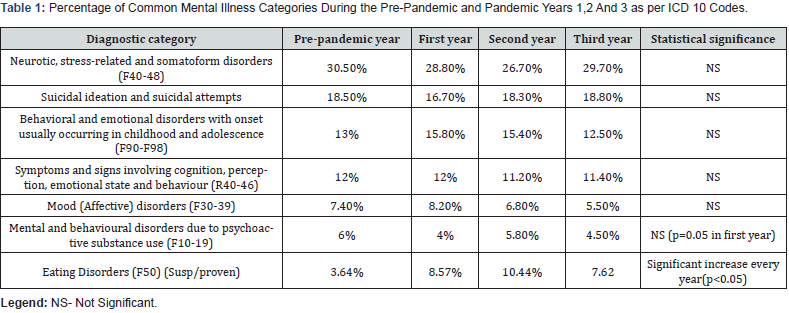
A total of 6431 ED visits related to mental health disorders were identified, which involved 5092 individuals. Most patients were biologic females (4084; 63.5%) with 97 (2.4%) identifying as non-binary or transgender. Among the 2347 (36.5%) children who were biologically males, 28 (1.2%) identified as non-binary or transgender. Mean age was 12.7 (SD ± 3.57) years. A total of 3173 (49.3%) had a known MI and 2333 (36.3%) were taking psychotropic medications. Compared to the pre pandemic year (2.04%), here was a significant increase in the proportion of MI related visits in the first (3.34%) second (3.5%) and third (2.92%) and third years of the pandemic (p < 0.001). Some of the common mental illness diagnosis in the pre-pandemic period and the first three years of the pandemic are listed in Table 1.

Period

New Onset MI

The proportion of new onset MI visits increased from 1.08% (852/78378) in the pre pandemic year to 1.63% (735/45158), 1.65 % (1054/63858) and 1.65% (617/37363) in the first second and third year of the pandemic respectively. Of all mental illness presenting to ED, new onset MI presentations were 53.3% in the pre pandemic year, which reduced to 49% and 47% in the two subsequent years of the pandemic and returned to 57% in the third year Table 2. New MI diagnosis increase was seen in the eating disorder category where it was 3.64% of all new MI diagnosis which went to 8.57%, 10.44%, 7.62% respectively in the first, second and third years of the pandemic (P< 0.001).
Patients With Known MI

The proportion of visits for known MI increased significantly from 0.96%(746/78378) in the pre-pandemic year to 1.71% (772/45158)and 1.85%(1180/63858) and 1.27%(475/37363) the first and second and third year of the pandemic. The percentage of patients with known MI presenting with exacerbations or new symptoms which was at 43.5% of all mental illness presentations increased significantly in the first (51.2%) and second (52.8%) years of the pandemic (p=0.01 and < o. ooo1 respectively). However, there was no statistically significant change compared to the pre-pandemic year in the third year of the pandemic (46.7%).

The common previous MI identified in children presenting with MI exacerbations and new symptoms were suicidal ideation and attempts (SI) (20.95%), neurotic, stress-related, and somatoform disorders (F40-48) (20.93%), mood disorders (F30-39) (13.28%) and eating disorders (F50) (11.48%). The percentage of patients with a history of SI/SA who presented with MI-related complaints significantly increased from 15.4% to 22.7% in the first year (p<0.0001,) and remained significantly increased at 23.4% in the second year (p<0.0001), and at 21.7% in the third (p<0.0001) of the pandemic. The percentage of patients with previously diagnosed history of mood disorders did not change significantly during the study period. The percentage of patients with neurotic, stress-related, and somatoform disorders (F40-48) also did not change significantly from pre-pandemic compared to the first 2 years of the pandemic. However, it was significantly reduced in the third year of pandemic from 21.2% to 16.7% (p=0.004)

The percentage of patients with a history of eating disorders significantly increased from 8.4% in the pre pandemic year to 15.9% in the first year of the pandemic (p<0.0001) and remained significantly increased at 11.9% in the second year (p=0.001). However, there was no significant change compared to prepandemic levels in the third year (9.1% vs 8.4%). There were no significant changes in other categories except for patients previously diagnosed with substance abuse where emergency visits significantly increased from 4.6% in the pre pandemic year to 7.1% in the first pandemic year (p=0.003) and remained significantly increased at 6.4% in the second year (p=0.013). In the third pandemic year, the difference was not statistically different (6.0% vs 4.6%; p=0.11).

Length of Stay (LOS) in the Emergency Department

Children with new onset mental illness had significant decrease in LOS (p<0.001) from pre-pandemic (292 ± 160) to first year (249 ± 149), followed by an increase in LOS in the next two years (326 ± 185) and (388 ± 187). A trend of decreased LOS in the first year of the pandemic, followed by an increase over the next two years, was observed in the patients with known mental illness but it was not statistically significant.

Hospital Admissions

Admission rates for all children with mental illness was at 21% in the pre pandemic year and increased significantly to 29% (p 0.001) in the first year and reduced to 25% and 22 % in the second and third years of the pandemic. In the pre pandemic year 30.6% of patients presenting with suicidal ideations or attempts were admitted which increased significantly to 41.3% in the first year(p=0.05) but was not statistically significant in the second (29.7%) and third years (32.3%) of the pandemic. Children with eating disorders had the highest admission rates in the pre pandemic year (63.6%) which was similar in the first (63.8%), second (57.8%) and third (62.2% years of the pandemic.

New MI presentations had no statistically significant change in admission rates compared to the pre pandemic rate of 2.1% during the first year (3.4%), but admission rates increased significantly to 5.5% in the second year (p<0.0001) and remained significantly increased at 5.2% in the third year of the pandemic (p=0.001). Patients diagnosed with MI presenting with exacerbation, had significant increase in the admission rate from the 49.3% in the pre pandemic year to 60.8% in the first year of the pandemic (p=0.0002). During the second and third pandemic year the rates were not significantly different from pre-pandemic year (50.7% and 54.1% respectively). For patients with MI presenting with new symptoms the pre pandemic admission rate of 30.41% did not change significantly in the three years of the pandemic (34.52%, 24.53% and 21.9% vs 30.41% respectively. Admission rates for patients with known MI on psychiatric medications were not statistically different from those who were not on psychiatric medications.

Discussion

Our findings support previous studies that have reported an increase in MI during the COVID-19 pandemic, as well as an increase in the proportion of MI-related visits to ED [6,7,13]. However, the distinction of new onset mental illness and exacerbation in children during pandemics has not been previously described well in the literature. Interestingly, during the first 2 years of the pandemic, a greater proportion of ED visits for MI were for exacerbation of symptoms rather than new onset mental illness compared to the pre-pandemic period. This suggests that the pandemic tends to exacerbate existing MI symptoms to a greater degree compared to causing new symptoms. However, this increase was not statistically significant in the third year of the pandemic, suggesting a shift back to the baseline of the prepandemic period.

It is worth noting that whilst some previous studies have also discussed the more severe effect of the pandemic on patients already suffering from MI disorders [10,11,13,20], others have indicated that children with high levels of mental health problems before and during the pandemic were less likely [20], or at least as likely [21] as their healthy peers to display an increase in mental health problems in response to the onset of the pandemic.

In our study we observed an increase in the proportion of patients presenting for MI complaints who had a history of suicidal ideations or attempts and those with eating disorders. This increase was more pronounced during the first two years of the pandemic, again suggesting a gradual return to baseline levels in the third year of pandemic. These findings are consistent with previous studies that have shown that individuals with these disorders are at higher risk of being affected by the COVID-19 pandemic [22].

Our study also found no significant increase in the proportion of patients presenting with mood disorders, which is consistent with previous reports suggesting that ED visits are increasingly reserved for more severe cases involving a risk of self-harm [7,16]. Therefore, the true rate of mood disorders in the community may not be accurately reflected by ED visits alone.

In our study, we observed that the highest admission rates were for those presenting with an exacerbation of a known MI. This finding was not surprising, as children who present to the ED with an already known condition may represent a situation where they have failed outpatient management or are unable to receive the help they need in the community setting, while those who present with new onset symptoms may simply require reassurance, education, and referral for appropriate services. While some previous studies have reported an increase in admission rates during the pandemic [6,12,13], not all studies have found this to be the case [16].

During the first year of the pandemic, the rate of admission increased in our institution and the reasons for this are challenging to discern. One possible explanation is that there were fewer community resources available, another explanation might be related to more severe presentations during the pandemic. The fact that children with a first-time presentation of MI symptoms were more likely to be admitted during the second and third years, but not during the first, is not well understood.

When children experience mental distress without access to evidenced-based interventions, their risk of relapse is seven times higher than those who receive treatment services [23], and as psychiatric hospitalization is a major contributor to high health care costs among children suffering from MI, providing a suitable work frame to address acute presentation of MI in a timely fashion and limiting hospitalization can help reduce costs [24].

Limitations

The study is a retrospective chart review in a single institution that relies heavily on the completeness of documentation in the charts and is limited by what is available on the charts. Data regarding previous mental health disorders, chronic medications, and previous diagnoses were extracted from the electronic medical records available at our site, and missing entries or visits to other hospitals may have affected our study results. However, we have no reason to suspect that the completeness of data has significantly changed between groups or over the study period. Therefore, we have assumed that any omission in the data entry would be random and equal over the various groups. As such, we do not anticipate any major errors in data collection.

Furthermore, our study is based on data from a single tertiary care paediatric hospital, and the findings may not be generalizable to other locations with different patient populations or healthcare systems. In addition, our study period was limited to the first three years of the pandemic, and future studies are needed to examine the long-term impact of COVID-19 on paediatric mental health. It should be noted that it was not possible to distinguish between the effects of the pandemic itself and the effects of the public health measures taken in response to the pandemic, such as school closures and social distancing. These measures may have had an impact on children’s mental health, but we were unable to quantify their effects.

Finally, our study was not designed to establish causal relationships between COVID-19 and mental health outcomes. While we observed associations between the pandemic and certain mental health conditions, further research is needed to determine the underlying mechanisms of these associations.

Conclusion

MI related visits to the Paediatric ED increased significantly during the pandemic, for both new onset MI presentations and exacerbation of previously diagnosed MI with increases being more pronounced in children presenting with exacerbations. The study results will hopefully aid in identifying populations that are at-risk for worsening MI and help us better target preventative strategies and guide resource utilization for the management and treatment of vulnerable individuals.


To Know more about Academic Journal of Pediatrics & Neonatology

To Know more about our Juniper Publishers

Monday, July 18, 2022

Visceral Leishmaniasis in a 3-Year-Old Girl with Recurrent Fever Episodes of Unknown Cause - Juniper Publishers

Juniper Online Journal of Case Studies - Juniper Publishers

Abstract

We describe the case of a previously healthy Norwegian 3-year-old girl who presented with recurrent episodes of high fever, poor appetite, vomiting and weight loss. For two months she had experienced episodes of high fever of unknown cause, especially at night. At admission the girl was pale with reduced general condition, and the liver was enlarged as well as the spleen. She complained of abdominal pain and was tender on palpation. Prior to her admission the family doctor had started oral antibiotics on suspicion of bacterial pharyngitis, but the symptoms and fever persisted. The girl had only been abroad once, last summer in the Mediterranean, two months prior to appearance of her symptoms. Fever in children can have many causes and this case highlights the diagnostic challenge of fever with unknown cause. The sum of thorough anamnesis, clinical suspicion, systematic and broad approach and specific examinations was decisive. PCR analysis of her bone marrow was positive for the parasite Leishmania, as well as findings of single-cell parasites by microscopy of blood and bone marrow aspirate. She was immediately provided liposomal Amphotericin B and recovered quickly. At 2 months follow-up she was well-functioning and healthy without fever episodes, organomegaly or anemia.

Keywords: Children; Leishmaniasis; Anemia; Hepatosplenomegaly; Fever

Abbreviations: AST: Anti Streptolysin Test; CRP: C Reactive Protein; IGRA: Interferon-Gamma Release Assay; HLH: Haemophagocytic Lymphohistiocytosis; NTD: Neglected Tropical Disease; VL: Visceral Leishmaniasis

Introduction

Fever and abdominal pain are common symptoms in children, even high fever with completely harmless viral infections. In general, the condition of the child is a more important indicator of how ill the child is than the temperature itself, but prolonged and recurrent fever, and lethargy, indicate underlying disease, especially if there is concomitant anemia and hepatosplenomegaly. Thorough medical records, systemic thinking and clinical examination are essential, and led to suspicion and specific investigation that revealed infection with the Leishmania parasite which has not previously been described in children in Norway. Immediate treatment probably saved her life.

Visceral leishmaniasis (VL) is a condition that can appear as a difficult differential diagnosis and a triggering cause of hemophagocytic lymphohistiocytosis (HLH) [1], which presents with fever, pancytopenia and organomegaly in endemic areas. The diagnosis of visceral leishmaniasis requires detection of the leishmania parasite in bone marrow or spleen aspirate, and possibly antibody detection.

VL is not endemic in Norway. The parasite is widespread in the southern Europe (4) including popular tourist destinations such as the Mediterranean basin [2,3]. Visceral leishmaniasis is a life-threatening infection with the protozoan Leishmania, transmitted by the bite of infected sandfly Phlebotomus argentipes [4]. The parasite affects internal organs such as lymph nodes, bone marrow, spleen, liver and the nervous system. Visceral leishmaniasis is classified as a neglected tropical disease (NTD) and 90% of the annual 1 million cases of the disease in the world occur in poor countries [3,5-7]. Only a small proportion of those infected develop VL and one may be infected without symptoms or signs of infection. VL is almost 100% fatal without treatment [3,7], and is considered the most serious of the so-called neglected diseases. Visceral leishmaniasis is relatively unknown to most medical practitioners in non-endemic areas such northern Europe and clinical vigilance is required to identify those who are infected. This aim of this case report is to highlight the importance of a detailed anamnesis, including travelling to endemic areas and popular tourist destinations as well as systematic thinking and follow-up.

Case Presentation

A 3-year-old girl, previously healthy, was referred to our hospital´s pediatric ward due to poor appetite, weight loss (from 12.4kg to 11.7kg) and vomiting over several days. Her anamnesis revealed recurrent episodes of high fever with temperatures above 40oC, especially at night.

Antibiotic treatment with penicillin provided by the family doctor who suspected airways infection, was discontinued at admission as bacterial infection was not suspected based on negative clinical effect and test results. At admission she was afebrile, pale and in poor condition. Physical examination revealed an enlarged spleen and liver, 3cm below the costal arch. C-reactive protein (CRP) was elevated (52 mg/L) with neutropenia (1.1 109/L) and borderline low platelet counts (158 109/L). She was respiratory and hemodynamically stable, and chest X ray was negative. The hemoglobin (Hb) concentration was 8.5 g/100ml (reference for age 11-13).

A week after admission her Hb had dropped to 7.7. Further investigations revealed elevated levels of ferritin (684mg/L (10-140)), transferrin receptor (13.1mg/L (1.5-3.3)), and haptoglobin MCV (70fL (75-87)), and microcytic anemia (MCH 20pg (22-30), MCHC 28g/dl (32-37), reticulocytes 206g/dL (20-95), immature reticulocytes of 0.30% (0.02-0.18)) and reticuloculocyte-Hb equivalent values 23pg (28-36), erythrocytes 3.9 1012/L (4.1-5.3).

Examination with blood smears and differential count performed 4 days after admission, showed no signs of malignancy.

She was discharged home after 12 days, when she had recovered well and been afebrile over the last 3 days. At discharge she agreed to take extra supplements of iron and come for an early outpatient check-up.

The girl was readmitted 4 weeks after discharge, due to febrile seizures. She was again strikingly pale and her Hb had dropped to 7.0g/dL, CRP was persistently elevated (58) and her pancytopenia had worsened; (leukocytes 1.0 109/L (4.4-12.5), neutrophils 0.2 109/L (1.0-8.5), lymphocytes 0.7 109/L (1.0-4.6), monocytes 0.1 109 / L (0.9-1.8) and platelets 62 109/L (145-390), all reference values per age). This was compatible with respiratory infection believed to be the cause of febrile seizures. She was in relatively good general condition and afebrile during the rest of her 2 days hospital stay, asked to recontact immediately if her condition worsened.

After another month the girl was readmitted. All family members were healthy except for the girl. Her mother was originally from Africa and pregnant in the last trimester when she came to Norway when she was tested negative for multi-resistant staphylococci (MRSA), HIV, toxoplasma and Triponema pallidum, and she was Rubella IgG positive. The girl was born in Norway. A delayed update of her anamnesis at this third admission revealed that she had been abroad once which was last summer in a country house up in the Spanish mountains.

At this last admission she had high fever with constant temperature of 40oC. She denied eating, was confused, exhausted and circulatory affected with tachycardia (160 beats /min). Liver and spleen were further enlarged by palpation, 6 and 9 cm below the costal arch, respectively. Organomegaly was also confirmed by ultrasound, which showed no other pathological findings. CRP was elevated to 78 and the pancytopenia was persistent.

Microbiological examinations showed undergone Cytomegalovirus and Epstein Barr virus infections (positive IgG), while Parvovirus B19, Hepatitis B and C, and HIV were all negative. The analyzes were also negative for anti-streptolysin (AST), Treponema pallidum and interferon-gamma release assay (IGRA, an ELISA Quantiferon analysis) showed sensitization to M. tuberculosis but is unaffected by BCG vaccine or common atypical mycobacteria. This indicated that there was no streptococcal infection, syphilis or tuberculosis. There was no growth in blood cultures, and no findings of intestinal pathogenic bacteria or parasites in faeces. Hypergammaglobulinemia (IgG 28.2 g/L (1.3-6.6) could indicate a chronic inflammatory condition, while IgA and IgM were normal (1.2 g/L (0.1-1.3) and (0.8 g/L (0-1.8), respectively). On suspicion of spherocytosis due to anemia and organomegaly, blood samples were taken showing positive eosin 5-maleimide (EMA) test, and the condition could therefore be compatible with spherocytosis.

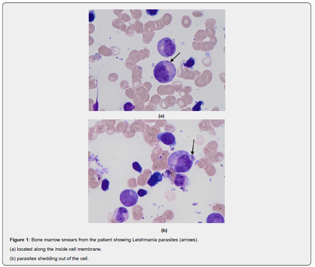
Since she and her family had visited Spain last summer, we assessed whether import fever and possible transmission during this stay could have caused the patient's condition. We checked for possible malaria disease due to the unclear disease picture and the periodic episodes of fever, but the girl had only stayed in Europe and the test was negative. We analyzed her bone marrow aspirate (Figure 1). Blood smears and now bone marrow showed no signs of malignancy. Blood and bone marrow specimens were also analyzed at the department of microbiology at Oslo University Hospital (OUS), with a request to analyze for import fever and neglectable infection diseases. This revealed a positive PCR in bone marrow for the parasite Leishmania, as well as findings of single-cell parasites by microscopy of blood and bone marrow aspirates. She was immediately put on liposomal Amphotericin B (3mg/kg) for 5 days. After 3 days, she was afebrile with improved general condition. After a week she was discharged home, and she received a final dose of Amphotericin 10 days after the 5-day treatment, at the out-patient clinic. At a follow-up check 2 months after discharge, the girl was well-functioning and healthy, and no longer anemic (Hb 11.9). There was a complete regression of her spleen and liver enlargement and she had not experienced episodes of fever after discharge.

Discussion

The 3-year old girl described in this report developed a severe form of visceral leishmaniasis (VL) with fever, anemia, hepatosplenomegaly, weight loss, and mild neurological symptoms. VL is a systemic disease that activates the blood-forming organs and the immune system and results in anemia and pancytopenia, hepatomegaly, splenomegaly and lymphadenopathy, and symptoms such as fever, weight loss, diarrhea, chills, anorexia and anemia [8]. Increasing anemia occurs due to progressive bone marrow failure, hemolysis and hypersplenism. Our patient had all of these symptoms, except for lymphadenopathy.

The case describes a protracted and atypical febrile illness. When her condition worsened, we suspected malignant disease or haemophagocytic lymphohistiocytosis (HLH) secondary to an underlying infectious disease, autoimmune or rheumatic disease [1]. Infection associated HLH disease may be elicited by virus, bacteria, protozoa or fungi, and infection with Leishmania may be a relatively frequent triggering cause [9,10]. HLH secondary to visceral leishmaniasis has been reported very rarely and there are only few cases with imported visceral leishmaniasis in Norway and none described in children [11]. The diagnosis is made by the patient meeting a set of clinical and biochemical criteria, and for primary HLH by detecting a mutation consistent with the diagnosis [12,13]. HLH is characterized by fever, pancytopenia and hepatosplenomegaly, as well as neurological symptoms that are seen in up to a third of patients, but the clinical picture and severity varies [12,14]. This girl had fever, cytopenia, hepatosplenomegaly, and elevated ferritin blood levels and she gradually developed diffuse neurological symptoms with dizziness, lethargy and fatigue. We suspected infectious HLH in this patient with a clinically sepsis-like disease who did not respond as expected to presumed adequate antibiotics treatment. An important marker of HLH disease is a highly elevated serum ferritin value, often far higher than what is otherwise seen in acute phase reactions [15]. This corresponds to this case where the girl had 5-fold elevated ferritin levels. In the event of a deviating clinical course or lack of treatment response, ferritin is a useful and available variable that can be quickly answered. The girl gradually developed severe anemia (Hb 7.0) and the investigation revealed possible hereditary spherocytosis. This is one of the most common hemolytic anemia forms in Norway and is autosomal hereditary in most people. The girl did not have a family history of hereditary spherocytosis, nor was there any information about jaundice, which may be an early sign of spherocytosis. However, spherocytosis can have a varying clinical course, from almost asymptomatic to severe anemia.

A delayed anamnestic work-up revealed that the girl had visited southern Europe three months prior to being admitted to our hospital. The girl was born in Norway, and her mother is ethnically African. Last summer, 2 months before onset of symptoms, she and her family visited the highlands of Spain, but except for this, none of her contacts or she had visited countries in subtropical or tropical regions. The girl often played in the sandbox outside the summer house, and with animals, as there were many stray dogs as an in the area [16]. This information led to extended investigations for import fever and neglected diseases, which revealed leishmania parasites in her bone marrow and blood smears.

Visceral leishmaniasis is a so-called neglected tropical disease (NTD) that, if left untreated, can be fatal. It is gradually spreading to larger areas, partly due to increased travel activity and global warming [7]. In endemic areas sandflies are found primarily in the countryside, in sandbanks in river beds, in dry and semi-arid areas in mountain villages, underground rodent passages, scrub forests and rock piles, also in tropical forests [17]. The transmission of the parasite is either zoonotically, with an animal as an intermediate host, or anthroponotically via an insect vector to human. The route of transmission of leishmania in this case is likely through sandflies or dogs as an intermediate host. The parasite is spread by the bite of female sandflies and the incubation period can range from 10 days to 24 months and may last for several years, but is usually 2-6 months, which fits well with this case as she visited Spain about 3 months prior to appearance of symptoms [8,18]. Dogs are the main host in Europe, but other animals, including rodents, are also intermediate hosts [16,17,19]. After transfer from the insect vector, the parasite is taken up by macrophages where it is transformed from promastigot to amastigot, multiplies and spreads to the entire reticuloendothelial system. The infection can be asymptomatic, and with a weakened immune system the probability of a serious course increases [8,20,21], but an underlying immune deficiency was not seen as there were no signs of malignancy and she was HIV unexposed and negative. The gradually more severe anemia, persistently elevated CRP and pancytopenia, as well as hypergammaglobulinemia fits with a chronic hyperinflammatory condition seen in this case.

Visceral leishmaniasis is caused by either Leishmania donovani eller L infantum, the last is prevalent in Europe and Spain. The infection is characterized by a wide clinical spectrum, which may range from mild to moderate and severe clinical manifestations [22]. The disease usually has an insidious course, with gradual onset of lethargy, fever, weight loss and hepatosplenomegaly, [23] as in this girl. Later in the course, liver failure may develop with icterus and ascites This girl showed no hyperbilirubinemia or ascites. Liver failure and gradually worsening thrombocytopenia may also cause bleeding, but no bleeding tendency was observed in this girl. Kala-azar (black fever) is another name used for Leishmaniasis and refers to a darkening of the skin, but this was not seen here. Of notice, her mother was ethnically African which may have complicated skin color judgement, but we did not prove darkening of the girl´s skin during the disease process.

Leishmaniasis is distributed worldwide and 13 million people are estimated to be infected by this parasite, with about 1.8 million new cases each year. When it comes to VL, the World Health Organization calculates approx. 500,000 new cases and 50,000 deaths, annually, and the disease is on the rise in southern Europe [6,17,19]. The increase is associated with expanded travel activity, more people with weakened immune systems and an increasing impact area for host animals and vectors such as the plenty of sandflies in forests of northern Spain and central France, probably without any connection to climate change [2-4]. VL should be suspected in children who present with specific manifestations and the diagnosis should be established, mainly by the demonstration of leishmania in tissue specimens [7,20]. In the Norwegian context, the disease is most relevant for those who have symptoms and findings compatible with VL and have been to the Mediterranean or in more remote areas during the past year.

Conclusion

This case study emphasizes the importance of a thorough medical history, which must be seen in connection with the disease picture and the clinical findings. A complete travel history is required in case of obscure febrile illness after a stay in subtropical or tropical regions. Leishmaniasis is endemic in southern Europe, including popular tourist destinations such as the Mediterranean basin. It is relatively unknown to most medical practitioners in non-endemic areas, including Norway, and clinical vigilance is required to identify those who are infected.

To Know more about Juniper Online Journal of Case Studies

Click herehttps://juniperpublishers.com/jojcs/index.php


To Know more about our Juniper Publishers
Click here: https://juniperpublishers.com/index.php 

Tuesday, July 12, 2022

The Role of Psychological Care in Children and Adolescents with Type-1 Diabetes - Juniper Publishers

 Current Research in Diabetes & Obesity - Juniper Publishers

Opinion

Diabetes is a major health-care problem all over the world with a high prevalence and incidence [1,2]. The approach and the management of the disease are challenging, especially in the early stages after diagnosis and in children. For this reason, a multidisciplinary diabetes team for the management of the disease is recommended [3]. This should include several professional figures, among which the pediatric endocrinologist, the psychologist the dietician and a specialized nurse. Psychological care for youth diabetic patients is of primary importance and recommended by guidelines [3]. As a matter of fact, children and adolescent with diabetes have a higher incidence of depression, anxiety, psychological distress, and eating disorders than patients without diabetes. These conditions should be early recognized by the psychologist, referred to the team, and treated.

In this regard, international guidelines, suggest that “the diabetes care team should receive training in the recognition, identification, and provision of information and counseling on psychosocial problems related to diabetes” and that “overt psychological problems in young persons or family members should receive support from the diabetes care team and expert attention from mental health professionals”.

Psychological assessment and counseling are also useful in patients without overt psychological problems since they are able to affect the quality of life and disease control [4-6]. A multicenter randomized controlled trial performed on a cohort of 66 teenagers with type 1 diabetes, randomly assigned to motivational interviewing or support visits, found that mean glycated hemoglobin (HbA1c) in the motivational interviewing group was significantly lower than in the control group (p=0.04), showing as this technique is effective method of inducing behavioral changes in teenagers with T1D and the improvement of their glycemic control.

Moreover, a meta-analysis of 21 randomized controlled trials (RCTs) assessing the effect of a psychological therapy on control of diabetes, 10 of them performed on children and adolescents, showed that psychological distress was significantly lower (estimates -0.46, 95% CI -0.83 to -0.10) and the percentage of HbA1c was significantly reduced (estimates -0.35; 95% CI 0.66 to -0.04) in patients receiving a psychological intervention compared to controls [7]. Psychological assessment and intervention should not be directed only to the patient but ideally to the whole family. Family therapy is focused to obtain an improvement of relatives’ interactions and changing problematic familial patterns and relationship hierarchies.

Evidence from literature clearly shows as the health of family relationships and the absence of family conflicts, as well as the cohesion and the collaboration of the family in the management of the disease, positively affect the glycemic control [8-12]. The effectiveness of family therapy for youth with type 1 diabetes (T1D) is confirmed by a systematic review of 25 RCTs showing that family-based interventions appear effective at improving diabetes and family-centered outcomes [13]. In addition, a study performed on 25 children with poorly controlled diabetes, showed that patients receiving family therapy improved their metabolic control and that family-based interventions enhance the health, quality of life, and family functioning of youth with T1D [14].

These evidence from literature, clearly support the importance of psychological care in children and adolescent with T1D and show as the regular intervention directed to the patients and his family positively affect, not only the distress and quality of life but, above all, the glycemic control. For these reasons, a multidisciplinary approach is recommended for children and adolescent with T1D and a regular phycological approach with family-based interventions during outpatient clinic visits is especially in patients with difficulty in adherence to therapy and glycemic control [3]. 

Click here: https://juniperpublishers.com/index.php  

Tuesday, May 24, 2022

Clinical And Evolutive Characteristics in Acute Toxic Methemoglobinemia in Children - Juniper Publishers

 Open Access Journal of Toxicology - Juniper Publishers

Abstract

Introduction: Acquired or toxic methemoglobinemia is the result of exposure by swallowing, inhalation or dermal absorbtion of medicines or non-drug chemicals with oxidizing properties. The main objectives of the study were to highlight the main causes of methemoglobinemia in children, to identify the associated factors and their influence on the clinical form of the disease as well as the response to antidote treatment with methylene blue.

Methods: All patients with acute toxic methemoglobinemia hospitalized over a period of six years were included in the study, the inclusion criteria being: age < 18 years, suggestive symptomatology (generalized cyanosis, asthenia, dyspnea) and methemoglobin values >3%.

Results: 82 patients with acute toxic methemoglobinemia were identified of which the majority (94%) were between 0-5 years. 69 cases were secondary to exposure to nitrates from water used to prepare food. The severity of methemoglobinemia was significantly higher among anemic and dehydrated patients. All patients received antidote treatment with methylene blue one or more administrations, and all had a favorable outcome.

Conclusions: Acute toxic methemoglobinemia is mainly observed in children up to 5 years of age, the majority being infants. The main cause of it in children is exposure to exogenous substances among which nitrates that contaminate well water occupy the first place. Age under 1 year, hemoglobin values lower than 11g/dl, and acute dehydration syndrome represent risk factors for severe forms. Methylene blue is the treatment of choice for acute toxic methemoglobinemia in children and its administration is effective in the majority of cases.

Keywords: Acute toxic, Methemoglobinemia, Children, Methylene Blue

Introduction

Acquired or toxic methemoglobinemia is defined as an increase in methemoglobin blood levels above 1%, as a result of exposure to medicines or chemicals with oxidizing properties. The medicinal etiologies of toxic methemoglobinemia include, acetanilide, phenacetin; local anesthetics (benzocaine, lidocaine, prilocaine, and articaine), chloroquine, dapsone, flutamide, metoclopramide, nitric oxide, nitroglycerin, nitroprusside of sodium, antibiotics of the class of nitrofurans and sulfonamides, sodium nitrate, silver nitrate, silver sulfadiazines, antiepileptics (sodium valproate, phenytoin), phenazopyridine, sulfasalazine, and zopiclone [1]. Among non-drug chemicals that can produce methemoglobinemia, the most common include aniline-based paints, silver nitrate, naphthalene, trinitrotoluene, nitrobenzene, Vicia faba, nitrates and nitrites from well water or contaminated vegetables, paraquat, phenol, or fires [1].

Acute toxic methemoglobinemia in children can be triggered by any of the above-mentioned xenobiotics It has the potential to cause severe injury or death but if recognized early, patients can significantly benefit from antidote treatment. The antidote of methemoglobinemia is methylene blue a thiazine coloring agent, which in the presence of NADPH-dependent methemoglobin reductase and nicotinamide adenine dinucleotide phosphate causes the reduction of methemoglobin and its subsequent transformation into hemoglobin. Antidote treatment with methylene blue is commonly indicated in all symptomatic patients or cases where methemoglobin concentration is above 30% [1].

In Romania acute toxic methemoglobinemia in children was recognized as a public health problem for many years [2]. A national report of the Ministry of Health published in 2012 revealed that Romania is characterized by the existence of numerous rural territories with frequent and important nitrogenous substances contamination [2]. Nitrates contaminate the soil from where water infiltrates into artisanal wells existing in rural areas. The toxicity of these salts is due to their transformation into nitrites under the action of bacteria at the level of the soil or gastrointestinal tract of the child. Nitrites exert a direct oxidant effect on hemoglobin, which subsequently loses its physiological capacity for carrying oxygen. The level of nitrates in the water depends on several elements, and hence it is difficult to quantify soil permeability, well depth < 15 m, agricultural activities, and inadequate measures for eliminating organic waste, making it difficult to identify populations at risk based only on geographic origin [3]. The World Health Organization has regulated the maximum permissible concentration of nitrates (NO) in drinking water to <50 mg/L [4]. Various local research has been proven that in countries like Romania, a country with poor infrastructure in rural areas, exposure to nitrates remains the main cause of acute toxic methemoglobinemia in children [5].

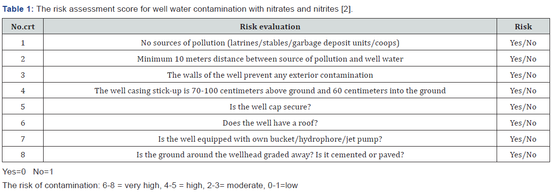
To come to health care specialists’ aid, the Ministry of Health of Romania has developed a score for assessing the risk for well water contamination with nitrates and nitrites (Table 1) [2].

The research presented in the study details a retrospective study including cases of acute methemoglobinemia that were hospitalized and treated at the Pediatric Poisoning Center of the Emergency Clinical Hospital for Children”Grigore Alexandrescu” in Bucharest Romania and analyzed , for a period of 6 years. The main objectives of the study were to highlight the main causes of acute methemoglobinemia in children, to identify the associated factors (age and associated pathology), and their influence on the clinical form of the disease as well as the response to antidote treatment with methylene blue. The study design will be of significant help to global researchers and pharmaceutical firms looking forward to analyze methemoglobinemia in study populations in their respective countries.

Methods

All patients with acute toxic methemoglobinemia hospitalized at the Pediatric Poisoning Center of the Emergency Clinical Hospital for Children “Grigore Alexandrescu” in Bucharest, Romania between January 01, 2015, and December 31, 2020, were included in the study. The inclusion criteria were age < 18 years, patients with suggestive symptomatology (generalized cyanosis, asthenia, dyspnea, and altered neurological status), and methemoglobin values > 3%. For this purpose, we used the computer data base of clinical consultation sheets and medical reports upon discharge of patients using for searching the ICD 10 code of diagnosis for acute toxic methemoglobinemia. The following data were analyzed: medical history data, demographic characteristics, data provided by clinical examinations from patient hospitalization and daily evaluations during hospitalization, the presence of methemoglobinemia (measured by spectrophotometric method), and associated conditions. The therapy regimens provided to the patients and the outcome of each patient were also analyzed, including the duration of hospitalization, the need to repeat the dose of the antidote, and admission to the intensive care department. The data obtained were statistically processed using the IBM® SPSS® Statistics 20program, considering significant results of statistical probability (p) less than 0.05.

Results

During the mentioned period, 82 patients with acute toxic methemoglobinemia were identified representing a percentage of 1.58% from the total of 5122 patients diagnosed with acute intoxication in the Pediatric Poisoning Center in Bucharest. Etiology Regarding etiology: clinicians ascribed 69 cases as secondary to exposure to nitrates from well water used to prepare food. The risk assessment score for well water contamination with nitrates and nitrites (Table 1) was used, all the 69 children having a risk score between 6 to 8. Ten cases were secondary to topical exposure to silver sulfadiazine used to treat severe burns, two cases were instances of medicines poisoning: one patient had accidental acute poisoning with pentaerythritol tetranitrate, (a coronary vasodilator) and another patient was undergoing chronic treatment with dapsone. In the study group, a case of acute toxic methemoglobinemia was identified, wherein the patient was a chronic consumer of drugs of abuse and was hospitalized following an overdose of volatile nitrites by inhalation (cigarettes soaked with a street substance known as “Liquid Gold”) (Table 2).

The average age of the patients was 23.11 months, and the median age was 6 months. The majority of patients were under 5 years old: 77(94%), The infants subcategory (children aged under 1 year) within this age group was most represented with 49 patients (53.7% out of the total of cases). At the same time the number of cases was much lower for other age groups (Table 3). Cases among patients of rural origin were significantly higher than those from urban areas (63 versus 19 cases, p<0.05). In addition, rural origin was significantly associated with exposure to nitrates contaminated well water (binomial test, p<0.0001). The study group included 42 boys and 40 girls, the difference in patient sex being insignificant (Table 3). The most common route of exposure was the oral route (87%), followed by cutaneous (12.2%), and inhalation (0.8%). In the majority of cases (85.3%), toxic exposure was accidental.

The mean methemoglobin concentration in the study group was 41.53%, with values ranging from 6.6% to 87% (Table 4). The mean value of methemoglobin for cases of exposure to nitrates -contaminated well water was 41.5%, and it was 39.2% for cases involving topical exposure to silver sulfadiazine, used in patients with severe burns. The methemoglobin values were: 41.9% for case of accidental acute poisoning with pentaerythritol tetranitrate, 44.2% for voluntary inhalation, with new substance of abuse “Liquid Gold “and 6.6% for a patient undergoing dapsone chronic treatment. Since the number of cases caused by exposure to dapsone, pentaerythritol tetranitrate, and new substance of abuse “Liquid Gold “(volatile nitrites) was small (one per etiology), we could not perform a statistical analysis of methemoglobin concentrations in these etiologies. Regarding the mean concentrations of methemoglobin secondary to exposure to nitrates contaminated well water and silver sulfadiazine, the difference was not statistically significant.

Clinical Manifestations:

Upon admission to the toxicology department, all patients showed clinical manifestations with different grades of severity. Clinical manifestations are summarized in Table 4.

The lowest concentration of methemoglobin recorded in this study was 6.6%. Regardless of the methemoglobin concentration, all patients experienced altered general conditions and cyanosis. For values between 6.6% and 20%, other clinical manifestations besides cyanosis, including dyspnea and agitation, were observed in 2 of the 10 identified cases (20%). The majority of patients with methemoglobin values between 20% and 50%, had dyspnea (24.4%), tachycardia (19.5%), psychomotor agitation (31.7%), and drowsiness (12.2%). Those with methemoglobinemia between 51% and 70% showed clinical manifestations, with a higher frequency of dyspnea (37%), tachycardia (29.6%), and altered sensory function (25.9%). Two patients in this group (7.4%) experienced generalized tonic-clonic seizures. These two patients had not preexisting neurologic disease and were 3 months respectively 7 months old. Seizures were ceased after 5 mg diazepam intrarectal administration and required one dose of 1 mg/kg of methylene blue.

All patients with a methemoglobin value above 70% experienced tachyarrhythmia, dyspnea, and impaired sensory function (Table 4). Evolution was favorable in all cases, and all patients were declared cured upon discharge.

Associated pathology We analyzed the associated pathology presented by patients, as shown in medical documents (Table 5). Of the 82 patients, 58 (70.7%) had a blood count at the time of admission, which revealed hemoglobin levels below 11g/dL. We found that the severity of methemoglobinemia was significantly higher among patients with hemoglobin levels below 11g/dl., (p=0.03). The concomitant presence of respiratory tract infections, cardiac pathology, or malnutrition was not significantly associated with a greater severity of methemoglobinemia. Eight patients (9.76%) experienced manifestations suggestive of an acute enterocolitis (fever, diarrhea, and vomiting). Of these in three cases, clinical and paraclinical changes of acute dehydration syndrome were obvious. The presence of dehydration syndrome was associated with a greater severity of methemoglobinemia (Mann-Whitney test, p<0.05).

A greater severity of methemoglobinemia was correlated with certain characteristics, including patients from rural areas, use of nitrates contaminated well water, age <1 year, presence of dehydration and hemoglobin values bellow 11g/dl, in a statistically significant manner. We analyzed the direct relationship between the severity of methemoglobinemia and the common presence of these parameters using multilinear regression. The results proved that the relationship between co-factors is complex and does not allow the establishment of a linear relationship between the severity of methemoglobinemia and a certain factor.

All patients received antidote treatment with methylene blue 5mg/mL solution intravenously, at a dose of 1mg/kg. The clinical status and methemoglobin value were analyzed during the first 24 hours post administration. In 60 cases (73.2%) cyanosis and the other symptoms disappeared in the first 60 minutes and no further administrations were required Of these 60 patients who received a single dose of antidote, 9 had methemoglobin values <20%, 31 had values ranging between 20 and 50%, and 20 had concentrations above 50% at the moment of diagnosis. Eighteen patients (21.9%) required two doses of methylene blue (1mg/kg), among whom 1 patient had an initial methemoglobin concentration <20%, 6 patients had values between 20% and 50%, and 11 patients had values >50%. Among the 4 patients (4.9%) requiring three doses of antidote, one patient had a methemoglobin concentration <20%, while the rest had concentrations above 50% before starting treatment.

Of the 22 patients who required additional doses of the antidote 20 (91%) had hemoglobin values bellow 11g/dl at the time of diagnosis. However, no significant association was reported between the need for additional doses of antidotes and the presence of hemoglobin values bellow 11g/dl. (Mann Whitney test, p=0.36). It was necessary to examine whether the lack of response to methylene blue administration is secondary to other causes. However, since this was a retrospective study, we did not have the values of glucose-6-phosphate dehydrogenase (G6PD) activity or Sul hemoglobin concentration. For all patients with acute dehydration syndrome, additional doses of antidote were required; in this situation, a statistically significant correlation (Mann-Whitney test p=0.009) was observed. All patients had a favorable outcome. In the analyzed group, there were no deaths and two patients required admission to the intensive care unit. The average duration of hospitalization was 3days; however, the majority of patients (51.2%) were discharged after 24h of staying.

Discussion

Acute toxic methemoglobinemia is a rare condition in pediatric toxicology, as evident in the present study, which revealed a prevalence of 1.58% of all intoxication cases in children. However, it can lead to severe functional anemia, especially in infants or in cases of associated pathologies. Early identification allows for quick initiation of antidote treatment with methylene blue, ensuring an excellent prognosis for patients.

Based on the data detailed above, the present study proposed a detailed analysis of cases of acute toxic methemoglobinemia from the point of view of etiology, clinical manifestations, associated factors (age, associated pathology), and their influence on the clinical form of the disease, as well as the outcome under treatment. The limitations of this analysis include difficulty in quantifying the extent to which each factor is independent of the severity of methemoglobinemia. the small sample size that hindered the predictive model, the difficulty associated with the evaluation nitrate concentrations in the wells of the patients and bias created by the type of admissions in an emergency hospital like the one we were referring to a large number of chemicals or drugs have been reported in literature as capable of producing oxidation of hemoglobin with the accumulation of methemoglobin. However, in this study, we identified only five categories of toxic substances inducing acute methemoglobinemia in children. Most of the cases in this study - 69 (84.1%), were secondary to exposure to nitrates contaminated well water used to prepare food in rural areas.

In a report published in 2009 by the National Institute of Public Health of Romania within the National Programme for Monitoring the Triggering Factors in the Life and Work Environment, 89 cases of toxic methemoglobinemia have been reported as a source of food contamination [5]. In the group of patients analyzed in the present study consisting exclusively of children, this source was not identified. In all cases, anamnesis referring the living conditions , using the risk assessment score for well water contamination with nitrates and nitrites [2]. indicated that the water used in the preparation of tea or milk was the source of the disease.

In 12 patients, the drug-induced cause of acute methemoglobinemia was identified. Of these, 10 patients (12.2%) were children with severe burns of at least II degree on extended body surfaces, who experienced episodes of acute methemoglobinemia during hospitalization. Analyzing the substances with the oxidant potential to which the children were exposed, we found that the common element was the repeated topical application of silver sulfadiazine on extended skin surfaces.

Silver sulfadiazine is an antibiotic of the sulfonamide class and exerts antibacterial and antifungal effects through the simultaneous action of silver ions and sulfadiazine. Its application on extensive skin areas and/or with significant destruction of superficial layers, such as burns, favors systemic action, leading to adverse effects similar to those of sulfonamides, including oxidation of hemoglobin with the formation of methemoglobin.

Another drug identified in our study as an inducer of methemoglobinemia was dapsone (dianinodiphenyl sulfone), a sulfone-class compound, considered in literature as one of the most common causes of toxic methemoglobinemia [6,7]. Dapsone is used in immuno depressed patients for the treatment of infection with Pneumocystis jiroveci or Toxoplasma gondii, in the treatment of leprosy, malaria, or some dermatological diseases, such as bullous pemphigoid and dermatitis herpetiformis.

In our study, dapsone was identified in a 4-year-old boy who was undergoing treatment for bullous pemphigoid. Clinical manifestations occurred at a methemoglobin concentration of 6.9%, which is consistent with the findings of other studies [8,9], confirming that therapeutic doses may trigger suggestive symptomatology at serum methemoglobin levels < 10%. Combination with other drugs with oxidant effects or concomitant presence of other diseases, such as heart, respiratory disease, and anemia, are considered to be promoting factors [9,10]. In the analyzed case, we only identified anemia with a hemoglobin level of 9.7 g/dL.

Another drug-induced cause of acute toxic methemoglobinemia was pentaerythritol tetranitrate.an organic nitrate that can induce methemoglobinemia in case of accidental or voluntary overdose. Methomoglobinemia occurs especially in cases of accidental or voluntary overdoses, with limited cases of methemoglobinemia occurring because of therapeutic doses.

In the analyzed group, we identified a female patient aged 2 years who presented with acute toxic methemoglobinemia due to accidental acute poisoning with pentaerythritol tetranitrate, with a methemoglobin concentration of 41.9%.

In one case, volatile substance were identified as etiological agents of methemoglobinemia. It was about a teenager aged 16 years, institutionalized, chronic consumer of substances of abuse, who presented with suggestive clinical manifestations: generalized cyanosis not responsive to oxygen administration, dyspnea, and tachycardia about an hour after smoking cigarettes soaked in a liquid street substance named “Liquid Gold”

Volatile abuse substances are another category with a well-documented role among the causes of toxic methemoglobinemia [11]. Volatile nitrites (amyl nitrite or isobutyl nitrite) are esters of nitric acid, which cause an intense vasodilating effect, with a rapid but short onset at the level of the central nervous system [12]. Although there are legislative regulations regarding the possession or sale of volatile nitrites in Romania as in the other European countries, these substances are available under various names: “Liquid Gold”, “Rush”, “Pig Black”, and are used both for inhalation and application in classical cigarettes. Most often, users of volatile substances are male adolescents, with poor socio-economic backgrounds, from disorganized families, and have low educational qualifications [13].

Although exposure to substances with oxidant potential may cause the accumulation of toxic concentrations of methemoglobin regardless of age, in the pediatric population, cases involving infants present the highest risk [14,15] due to functional particularities. The main peculiarity is the predominance of fetal hemoglobin, especially up to the age of 3 months, which can be oxidized much more easily than in adult form, which becomes predominant after 18 months of age. At this age, cytochrome b5 methemoglobin reductase activity is reduced [16], and gastric pH is less acidic than in older children or adults, thus facilitating bacterial proliferation, which plays an important role in converting nitrates in food into nitrites [17,18]. The analysis of the group of patients provided consistent results; the age subcategory of under 1 year being the best represented with 49 infants (59,7%) ,and the median age in the studied group was of 6 months.

Clinical manifestations are similar regardless of the incriminated oxidant substance and depend primarily on the concentrations of methemoglobin. In the vast majority of cases, levels below 10% are well tolerated, and patients are asymptomatic [15,19]. Cyanosis is the main clinical manifestation and is an indispensable clinical element for positive diagnosis along with an increase in methemoglobin values, as evidenced by literature. Most previous studies have shown that cyanosis becomes obvious at methemoglobin concentrations of more than 10%. However, in our study, the lowest value at which cyanosis was noted was 6.6%. As methemoglobin concentrations increase, the clinical picture is supplemented with changes secondary to hypoxia: cerebral symptoms (agitation, restlessness, irritability, headache and subsequently drowsiness, obnubilation, seizures, or coma), cardiac symptoms (tachyarrhythmias, circulatory failure), or respiratory symptoms (dyspnea, tachypnea, respiratory failure). Values above 70% are generally associated with an increased risk of death. In our study, the severity of clinical manifestations varied with the concentration of methemoglobin, but cases with values above 70% (72%–86% in our study) had a favorable outcome and no death was reported. There were 4 cases with methemoglobin values above 70%. All of them were infants and in addition to cyanosis presented tachycardia dyspnea and neurologic symptoms (drowsiness or agitation) Of these, 3 required three administrations of methylene blue and 1 required two doses.

In case of association with other pathologies that cause cell hypoxia (respiratory, cardiac, hematological conditions), the symptomatology is more severe at reduced concentrations of methemoglobin [15,20]. The analyzed group included a large number of patients with such associated pathologies, identifying a significant correlation between the presence of hemoglobin levels below 11g/dl, and the severity of clinical manifestations in patients with methemoglobinemia. Another situation encountered in the study, which has been significantly associated with a higher severity of the clinical picture of methemoglobinemia, was acute dehydration syndrome in patients with acute enterocolitis, most likely secondary to associated metabolic acidosis.

Exposure to methemoglobin zing substances can be life-threatening, so that the antidote treatment with methylene blue can be administered as soon as possible. Methylene blue is a thiazine coloring agent, which in the presence of NADPH methemoglobin reductase and NADPH is reduced to an intermediate metabolite, methylene blue leuco, capable of donating an electron to methemoglobin to reduce it to oxyhemoglobin.

The indication to initiate antidote treatment is symptomatic methemoglobinemia, since there are situations where the concentration of methemoglobin does not correlate with the severity of clinical manifestations [21]. The dose is 1-2mg/kg, administered intravenously over 3-5min, and repeated, if necessary, when the symptomatology does not cease after 60 min, up to a maximum dose of 7 mg/kg [9,15].

Although extremely effective in a vast majority of situations, the administration of methylene blue is not without risk. Digestive side effects (nausea and vomiting), electrocardiographic abnormalities (inversion of T wave, flattened R waves), profuse sweating, dyspnea, retrosternal pain, and oral dysesthesia usually occur at doses above 7 mg/kg [9]. It can also cause a paradoxical increase in methemoglobin through its oxidant effect, especially in patients with glucose-6 phosphate dehydrogenase deficiency. In the study group, methylene blue administration was limited to a single dose in most situations, 73.2% (60 patients), and the majority (51 of 60) had methemoglobin concentrations over 20%. In 22 cases, it was necessary to administer one or two additional doses, most of them [16] having hemoglobin levels lower than 11g/dl. Even that there were not possible to establish a significant association between the need to supplement the initial dose and the presence of hemoglobin levels lower than 11g/dl. The total dose of methylene blue used in the treatment of patients in this group did not exceed 3 mg/kg, and no adverse effects of the treatment were reported.

Conclusion

Acute toxic methemoglobinemia is mainly observed in children up to 5 years of age. This age group represents 94% of all cases in our study, of which the majority belong to the subcategory of 0-1 year. In countries as Romania where the rural infrastructure is less developed the main cause of acute toxic methemoglobinemia in children is the exposure to nitrates contaminated well water used for food preparation. Silver sulfadiazine cause acute toxic methemoglobinemia when applied to extensive areas, repeatedly and/or under the condition of significant destruction of the surface layers of the skin. Methemoglobinemia secondary to exposure to volatile substances, such as amyl nitrile or isobutyl nitrile, used as new psychoactive substances, is a newly described condition in recent years and must remain under the attention of specialists.

Although the relationship of co-factors is complex and is difficult to quantify to what extent the severity of the clinical picture is in linear correlation to a certain factor. We believe that the age under one year, hemoglobin values lower than 11g/dl, and acute dehydration syndrome are factors that can aggravate clinical symptomatology and outcome in acute toxic methemoglobinemia.

Methylene blue is the treatment of choice for acute toxic methemoglobinemia in children, and its administration is effective in a single dose of 1mg/kg in 73.2% of cases without adverse reactions.

Click here: https://juniperpublishers.com/index.php  

Tuesday, August 24, 2021

The Relationship between Anxiety and Executive Functioning in Children with Williams Syndrome - Juniper Publishers

 Intellectual & Developmental Disabilities - Juniper Publishers

Abstract

Objective: Anxiety has a major impact on children with William syndrome and their families, as does executive dysfunction. Poor executive functioning may play a role in causing or maintaining anxiety. However, little research has investigated this relationship in individuals with WS. The primary aim of this study was to investigate whether impairments in executive functioning are associated with greater anxiety in pre-schoolers and school-aged children with WS (N = 28; age range = 3 - 9 years).

Method: Parents completed the Spence Children’s Anxiety Scale or Spence Preschool Anxiety Scale and the Behaviour Rating Inventory of Executive Functioning (preschool or school-age version).

Results: Correlations revealed several significant, positive and moderate relationships between generalised anxiety and the executive functions of interest (inhibition, shifting, working memory, and emotional control), controlling for age and IQ.

Conclusions: This study provides some support for theoretical models that suggest a role of executive functioning in anxiety. Findings have important clinical implications in relation to the management of anxiety and executive dysfunction in WS.

Keywords: Williams syndrome; Executive functioning; Anxiety; Children

Abbreviations: WS:Williams Syndrome; ADHD: Attention Deficit Hyperactivity Disorder; GAD: Generalised Anxiety Disorder; PAS: Preschool Anxiety Scale; BRIEF: Behaviour Rating Inventory of Executive Functioning; SCALE-P: Spence Children’s Anxiety Scale - Parent Form; GEC: Global Executive Composite; ELC: Early Learning Composite; ODQ: Overall Developmental Quotient; DQ: Developmental Quotients; ASD: Autism Spectrum Disorder; GCA: General Conceptual Ability: DAS-II: Differential Ability Scale (2nd Edition); CBT: Cognitive Behaviour Therapy

Introduction

Williams Syndrome (WS) is a neurodevelopmental condition with anxiety as a hallmark feature [1]. While our knowledge of anxiety in WS has grown substantially in terms of its prevalence and trajectory from childhood to adulthood, less is known about the mechanisms that cause and maintain anxiety in WS. A clearer understanding of such mechanisms in WS will aid in developing targeted interventions in order to reduce the impact of anxiety in this disorder. This study explored the relationship between anxiety and executive functioning in a paediatric WS sample.

WS is caused by a deletion of approximately 26 to 28 genes on chromosome 7 at the location 7q11.23 [2] and has a reported prevalence of somewhere between 1:7,500 [3] and 1:20,000 [4]. The condition is characterised by a distinct physical, cognitive, psychological and behavioural phenotype. Physical characteristics include dysmorphic facial features; short stature, cardiovascular and renal abnormalities, and hyperacusis. Behaviourally, WS is characterised by hyper-sociability and a high incidence of co-morbid Attention Deficit Hyperactivity Disorder (ADHD). Cognitively, individuals with WS typically display a mild to moderate intellectual disability, with a relative weakness in spatial construction abilities [5,6]. In addition to their intellectual impairment, deficits have been found in shifting or cognitive flexibility [7-9], working memory [8-10], and inhibition [7,10].

Anxiety is frequently present in individuals with WS [1]. Royston et al. [1] conducted a meta-analysis and reported that specific phobias and generalised anxiety disorder (GAD) were the most prevalent anxiety subtypes in WS, with a 39% and 10% quality effects pooled prevalence, respectively [1]. However, Dykens [11] noted that 51% of their WS sample endorsed the item of being a “worrier”, and 96% indicated having “marked, persistent, anxiety-producing fears” on the Child Behaviour Checklist for ages 4 to 18 [12]. Hence, the majority of individuals with WS suffer from symptoms of anxiety, with a smaller, but still high proportion, meeting criteria for an anxiety disorder diagnosis.

The Relationship between Executive Dysfunction and Anxiety

Attempts to understand the aetiology and maintenance of anxiety in the general population have generated considerable research interest and have led to the development of anxiety models. Of particular note, the Attentional Control Theory postulates that anxiety is associated with deficits in inhibiting task-irrelevant information, shifting attention, and updating working memory [13]. Eysenk et al.’s [13] model has been supported by numerous studies in the general population [14,15], which often utilise Stroop tasks. In one version of the Stroop task, anxious individuals (those with worries of a social or physical nature) were slower at naming the colour of the text, if the written word was threatening (e.g., ‘cancer’ representing physical threats, and ‘failure’ for social threats) compared to neutral (e.g., ‘holiday’) [15]. Derryberry and Reed [16] asserted that the negative attention bias in anxious individuals makes inhibiting the task-irrelevant information difficult. As the attention of the anxious individual is drawn to the negative/threatening word, they are slower at shifting their attention away from the threatening stimulus to the colour-naming task. Hence, anxious individuals experience difficulties in inhibiting and shifting away from negative/threatening information/stimuli. As anxious individuals are unable to filter out distractors (i.e., threatening stimuli) and focus on task-relevant information, their ability to update and manipulate information in the working memory system is impaired [17].

Few studies have investigated this relationship between executive functioning and anxiety in WS. Ng-Cordell, Hanley, Kelly & Riby [18] studied 26 individuals with WS aged 5 to 37 years and found that greater anxiety symptomology was associated with more severe executive dysfunction in shifting, inhibition and emotional control. A regression model found that shifting was the only significant predictor of anxiety severity, when controlling for the other executive functions.

Woodruff-Borden, Kistler, Henderson, Crawford & Mervis' [19] study was the first longitudinal examination into anxiety and its relationship to executive functioning in WS. Multilevel logistic regression models showed that those with an anxiety diagnosis had significantly higher Behavioural Regulation Index scores (measured on the Behaviour Rating Inventory of Executive Functioning [BRIEF], an index comprised of Inhibit, Shift, and Emotional Control; Gioia, Isquith, Guy, Kenworthy & Baron [20].

The above studies [18,19] explored the anxiety as a single construct, and this has precluded investigations into the relationship between the different subtypes of anxiety and executive functioning. To the author's knowledge, there is only one study to date that has examined the relationship between an anxiety subtype and executive functioning in WS. Pitts, Klein-Tasman, Osborne and Mervis [21] investigated specific phobias and found that more severe behavioural and emotional self-regulation difficulties predicted a greater likelihood of specific phobia diagnosis in participants with WS aged 6 to 17 years. Investigations into other anxiety subtypes are necessary in order to identify differences between anxiety subtypes.

The present study

The aim of the present study was to investigate the relationship between various executive functioning sub-domains and the severity of symptoms in various anxiety subtypes in children with WS. Anxiety subtypes included: physical injury fears, separation anxiety, social anxiety, obsessive-compulsive disorder and generalised anxiety disorder. In line with previous findings, the executive functions investigated were shifting, inhibition, working memory, and emotional regulation. It was predicted that all the executive functioning subdomains would be significantly and positively correlated with increased anxiety symptoms for all anxiety subtypes, with the exception of inhibition, which would be negatively correlated with social anxiety in WS.

Method

Participants

Participants were recruited through the Williams Syndrome Australia Limited and the Williams Syndrome Association of New Zealand. Twenty-eight participants with WS (14 males) aged from 3 to 9 years (M = 5.21 years, SD = 1.47 years) were included. Diagnosis of WS was confirmed through genetic testing, which showed a microdeletion at 7q.11.23. During recruitment, all WS participants were interviewed for a history of other neurological anomalies unrelated to the syndrome. Any such anomalies, which may impact executive functioning or anxiety, served as exclusion criteria from the study. No participants were excluded based on this criterion. All participants underwent cognitive/developmental assessment. Sixteen participants were administered the Mullen’s Scale of Early Learning [22]. Twelve participants were administered the Differential Ability Scale, Second Edition [23]. Parents of the participants completed either the Preschool Anxiety Scale-Parent Version [24] or the Spence Children’s Anxiety Scale - Parent Form [25], as well as either the preschool or child version of the Behaviour Rating Inventory of Executive Function [20,26]. Measures were chosen according to the child’s chronological age. Sample characteristics are reported in Table 1.

Measures

Preschool anxiety scale - parent version (PAS): The PAS [24] is a 28-item measure of anxiety symptoms for children aged 3 to 5 years old. There are 5 subscales–physical injury fears, separation anxiety, social phobia, OCD, and GAD. Parents are asked to rate the items on a 5-point scale (0 = not true at all to 4 = very often true). Standardised scores can be calculated using the mean scores and standard deviations provided by Spence et al. [24].

Spence children’s anxiety scale - parent form (SCAS-P): The SCAS-P [25] is a 38-item measure of anxiety symptoms for children aged 6 to 18 years. There are 6 subscales—panic/agoraphobia, physical injury fears, separation anxiety, social phobia, OCD, and GAD. Parents are asked to rate the items on a four-point scale (i.e., never, sometimes, often, always). Standardised scores can be calculated using the mean scores and standard deviations provided by [27].

Behaviour rating inventory of executive function - preschool (BRIEF-P): The BRIEF-P [26] is a 63-item informant-report questionnaire examining executive functioning in children aged 2 years 0 months to 5 years 11 months. Parents rate the frequency of particular behaviours over the past 6 months on a 3-point scale (1 = never, 2 = sometimes, and 3 = often). The five clinical subscales are Inhibit, Shift, Emotional Control, Working Memory, and Plan/Organise. There are three indexes: Inhibitory Self-Control Index, Flexibility Index, and Emergent Metacognition Index. The five clinical scales sum to create a total composite score (i.e., the global executive composite [GEC]).

Behaviour rating inventory of executive function - parent form (BRIEF): The BRIEF [20] is an 86-item parent-report questionnaire examining executive functioning in children aged 5 to 18 years. Parents rate the frequency of particular behaviours over the past 6 months on a 3-point scale (1 = never, 2 = sometimes, and 3 = often). The eight clinical subscales are Inhibit, Shift, Emotional Control, Initiate, Working Memory, Plan/Organise, Organisation of Materials, and Monitor. There are two indexes: Metacognition Index, and the Behavioural Regulation Index. All eight scales sum to create a total composite score (global executive composite [GEC]).

Mullen’s scale of early learning (Mullen’s): The Mullen’s is a performance-based test measuring development in children, aged 0 to 68 months. It contains 124 items that cover the five domains of development: gross motor, fine motor, visual reception, receptive language, and expressive language (for further details, see Mullen [22]). Four of the domains (fine motor, visual reception, receptive language, and expressive language) are combined to create the Early Learning Composite (ELC) score, a standardised score equivalent to an IQ (M=100, SD=15).

Differential ability scale (2nd Edition) (DAS-II): The DAS-II is a performance-based test of cognitive ability for children aged 2 years 6 months to 17 years old (for further details, see Elliot [23]). A global cognitive ability score, General Conceptual Ability (GCA), can be derived from the core subtests of a battery (M=100, SD=15).

Scoring

Raw scores were converted into standard scores (T scores for the BRIEF-P and BRIEF; z-scores for the PAS and SCAS-P, cognitive ability quotients for the DAS-II), in accordance with the tests’ respective manuals. For the Mullen’s, an overall developmental quotient (ODQ) was calculated by averaging the developmental quotients (DQ) from the fine motor, visual reception, receptive language, and expressive language subscales. A DQ is calculated using the following formula: DQ = age-equivalent/chronological age x 100. The calculated ODQ was used instead of the ELC, as many participants performed at floor with a T-score of 20 [28,29]. The DAS-II GCA and Mullen’s ODQ were collapsed into a single variable, henceforth called IQ. Previous studies have also collapsed the DAS-II and Mullen’s as the two tests have been shown to have good convergent validity in Autism Spectrum Disorder (ASD) and non-ASD populations [30], and good concurrent validity in typically-developing individuals, and individuals with ASD or developmental delays [31].

Data analysis

Partial correlations were used to examine the relationship between anxiety and executive functioning, while controlling for age and IQ. All tests were two-tailed. Non-parametric tests were employed where appropriate. Due to the small sample size, and in order to reduce the likelihood of Type-II error, the p value was set to 0.05 for all inferential statistical tests [32].

Results

Partial correlations showed that GAD scores are significantly correlated with inhibit scores (rs = .42; p = .031), shift scores (rs = .43; p = .024), emotional control (rs = .53; p = .005) and working memory (rs = .44; p = .023), controlling for IQ and age. All significant correlations were of a moderate size and in the positive direction, such that greater executive dysfunction was associated with more severe generalised anxiety symptoms (see Table 2).

Discussion

This study expanded on findings from the existing literature on the relationship between anxiety and executive functioning in a sample of children with WS. Past research had tended to look at total executive functioning and total anxiety rather than exploring executive functioning subdomains and anxiety subtypes. In addition, anxiety and executive functioning have remained relatively unexplored in an exclusively young paediatric WS sample.

As predicted, results showed that increases in generalised anxiety symptomology were significantly associated with greater difficulties in inhibition, shifting, working memory, and emotional control. This is consistent with cognitive models of anxiety-such as Eysenk, Derakshan, Santos & Calvo’s [13] Attentional Control Theory-which describe how anxiety impairs shifting, inhibiting, and working memory abilities, and propose that executive dysfunction maintains anxiety.

Clinically, the GAD–executive functioning relationship suggests that early intervention targeted at either executive functioning or anxiety may generalise to improve the other domain. Findings also suggest that a combination of anxiety management, such as cognitive behaviour therapy (CBT) and cognitive/executive functioning intervention may be particularly efficacious, however further research is needed before any firm claims can be made.

Contrary to expectations, social phobia, OCD, physical injury, and separation anxiety were not significantly correlated with executive functioning. This suggests that the executive functioning–anxiety relationship in WS may not be as consistent or robust as suggested by previous research, and that there may also be other factors at play that are confounding this relationship. However, more research in this area is warranted.

The study’s limitations need to be noted. First, the use of informant-report questionnaires is limited, as they are prone to informant bias. To reduce this bias, future studies should aim to collect information from multiple informants (e.g., both parents/caregivers and teachers). Researchers may also wish to supplement these questionnaires with diagnostic interviews of anxiety and performance-based measures of executive functioning. With regards to the latter, one study, albeit in adults with WS, found a significant and strong correlation between the BRIEF and performance-based measures of executive functioning [33], so the use of performance-based measured are not as crucial as the utilisation of diagnostic measures of anxiety. Second, the anxiety questionnaires utilised in this study-the SCAS-P and PAS-are limited in that they do not investigate specific phobias, the most common anxiety in WS [1].

The final limitation is the small sample size and associated lack of statistical power. Larger samples would enable researchers to determine which executive functions are most related to anxiety. However, WS has a low prevalence rate and obtaining large samples can be difficult, especially in a short time frame and in samples of a restricted age range. Indeed, the current study recruited children nationally. Future researchers need to work together to build larger, multi-site datasets across countries. This will also provide an opportunity to investigate cultural differences.

In addition to improving the methodology and addressing future directions mentioned above, future studies may delineate the directionality of the executive functioning–anxiety relationship using a longitudinal study design. While several studies have found associations between executive functioning and anxiety, including the findings of the present study, the directionality of this relationship remains unclear. Some researchers claim that executive functioning deficits are risk factors for anxious psychopathology (e.g., difficulty regulating intrusive thoughts, catastrophising and ruminating; Han et al. [34]; Nelson et al. [35]), while others believe that chronic anxiety leads to the executive functioning deficits [36-38]. This necessitates further research to allow clinicians to make informed decisions regarding which deficit (i.e., executive functioning or anxiety) to prioritise during intervention, in order to enhance the therapy’s efficacy.

In conclusion, the current study highlights the contribution of executive dysfunction in generalised anxiety. These findings have theoretical and clinical implications that may help lessen the burden of generalised anxiety in young children with WS and their families.

To Know more about Intellectual & Developmental Disabilities

Click herehttps://juniperpublishers.com/gjidd/index.php


To Know more about our Juniper Publishers

Click here: https://juniperpublishers.com/index.php    


Artificial Intelligence System for Value Added Tax Collection via Self Organizing Map (SOM)- Juniper Publishers

  Forensic Sciences & Criminal Investigation - Juniper Publishers Abstract Findings:  Based on our experiments, our approach is an effec...