Showing posts with label Cardiac arrest. Show all posts

Thursday, June 20, 2024

Does the Use of Auto-Transfusion Tourniquet (A-TT) during CPR Drill Influence its Quality Parameters? - Juniper Publishers

 Anesthesia & Intensive Care Medicine - Juniper Publishers


Abstract

Introduction: The Auto-Transfusion Tourniquet (A-TT®) shifts the blood from the legs to the central circulation and blocks its return to the legs. The use of the A-TT has previously been shown to be effective during CPR and was shown in a pig study [1] to increase systolic and diastolic blood pressures, coronary perfusion pressure, cerebral perfusion and end-tidal PCO2. The present study was done to assess if using the A-TT as part of CPR protocol in pre-hospital cardiac arrest patients treated by a team of 4 caregivers interferes with the quality parameters of the CPR.

Methods: Twenty-six cardiac arrest scenarios were performed by teams of 4 paramedics-in-training where half [13] were done in the standard way and in 13 A-TT devices were applied on both legs of the training mannequin. CPR continuity, time to onset of CPR, time to first defibrillation, time to first dose of adrenaline, time to first and subsequent changes of massagers and A-TT application timing and duration were measured.

Results: in all scenarios randomized to A-TT use, it was applied correctly by one person with no difficulty. There were no statistically significant differences between the measured quality parameters of the A-TT vs. No-A-TT scenarios.

Conclusion: Placing A-TT on the legs during cardiac arrest managed by a team of 4 caregivers does not interfere with the quality of the delivered CPR.

Keywords:Cardiac Arrest; Paramedic Training; Cardiopulmonary Resuscitation; CPR Quality

Introduction

The outcome of CPR performed in order to treat patients in out-of-hospital cardiac arrest (CA) continues to be low when the AHA protocol is used (2; 3). While Return of Spontaneous Circulation (ROSC) upon hospital arrival is achievable in over 1/3rd of witnessed CA patients undergoing by-stander CPR and administration of adrenaline by paramedics, the ultimate outcome of hospital discharge in acceptable neurological functionality is very poor (~2%). The neurological outcome when adrenaline is not used is not different (~2%), despite a 70% lesser rate of ROSC upon ED arrival. It is suspected that adrenaline administration during CPR contributes to the poor neurological outcome due to constriction of the cerebral circulation leading to further diminished O2 supply to the brain [3].

In recent studies a novel exsanguination tourniquet device (Auto-Transfusion Tourniquet (A-TT®), “Hema Shock”, Oneg Ha Karmel Ltd. Tirat Carmel, Israel) has been shown to displace over 1000 cc of blood from the legs to the central circulation while blocking the re-entry of the blood into the legs [4]. The A-TT consists of two elastic rings, each wrapped by an elastic sleeve and pull-straps with handles (Figure 1a-b). The A-TT is vacuum-packed in a durable pouch. To apply, the patient’s shoes are removed, but not the pants. The A-TT is then placed on the toes and the ring is rolled up over the foot, the heel and up the leg all the way to the groin area by pulling the handles and straps along the axis of the limb. The A-TT is first placed on one leg and then, in cardiac arrest, immediately on the other. Upon achieving steady ROSC, the A-TT is removed by manually rolling it down the leg in short steps of 20-25 cm at a time. Vital signs should be monitored in each step.

Applying the A-TT caused increase in systolic and diastolic blood pressures in normal volunteers [4], increase in coronary perfusion pressure (CPP), cerebral blood flow (CBF), and end-tidal CO2 (ETCO2) in cardiac arrest pigs undergoing CPR with A-TT on all 4 legs [1]. There were no negative effects on gas exchange or on biochemical markers. In another study in volunteers, it was found that in addition to increased blood pressures, there was also higher cardiac output and stroke volume when A-TT was placed on both legs [5]. In another series [6] A-TT induced ROSC in 7 of 10 terminal cardiac arrest cases arrived at the emergency department after prolonged out-of-hospital CPR by paramedics. 5 of them survived ICU care and one was discharged with fully preserved mental/cognitive functions.

The study reported here was performed in anticipation of a wider clinical use of A-TT as part of CPR protocol by paramedics in pre-hospital cardiac arrest. We tested if field application of A-TT by a team of 4 paramedics-in-training performing CPR has any negative effect on the standard parameters of CPR quality [7]. We tested if A-TT application causes delays in onset of CPR chest compressions, onset of defibrillations in scenarios where the rhythm was shockable, onset of Epi administration or increased number of >10 sec pauses of chest compressions. We also measured the time it took to apply A-TT on both legs. The quality parameters values were compared to the no-A-TT scenarios and to accepted standards of care [8].

Methods

The study was approved by MDA's research committee. Paramedics-in-training who participated in the study first received a lecture on the physiology and clinical use of the A-TT in cardiac arrest and severe shock, signed a participation consent form and practiced applying the A-TT on a mannequin leg under supervision. The participants were then divided into teams of 4 paramedics-in-training of mixed gender. Every team performed 4 CPR scenarios lasting 10 minutes each. Each scenario started with a brief description of the patient’s whereabout and status, a quick evaluation of vital signs followed by immediate initiation of manual chest compressions of the training mannequin paced by a metronome. In each scenario one of the team members acted as a team leader, another one was assigned to apply the A-TT and the other two alternated giving CPR, starting IV and preparing and administering medications.

A-TT was applied in two of the four scenarios performed by each team, selected by the moderator in a random order. (Figure 2a) shows the application of an A-TT on a limb and (Figure 2b) shows a photograph of the A-TT when up on both legs.

During each scenario we monitored and documented the standard parameters of CPR quality [7,8]:

1. Time to onset of CPR chest compressions

2. Time to first and subsequent defibrillations

3. Time to first and subsequent doses of adrenaline

4. Time to onset of A-TT placement (in scenarios where A-TT was indicated)

5. Duration of A-TT placement (from onset of placement on first leg to completion of second leg)

6. Number and duration of chest-compressions interruptions for more than 10 sec. for any reason.

The data were tabulated in excel which was used for descriptive statistics (mean +/- SD). Comparison between yes-A-TT and no-A-TT scenarios was done using Student’s t-test with two tails assumption. P < 0.05 was considered statistically significant.

Results

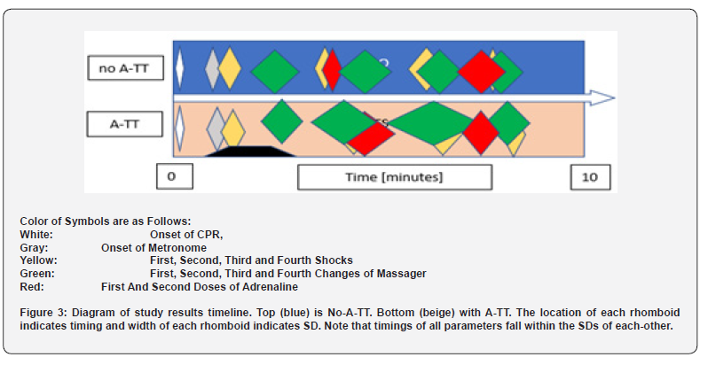
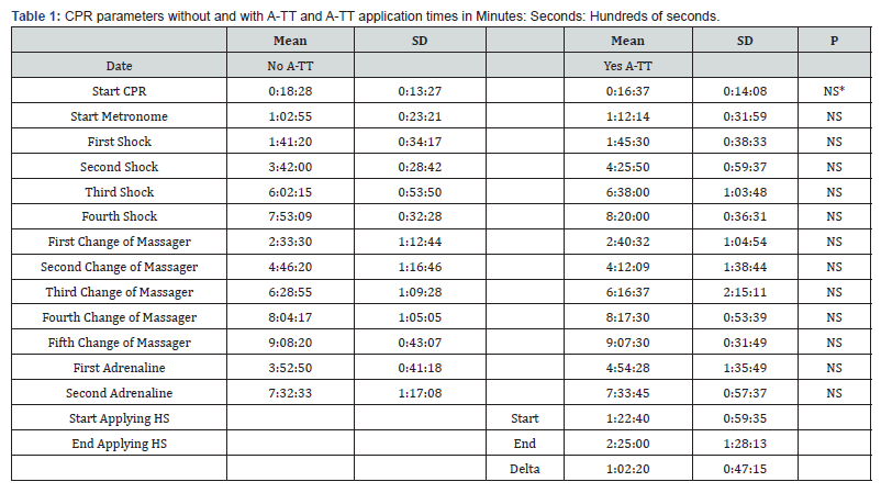
Twenty-six scenarios were performed by the teams, where 13 scenarios were done with A-TT and 13 without. CPR was started within 18.3 +/- 13.3 sec (mean +/- SD) from the onset sign in the no-A-TT scenarios and within 16.4 +/- 14.1 sec in the yes-A-TT scenarios (NS – not a statistically significant difference). (Table 1). In two cases more than 60 sec elapsed from the beginning of the scenario until chest compressions were started: one case with NO-A-TT and one case with A-TT. Time to first cardioversion was 101.2 +/- 34.2 sec in the NO-A-TT scenarios and 105.3 +/- 38.3 sec in the A-TT scenarios (NS). Time interval to first change of massagers was 2:33 +/- 1:12 min in the NO-A-TT scenarios and 2:44 +/- 1:06 min in the A-TT scenarios (NS). Time interval to first IV Epi injection was 3:52 +/- 0:41 min in the NO-A-TT scenarios and 4:44 +/- 1:33 min in the A-TT scenarios (NS). See (Table 1) and (Figure 3) for details.
CPR Quality

There was a total of 6 interruptions of CPR chest compressions that lasted more than 10 sec in 5 of the No-A-TT scenarios (in one scenario there were two interruptions) and 2 CPR interruptions for >10 sec in the A-TT scenarios, both occurring in a single scenario (NS).

A-TT Placement

A-TT was successfully placed by a single paramedic-in-training in all the scenarios that were randomized to A-TT placement. The A-TT placement started 82.4 +/- 59.4 sec after the onset of the scenario and application to both legs was completed 145.0 +/- 68.1 sec after scene arrival. The mean placement duration was 62.2 +/- 47.2 sec (range 30-180 sec) with all but two placements taking less than 60 sec. A-TT placement occurred before first massager replacement in 9 scenarios, before the second replacement in 3 scenarios and before third massager replacement in 1 scenario. All the participants rated the A-TT placement as “Easy”.

Discussion

The purpose of this study is to address a concern that the use of the Auto-Transfusion Tourniquet (A-TT) during performance of CPR in out-of-hospital cardiac arrest may interfere with the quality of CPR. The current AHA directive for high quality CPR requires early onset of uninterrupted and effective chest compressions, early defibrillation if rhythm is shockable, repeated in intervals if ROSC was not achieved, and quick administration of epinephrine with repeated doses in intervals [7,8]. It is now standard to monitor administration of CPR during training and in practice to assess the quality of the care and correct it when needed. As such, this study addresses the question: Does the use of A-TT during CPR interfere with its quality? To do so, we conducted CPR drills with and without application of the A-TT, while measuring the quality parameters. This prospective, non-blind controlled study was performed by trainees in MDA’s paramedics course and their trainers during Q1 2023. The paramedics-in-training were half-way of their course and have all performed CPR drills beforehand.

The physiological rationale for using A-TT during CPR consists of the following factors:

1. Rolling the A-TT on each leg pushes >500 cc of the patient’s own fresh blood to the core and >1000 cc from both legs [4]. In fact, it is postulated that during cardiac arrest the blood volume in the legs vessels is even higher due to the paralysis of the sympathetic nervous system during cardiac arrest, thereby significantly increasing the squeezable blood quantity relative the amount in normal adults. This shift of blood to the central circulation increases the end-diastolic volume of the heart chambers thereby amplifying the effectiveness of chest-compressions and the resulting stroke-volume [5].

2. The positioning of the A-TT ring in the upper thigh blocks the flow of blood into the legs. [The legs blood supply in a normal adult is approximately 24% of the total cardiac output [9]. As a result, the entire CPR-induced cardiac output is distributed to the essential organs (brain, heart, liver, gut, and kidneys) and is not “wasted” to perfuse the legs which can withstand 2 hours of ischemia with no risk of damage [10-12]. If simple arithmetic is used, this increases the share of the CPR cardiac output to the core from 75% to 100% or by approximately 25/75=0.33.

3. The blocking of blood-flow into the legs substantially increases the total vascular peripheral resistance. Therefore, the blood pressure during diastole, which is very low during CPR, increases substantially causing a major rise in coronary perfusion pressure (CPP). It was shown in a porcine study [1] that CPP nearly doubled when A-TTs were applied to the 4 legs of the experimental animals during induced cardiac arrest. Given the fact that most coronary perfusions are diastolic, having a sufficient level of CPP is critical for cardiac O2 supply and the likelihood of successful cardioversion.

4. The application of A-TT is quick, can be done by a single person with minimal training and does not interfere with other treatments given to the patient around his/her upper body (e.g. CPR, insertion of lines, intubation etc.). When ECMO is used as part of CPR [13], A-TT may facilitate cannula placement by increasing large vessels volume and diameter and increase the ECMO rate of suctioning the blood into the oxygenator for the same reason and by prevention of blood vessels flutter. Care must be taken to leave enough space for the sterile insertion of the cannulas.

5. The A-TT completely constricts the blood vessels of the legs. As such, it is an ultimate mechanical vasoconstrictor matching or surpassing the effect of adrenaline on the periphery without any detrimental effect on brain blood supply [14]. It is hypothesized that the use of A-TT in CPR may be able to eliminate the need for adrenaline with similar outcome on ROSC and better cognitive outcome when it is used instead of adrenaline. This hypothesis needs to be tested in prospective clinical studies.

Discussion of the Results of this Study

In all the categories of the CPR quality parameters, no statistically significant differences were observed when A-TT was used compared to when A-TT was not used. Onset of CPR, timing of massager replacement, time of first and subsequent shocks, first and subsequent Epi and the number of CPR interruptions were not statistically different between the scenarios with and without applying A-TT. In the A-TT scenarios the rings were applied approximately 82 seconds after arrival and the application took 62 sec, where all but two applications took less than 60 sec with the shortest application done in 30 seconds and the longest 3 min (Table 1). There were no difficulties in applying the A-TT in any of the scenarios and the paramedics-in-training did so without a problem after a brief practical training on a mannequin leg. The paramedics-in-training consisted of both males and females in equal numbers and there was no advantage to physical strength or body mass in achieving successful applications.

Study limitations

This study was done in teams of 4 paramedics-in-training and its results are only applicable to the situations where 4 or more providers are available on the scene. The number of scenarios with and without A-TT was 13 in each. This number is relatively small but there were no trends in the measured valued that could have become significant had the groups been bigger except, perhaps, the timing of first epi that was 52 sec longer at 4:44 min vs. 3:52 min (p = 0.078) when A-TT was used. Clearly, additional evaluations can and should be done in real-life cases of cardio-pulmonary resuscitations.

Applicability to Clinical use and Conclusions

We conclude that applying A-TT during CPR administered by a team of 4 trained persons does not impose a detrimental interference on the quality of CPR. Other safety and effectiveness aspects of A-TT use in CPR should be studied in real-life cases.


To Know more about Journal of Anesthesia & Intensive Care Medicine
Click here: https://juniperpublishers.com/jaicm/index.php

To Know more about our Juniper Publishers
Click here: https://juniperpublishers.com/index.php

Wednesday, October 18, 2023

Role of Cardiovascular Magnetic Resonance Imaging in Early Detection of Myocarditis in Patients Recovered from COVID-19 - Juniper Publishers

 Cardiology & Cardiovascular Therapy - Juniper Publishers


Abstract

Objective: To assess cardiac involvement in patients recovered from COVID-19 with no clinical evidence of myocarditis, using various non-invasive parameters including Transthoracic Two-dimensional Speckle Tracking Echocardiography (STE) and cardiac magnetic resonance imaging (CMR).

Methods: This prospective study was conducted in the Cardiology Departments of Maadi Military Hospital and Benha University Hospitals. A total of 74 patients were initially enrolled during their hospital stay, but only 50 of them met the inclusion and exclusion criteria. The CMR examination was performed in conjunction with echocardiography, ECG, and laboratory investigations on the same day, which occurred 2-12 weeks after recovering from confirmed COVID-19 infection. Patient scheduling for CMR and other examinations depended on the availability of the CMR machine.

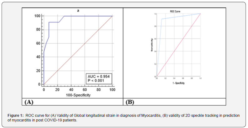
Results: According to CMR diagnosis, 23 (46%) patients were diagnosed to have myocarditis by CMR, the patients were categorized into two groups based on these results: normal group (27 patients) & myocarditis group (23 patients). According to GLS, 21 (42%) patients were diagnosed to have myocarditis by STE (diagnosis of Myocarditis with Global longitudinal strain was considered with cut-off point of >-21.33 with Sensitivity of 91.30% and Specificity of 92.59% (P <0.0001). 2D STE showed 87.50% sensitivity, 92.31% specificity and 90.00% accuracy to diagnose myocarditis, while CMR showed a 95.83% sensitivity, 96.15% specificity of and 96.00% accuracy.

Conclusion: Myocarditis was found in 46% of asymptomatic COVID-19-recovered patients. CMR is a valuable tool for early myocarditis detection when combined with 2D STE, offering high accuracy. Significant differences were observed between suspected myocarditis patients and those with normal results on both 2D STE and CMR.

Keywords: CMR; 2D STE; Myocarditis; Post COVID-19

Introduction

COVID-19 is a contagious illness caused by the SARS-CoV-2 virus, which was initially identified in Wuhan, China, in December 2019 and subsequently spread worldwide [1].

The COVID-19 pandemic has had far-reaching effects on global health, society, and economies [2]. The primary target of COVID-19 is the lungs, as the SARS-CoV-2 virus invades pulmonary tissue through the angiotensin-converting enzyme 2 (ACE2) receptor [3].

ACE2 is not limited to the pulmonary system but is also found in various other organs, including the cardiovascular system [4]. There is evidence linking COVID-19 to various cardiovascular complications, such as arrhythmias, myocarditis, acute coronary syndrome, acute onset heart failure, and cardiac arrest. Additionally, a strong association has been observed between acute cardiac injury and increased mortality rates among COVID-19 patients [3].

Previous research has indicated that a significant proportion of COVID-19 patients, ranging from 12% to 15%, exhibit elevated levels of high-sensitive cardiac troponin I (hs-cTnI) during their hospitalization, indicating myocardial injury. Furthermore, severe cases of COVID-19 may have cardiac involvement in as many as 31% of patients, but it remains uncertain how long this cardiac involvement persists after recovery [5,6].

SARS-CoV-2 has the potential to induce the release of chemokines and cytokines, which can lead to vascular inflammation, destabilization of atherosclerotic plaques, and myocardial inflammation. Consequently, elevated troponin levels in these patients could be attributed to stress cardiomyopathy, microvascular thrombosis, demand ischemia, and the secondary effects of systemic inflammation. Another possible cause of myocardial damage associated with COVID-19 is direct viral infection of the myocardium, potentially indicating viral myocarditis. While myocarditis-like clinical presentations have been reported in a small number of COVID- 19 patients, suggesting that fulminant myocarditis is uncommon [7].

Cardiovascular magnetic resonance (CMR) has emerged as the preferred non-invasive imaging technique for assessing heart volume, function, and myocardial tissue characteristics in cardiovascular medicine. Abnormalities in cardiac magnetic resonance (CMR) related to COVID-19 have been identified in 78% of predominantly ambulatory adults [8].

The main aim of our investigation was to evaluate the degree of cardiac engagement in individuals who have recuperated from COVID-19. We achieved this by employing cardiac CMR as a highly sensitive imaging method and comparing it to Transthoracic Echocardiography with 2D STE.

Patients and Methods

This prospective study was done in cardiology department of Maadi Military hospital & Benha University hospitals over 74 patients who were enrolled for this study during their hospital stay. Only 50 patients were fulfilling the inclusion and exclusion criteria and were enrolled in the study after 2-12 weeks after discharge from hospital with diagnosis of (SARS-CoV-2) by PCR on swab test of the upper respiratory tract.

The Banha Faculty of Medicine Human Research Ethics Committee approved the study protocol, and informed consent were obtained from Benha University prior starting data collection. This study was conducted over a 1-year duration, spanning from January 2021 to January 2022.

Inclusion criteria

Patients who previously confirmed with SARS-CoV-2 infection using (RT-PCR) swab test (who needs hospital admission due to significant lung affection or decreased oxygen saturation > 90%) and considered recovered by the discharging criteria ( resolved respiratory symptoms, normal temperature lasting longer than 3 days, 2 consecutive negative RT-PCR test results separated by at least 24 h and substantially improved exudative lesions on chest CT images) and were isolated for two weeks and have no any cardiac symptoms. All patients agreed to participate and sign informed consent.

Exclusion criteria were active Covid-19 infection. Previous myocardial infarction or coronary artery disease (evidence of coronary artery stenosis > 50%) , history of ischemic heart disease, previous myocarditis or heart failure, or known cardiomyopathy, uncontrolled hypertension, extremely irregular heart rates or previous atrial fibrillation, moderate to severe valvular heart disease, inability to effectively take part in breath-holding procedures and is thus unsuitable for undergoing CMR assessment, intra-cardiac devices non-MRI compatible (ICDs– Pacemakers), significant renal impairment ((CrCl rate < 30 mL/min/1.73 m2), contraindications to gadolinium contrast, disagree to participate or sign informed consent and insufficient CMR image quality for analysis.

All cases underwent a full history taking, complete physical examination, 12 leads Electrocardiogram, laboratory investigations (Creatinine, Troponin I & CBC), trans-Thoracic Echocardiography which was done for all patients according to EACVI Echocardiography guidelines [9] (Conventional study & 2D Speckle tracking), and CMR.

Protocol for CMR scanning included the following

conventional sequences: long-axis and short-axis cine, late gadolinium enhancement (LGE) and T2- weighted imaging (T2WI).

Quantitative mapping sequences: post-contrast T1 mapping and native T1/T2 mapping.

The diagnosis of myocarditis was confirmed by Updated Lake Louis criteria (2018) for diagnosis of Myocarditis which consist of two main criteria (T1-based criterion and T2-based criterion). The T1-based criteria is regarded as positive when there are increases in native T1 relaxation durations, increases in extracellular volume (ECV), or the presence of positive LGE.

The T2-based criterion is positive when there is an increase in T2 relaxation times, regionally high T2 signal intensities on T2- weighted images, or an elevation in the global T2-signal intensity ratio.

Supportive Criteria: pericardial effusion, pericardial signal abnormality, systolic LV dysfunction. All CMR images were evaluated by consultant cardiologist with 6 years of CMR diagnosis experience.

Sample size

Using PASS software, the minimum sample size required for specificity and sensitivity tests was calculated (PASS 11 citation: Hintze J (2011). PASS 11. NCSS, LLC. Kaysville, Utah, USA). The required sample size for the study is determined to be 50 patients without accounting for dropout rate.

Statistical Analysis

We conducted the statistical analyses using SPSS software (Statistical Package for the Social Sciences, version 24, SSPS Inc, Chicago, IL, USA). Categorical variables were presented in frequency tables with corresponding percentages, whereas descriptive statistics like standard deviation and mean were used to describe numerical data. Data entry, processing, and statistical analysis were performed using MedCalc ver. 20 (MedCalc, Ostend, Belgium). We employed various tests of significance, including logistic multiple regression analysis and ROC Curve analysis. The choice of analysis method was based on the nature of the data, whether it was parametric or non-parametric. We considered p-values less than 0.05 (5%) to be indicative of statistical significance [10].

Results

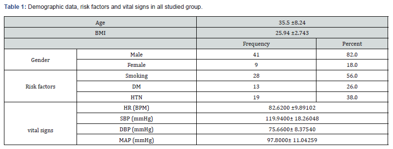
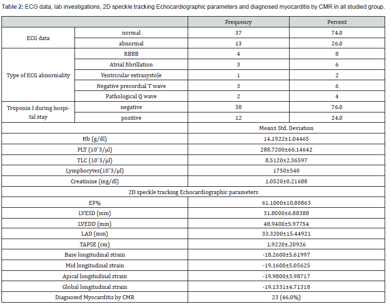
Table 1 shows demographic data, risk factors and vital signs in all studied group. Table 2 shows ECG data, lab investigations, 2D speckle tracking Echocardiographic parameters and diagnosed myocarditis by CMR in all studied group.

BMI: body mass index, HTN: hypertension, DM: diabetes mellitus, HR: heart rate, SBP: systolic blood pressure, DBP: diastolic blood pressure, MAP: Mean Arterial pressure.

EF: Ejection Fraction, LVESD: Left Ventricular End-Systolic Dimension, LVEDD: Left Ventricular End-Diastolic Dimension, LAD: Left Atrial Dimension, TAPSE: Tricuspid Annular Plane Systolic Excursion.

According to CMR diagnosis, 23 (46%) patient was diagnosed to have Myocarditis by CMR using Updated Lake Louis criteria (2018) and patients were categorized into two groups based on these observations: normal group (27) patients & myocarditis group (23) patients.

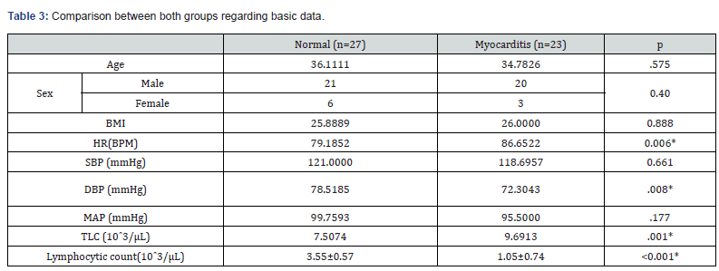
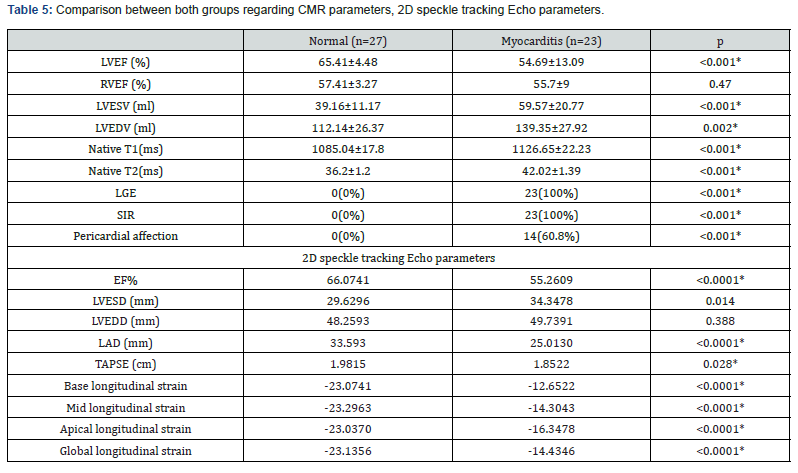
There was no statistically significant difference between both groups regarding age, sex, BMI, SBP and MAP. While the HR, DBP & TLC were higher in myocarditis group than in normal group with statistically significant difference (P=0.008, p=0.006 & p= 0.001 respectively). The lymphocytic count was lower in myocarditis group with statistically significant difference (p<0.001). Table 3 A statistically significant difference was seen between the two groups regarding ECG data and Troponin I during hospital stay with higher incidence of abnormal ECG & elevated Troponin I in myocarditis group than the other group. (P<0.0001). Time interval per weeks was about 4.5 weeks between hospital discharge & CMR & Echocardiographic examination with no significant difference between both groups. Table 4 Average left ventricular (LV) systolic function in myocarditis group was significantly lower than the normal group (P <0.001). Differences between the two groups were statistically significant concerning the LVESV (P = 0.012) & the LVEDV (P = 0.002). The average right ventricular ejection fraction (RVEF) was no significant difference between both groups. Global native T1, T2 values showed significant elevation in myocarditis group compared with the normal group (P. <0.001). Global Native T2 was significant high in the myocarditis group than in the normal group (P.<0.001). Pericardial enhancement and pericardial effusion were found in 14 patients (60.8%) of myocarditis group and was absent in normal group (P. <0.001). According to 2D STE parameters, there was a statistically significant difference between both groups regarding EF (P<0.0001), LVESD (P=0.014) TAPSE (P=0.028) and LAD (P <0.0001).

*: significant P value, BMI: Body Mass Index, DM: Diabetes Mellitus, HTN: Hypertension, HR: Heart Rate, SBP: Systolic Blood Pressure, DBP: Diastolic Blood Pressure, MAP: Mean Arterial Pressure.

Global longitudinal strain was significantly lower in myocarditis group (-16.34% vs -23.07%). Also, base longitudinal strain, mid longitudinal strain and apical longitudinal strain were lower in myocarditis group (P<0.0001) (Table 5).

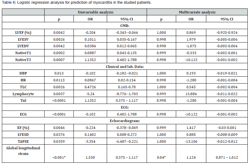
2D speckle tracking Echo parameters were used for diagnosis of myocarditis with global longitudinal strain cut-off point of >-21.33 with Sensitivity of 91.30% and Specificity of 92.59% (P <0.0001). 2D STE showed a Sensitivity of 87.50%, Specificity of 92.31% and Accuracy 90.00% for detection of myocarditis, while CMR showed a Sensitivity of 95.83%, Specificity of 96.15% and Accuracy 96.00%. Figure 1 Logistic regression analysis was conducted for the prediction of myocarditis in the studied patients. Global longitudinal strain, LVEF, LVESV, LVEDV, NativeT1, NativeT2, DBP, HR, TLC, Lymphocyte, troponin I, ECG abnormality, EF, LVESD & TAPSE were associated with the risk of myocarditis in univariate analysis. Only global longitudinal strain was associated with risk of myocarditis in both univariate and multivariate analysis Table 6.

*: significant as P-value.

*: significant as P-value, LVEF: Left Ventricular Ejection Fraction, RVEF: Right Ventricular Ejection Fraction, LVESV: Left Ventricular End-Systolic Volume, LVEDV: Left Ventricular End-Diastolic Volume, LGE: Late Gadolinium Enhancement, SIR: Signal Intensity Ratio.

Discussion

Myocarditis is a condition that results from inflammation of the heart muscle, usually caused by a viral infection or an autoimmune disease. With the outbreak of COVID-19, reports of myocarditis cases in patients who have recovered from the virus have emerged. Given the severity of the pandemic, the early detection and treatment of myocarditis are crucial to prevent longterm cardiac damage and potential complications.

The mean age in our study group was 35.5 ± 8.24 years, this was concordant with Huang et al. [11], who studied 26 patients who recovered from covid-19 and stated that patients with a mean age of 38. In contrast Wang et al. [12], who investigated cardiac consequences of COVID-19 in 47 recovered patients using CMR, reported higher mean age as they stated that mean age of their patients was 47.6 ± 13.3 years, In our study 41(82%) were males which was consistent with Ulloa et al. [13] Who studied 57 recovered patients searching for myocardial affection in recovered patients from covid-19 with male predominance with 80% of study group were males. In contrast, Huang et al. [11] showed female predominance as only 38% of study group were males.

In our study, 28 (56%) of all studied group were smokers, 13 (26%) patients had DM and 19 (38%) had hypertension. This was comparable with Wang et al. [12] Who reported that 18% of their patients had DM and 25% of them had hypertension. In contrast, Huang et al. [11] in which only 8% of patients were hypertensive and none had DM.

According to CMR parameters, 23 (46%) patients were diagnosed to have myocarditis by CMR, and patients were categorized into two groups based on these results: normal group (27 patients) & myocarditis group (23 patients).

There was no statistically significant difference between both groups regarding age, sex, BMI, SBP and MAP. Differences between the two groups were statistically significant regarding DBP, HR, TLC and lymphocytic count. In contrast, Huang et al. [11] reported that there was no statistically significant difference between patients with normal and abnormal CMR finding regarding age, sex, SBP, DBP, HR, TLC and lymphocytic count.

Our study showed statistically significant difference between both groups regarding level of Tn I as we found 11 (47%) patients with elevated Tn I in myocarditis group versus only 1 (3.7%) patient in the normal group. This was in line with Wojtowicz et al. [14] who reported statistically significant difference between the two groups of patients with and without non- ischemic cardiac injury using CMR regarding Tn I level. In the current study, there was a statistically significant difference between both groups regarding ECG abnormality as we found 12 (52%) patients with ECG abnormality in myocarditis group versus only 1 (3.7%) patient in the normal group. This was supported by a study by Ulloa et al. [13] which was conducted in Spain and studied myocardial affection in post covid-19 patient over 57 patients and showed statistically significant ECG abnormality which was found in 24 post covid patients (42%) inform of RBBB (15.8%), Atrial fibrillation (5.3%), Ventricular extrasystole (1.8%), Negative precordial T wave (12.3%) and Pathological Q wave (7%).

In our study, Echocardiographic examination showed statistically significant difference between both groups regarding EF, LVESD, TAPSE and LAD. In comparison, Özer et al. [15] who studied 74 patients with previous Covid-19 infection after 1 month from recovery reported also statistically significant difference between patients with elevated troponin group and the other group without elevated troponin regarding LAD, LVEF but no statistically significant difference between both groups regarding LVESD & LVEDD.

Our study found 21 (42%) recovered Covid-19 patients of all studied group had reduced GLS. This was in line with Mahajan et al. [16] who found reduced GLS in 40 (29.9%) recovered Covid-19 patients of his study group.

In our study, Global longitudinal strain was significantly lower in myocarditis group. Also, Base longitudinal strain, mid longitudinal strain and apical longitudinal strain. In line with our study, Mahajan et al. [16] showed statistically significant difference between reduced LV GLS group and Normal LV GLS group regarding LV GLS.

In contrast, Özer et al. [15] reported no statistically significant difference between group with myocardial injury and the other group without myocardial injury regarding LV GLS (-17.7 ± 2.6 Vs -18.9 ± 1.8, P=0.051). In our study we found 23 out of 50 (46%) patients had abnormal CMR parameters including myocardial oedema and LGE and diagnosed as myocarditis by updated Lake Louise criteria with mean time between hospital discharge and CMR examination of 34 days. This was in line with Huang et al. [5] who found median (IQR) time between COVID-19 diagnosis and CMR was 47 (36-58) days, and 15 of 26 patients (58%) reported abnormal CMR results on conventional CMR sequences.

In our study, myocarditis group were significantly different from healthy one in CMR examination regarding LVEF, LVESV, and LVEDV. But no statistically significant difference regarding RVEF. It was in concordance with Wojtowicz et al. [14] who reported statistically significant difference between the two groups of patients with and without Non-Ischemic Cardiac Injury regarding LVEF (57% vs 61%, P<.001), but no statistically significant difference regarding LVEDV and RVEF.

Our study showed increased myocardial oedema parameters with statistically significant difference between myocarditis and normal groups in form of increased T2 signal intensity ratio (T2 SIR), increased T2 relaxation time and increased native T1 respectively. In concordance to our study, Huang et al. [5] revealed that global native T1, T2, and ECV values were significantly greater in recovered COVID-19 patients with positive conventional CMR findings than in patients without positive findings (native T1 1,271 ms versus 1,237 ms, P=0.002), (T2 42.7± 3.1 ms vs 38.1± 2.4 ms, P=0.005), (ECV 28.2% vs 24.8%, P=0.001).

Also, Kunal et al. [17] reported significantly elevated native T1 (1301ms Vs 1264 ms, P =0.022) and native T2 (55.62 vs 45.25 ms, P =0.004) in post Covid-19 patients with abnormal CMR group compared to the other one with normal CMR.

In our study, non-ischemic pattern of LGE was found in 23 patients (46%) mainly sub- epicardial and mid wall enhancement affecting the apical and mid cardiac muscle segments with P value <0.001, denoting presence of myocardial fibrosis and/or necrosis. Wang et al. [12] reported non-ischemic sub-epicardial and mid wall LGE in 13 of 44 (30%) of the post-COVID- 19 patients’ group (a finding similar to our cohort results). Then they performed a strain study revealing that patients with LGE exhibited poorer LV and RV Performance.

In our study, Pericardial involvement in form of pericardial effusion and pericardial LGE which develops as a consequence of myocardial damage, was also found in 14 patients (28%) of all studied patients and was found in 60% of myocarditis group. In concordance, Huang et al. [5] reported that 7 (28%) of 26 patients were positive for pericardial LGE and have a minor pericardial effusion.

Regarding the validity of 2D speckle tracking Echocardiography (STE) and CMR in final diagnosis of myocarditis, In previous study by Luetkens et al. [18] found that the diagnosis of myocarditis using Updated Lake Louise Criteria yielded a sensitivity of 87.5% and a specificity of 96.2%.

In our study, speckle tracking echocardiography showed a sensitivity of 87.5% and specificity of 92.3 %. A study by Sharifkazemi et al. [19] compared the diagnostic performance of CMR and 2D STE in 57 patients with suspected acute myocarditis. They found that CMR had a higher sensitivity (96.8%) and specificity (93.5%) compared to 2D speckle tracking Echo (78.4% and 77.4%, respectively) in the diagnosis of acute myocarditis. In our study logistic regression analysis showed that only global longitudinal strain was associated with risk of myocarditis in both univariate and multivariate analysis.

Finally, this study had some limitations, as study did not include long-term follow-up to assess the outcomes of patients with myocarditis. Also, the study did not provide detailed information on the severity of COVID-19 in the patients, which may have influenced the development of myocarditis.

Case Presentations

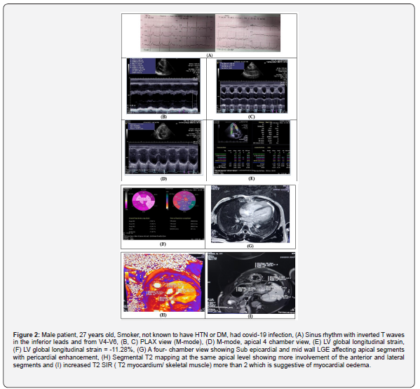
Case 1: Figure 2.

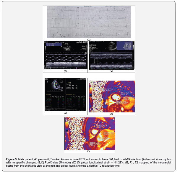
Case 2: Figure 3.

Conclusion

Evidence of myocarditis in asymptomatic patient recovered from COVID-19 was present in 46% of patients. Non-invasive predictors of myocarditis are Global longitudinal strain, LVEF, LVESV, LVEDV, NativeT1, NativeT2, DBP, HR, TLC, Lymphocyte, TnI, ECG abnormality, EF, LVESD and TAPSE while Global longitudinal strain was the only independent predictor. According to this research’s findings, it can be concluded that CMR is a valuable tool in the early detection of myocarditis in those who have achieved recovery from COVID-19. The use of CMR in combination with 2D STE has shown a notable level of accuracy in the detection and assessment of myocarditis. The findings of this study showed that there were significant differences between patients with suspected myocarditis and those with normal results on both 2D STE and CMR. The results also showed that CMR had higher sensitivity, specificity, and accuracy compared to 2D STE in the diagnosis of myocarditis. Further studies with larger sample sizes and multi-center cooperation are needed to validate the diagnostic accuracy of CMR and 2D STE in the early detection of myocarditis in patients recovered from COVID- 19.

To Know more about  Journal of Cardiology & Cardiovascular Therapy
Click here: https://juniperpublishers.com/jocct/index.php

To Know more about our Juniper Publishers
Click here: https://juniperpublishers.com/index.php

Typhlitis (Neutropenic Enterocolitis): A Multidisciplinary Approach- Juniper Publishers

   Reviews and Research- Juniper Publishers Abstract Typhlitis, or neutropenic enterocolitis, is a life-threatening gastrointestinal complic...