Showing posts with label Anorexia. Show all posts

Thursday, July 23, 2020

A New Challenging Strategy in the Prevention and Management of Tumor Lysis Syndrome in Patients with Chemo-Sensitive Hematological Malignancies - Juniper Publishers

Blood Research & Transfusion - Juniper Publishers

Summary
Introduction Tumor lysis syndrome (TLS) is a metabolic derangement that results from rapid destruction of cells. It happens frequently in cancers receiving chemotherapy, particularly hematological malignancies. It can lead to death in severe cases. Tumor lysis syndrome that leads to acute renal failure requiring dialysis and/or ICU admission can be associated with a higher rate of complications and mortality.
Case Report: We present a 24-year-old male patient with Burkitt’s lymphoma. After receiving one cycle therapy, He developed severe kidney injury from TLS. We initiated renal replacement therapy soon after his admission to the ICU, with marked response to therapy. This led to early discharge from the ICU.
Conclusion: Early initiation of renal replacement therapy After TLS- AKI Can improve the severity of AKI and hasten recovery and prevent complications. This can lead to earlier discharge from the hospital and better outcomes.

Introduction
Tumor Lysis syndrome (TLS) is a serious complication of chemotherapy in rapidly progressive cancer. It presents clinically with nausea, vomiting, fatigue, acute kidney injury, seizure, arrhythmia, and even death. It is caused by the release of electrolytes from the damaged cells. Characteristic electrolyte disturbances include hyperuricemia, hyperkalemia, hyperphosphatemia, and hypocalcemia [1]. The National Comprehensive Cancer Network (NCCN) guidelines recommended prophylaxis and treatment with aggressive hydration and control of hyperuricemia with Allopurinol (xanthine oxidase inhibitor) 2-3 days prior to chemotherapy, or Rasburicase (urate oxidase) which is highly effective in prevention and treatment of TLS, it’s indicated for high-risk patient, patient with adequate hydration not possible to achieve, urgent therapy in high bulk patient, and acute kidney injury [2]. Acute Kidney Injury (AKI) may frequently complicate TLS. It is caused by precipitation of uric acid, calcium phosphate or hypoxanthine in the renal tubules [3]. It may lead to renal failure requiring renal replacement therapy (RRT) in its most severe forms.

Case Scenario
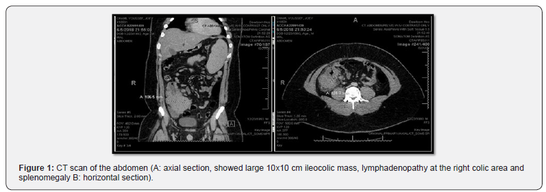
A 24 years old male with no significant past medical history presented to ED with gradually worsening abdominal pain for one month. His symptoms were associated with anorexia, black tarry stools, intermittent fever, sweating, vomiting, dizziness, and significant unintentional weight loss (40Ibs in 3 months). On admission, his vital signs were stable. He was anxious and clammy, and his physical exam was significant for splenomegaly. Initial blood work showed leukocytosis with a white cell count of 33.9 K/Ul, Hemoglobin was 15.3 g/dl, Platelet count was 67 K/Ul with Blast cells of 30%. The liver panel showed aspartate aminotransferase (AST) of 161 IU/l, alanine aminotransferase (ALT) of 65 IU/l, alkaline phosphatase (ALP) of 64.3 IU/l. Total bilirubin was 0.9 mg/dl. Lactate dehydrogenase (LDH) was 6000 U/l, and uric acid was 11.3 mg/dl. Urea was 14 mg/dl, and serum Creatinine was 1.24 mg/dl. Na was 139 mmol/l, K 3.9 mmol/l, and Ca was 9.6 mmol/l. An abdominal CT scan showed large ileocolic mass with enlarged lymphadenopathy at the right colic area (Figure 1). Peripheral smear showed immature cells with increase blast suggestive of acute leukemia. Biopsy of the colonic mass showed lymphoma. Flow cytometry confirmed the diagnosis of Burkitt’s lymphoma. CT head, neck, and thorax no other lymph nodes involvement (Figure 2).
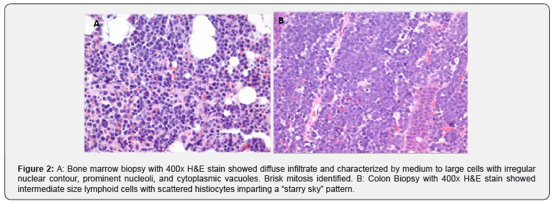
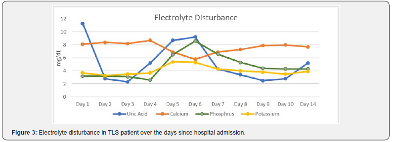
Bone marrow biopsy with 400x H&E stain showed diffuse infiltrate and characterized by medium to large cells with irregular nuclear contour, prominent nucleoli, and cytoplasmic vacuoles Figure 2A. Brisk mitosis identified. B: Colon Biopsy with 400x H&E stain showed intermediate size lymphoid cells with scattered histiocytes imparting a “starry sky” pattern. The patient was diagnosed with stage IV Burkitt’s lymphoma with leukemic phase. Treatment was initiated with R Hyper- CVAD (hyper-fractionated Cyclophosphamide, Vincristine, Doxorubicin, and Dexamethasone), he received one cycle of part A, then switched other treatment regimen DA EPOCH-R (Doseadjusted Etoposide, Prednisone, Vincristine, Cyclophosphamide, Doxorubicin, and Rituximab), and prophylactic intrathecal chemotherapy (Methotrexate, Cytarabine and Hydrocortisone). On day 2 of admission, the patient developed TLS-AKI. His glomerular filtration rate (GFR) dropped to 22 ml/min/1.73 m2. and serum Creatinine increased to 4 mg/dl (Figure 3). Treatment was started with aggressive hydration, rasburicase, and allopurinol. on day 5 all of the previous measures failed, and his kidney functions were deteriorated, so renal replacement therapy (RRT) was initiated and continued for a total of 14 days. He responded well to therapy and was discharged from the hospital after 25 days of management with recommendations for outpatient follow up.

Discussion
The overall incidence of TLS was reported at 4.4%in two large multicenter studies of Non- Hodgkin lymphoma (NHL). Of these, TLS occurred in 8.4% of the patients diagnosed with Burkitt lymphoma/leukemia or B-cell acute lymphocytic leukemia (B-ALL) [4]. In a large retrospective study in the United States, the nation-wide estimate of patients admitted with TLS was 22,785. 12.8% of them developed acute kidney injury requiring dialysis (AKI-D) [5]. AKI-D patient has higher mortality (41.9% vs. 19.1%; P<0.01), and longer hospital stay than patient with AKI without dialysis requirement (19 vs. 14.9 days; P<0.01). [5] In a single-center study at Saint- Louis University Hospital in France that included 63 patients, the adjusted mortality at hospital discharge was higher in patients with TLS who developed AKI (odds ratio, 10.41; 95% confidence interval 2.01- 19.17; p=0.005). Interestingly RRT was performed in 17 patients without AKI, and in 31 patients with TLS induced AKI [6]. TLS induced AKI may decrease the probability of getting a long-term remission of the malignancy therefor prevention of development of AKI to play an essential role in the management of TLS, and consequently primary malignancy outcome [6,7]. In patients with hematological malignancies who require chemotherapy, anticipation and early recognition of TLS and intervention play a crucial role in the treatment and prevention of complications. Although RRT by self can’t prevent AKI from occurrence in a patient with TLS, we challenge early initiation of RRT could play a pivotal role in preventing complication associated with TLS, even in early stages of AKI. Further clinical trials are needed to evaluate early intervention using RRT to assess prognosis as well as mortality and morbidity in the patient with chemo-sensitive hematological malignancies who developed TLS.


Wednesday, July 8, 2020

Periorbital Necrotizing Fasciitis in a Young Woman with Anorexia Nervosa-the Management and Considerations of Cosmetic and Functional Outcomes of Fulminant Disease in a Critically Unwell Patient - Juniper Publishers

Head Neck & Spine Surgery - Juniper Publishers  


Abstract

Necrotizing fasciitis is a rapidly progressive and potentially deadly bacterial infection of the superficial fascia. The horrific and grossly debilitating nature of necrotizing fasciitis has led to an increased prominence of its reporting in mainstream media however the disease entity has not been historically well understood, classified or reported in medical literature. While capable of manifesting in numerous bodily locations following inoculation during trauma, the periorbital region is the most commonly involved facial site and presents unique challenges in treatment of the disease due to the unique anatomical and functional attributes of this area. This case report describes the diagnosis, surgical and medical management in a young woman with severe malnutrition due to anorexia nervosa.

Introduction

Necrotizing Fasciitis (NF) is a devastating rapidly progressive bacterial infection involving the superficial fascia leading to necrosis of overlying skin and systemic toxicity [1,2]. We present a case of periorbital NF in a chronically malnourished and unwell patient following facial trauma.

Case Report

A 36-year-old female presented to the emergency department following a mechanical fall with extensive left facial bruising extending to the neck and chest wall. The patient featured a complex medical history including severe anorexia nervosa. Pathology revealed significant electrolyte derangement, hypothermia, osteoporosis, bone marrow suppression and synthetic hepatic impairment. A clinical diagnosis of NF was made 6 days into her ICU admission following the development of anaesthetic, necrotic-appearing skin associated with purulent exudates from the palpebral fissure and a corresponding rise in the patient’s White Cell Count (WCC) and C-Reactive Protein (CRP). Examination of the eye was unremarkable, with the exception of a subconjunctival hemorrhage and nasally oriented corneal dellen. Initial emergency debridement was undertaken as a joint operation between ophthalmology and ENT teams, stripping back necrotic periorbital tissues down to the lower cheek and performing a lateral tarsorrhaphy to prevent exposure keratopathy. ENT undertook further exploration and debridement down to the neck and submental region, leaving a VAC dressing in-situ and harvesting multiple tissue samples for histopathological evaluation. Histology revealed marked suppurative inflammatory changes in the reticular dermis, skeletal muscle and subcutaneous fat in association with venous congestive changes and large regions of muscular and dermal necrosis (Figure 1).

Pan-sensitive Streptococcus pyogenes and penicillinresistant Staphylococcus aureus (MSSA) were isolated from the excised tissues, while Pseudomonas species were isolated from the superficial left facial and eye swabs. Pending sensitivities, targeted antimicrobial therapy was instituted including the use of intravenous flucloxacillin, vancomycin, piperacillin-tazobactam and meropenem. Subsequent operative debridement and exploration was undertaken two days later, excising further nonviable periorbital tissues down to the buccal fat pad. Inspection of oral tissues was performed under direct laryngoscopy along with extension of tissue debridement from the neck area, noting that progression did not appear to track along fascial planes. Two months later, initial reconstruction surgery was performed. After washout of the left cheek and periorbital wounds, a Split Skin Graft (SSG) was applied to the left cheek (from a left thigh donor site) and Full-Thickness Skin Graft (FTSG) was applied to the superior and inferior left eyelids (abdominal donor site). The left tarsorrhaphy was also revised. Six months following her initial presentation, horizontal lateral canthotomy and division of tarsorrhaphy was performed to allow for a wider palpebral fissure. Finally, nine months after the initial diagnosis of NF, debulking of the upper lid was performed in conjunction with suturing of trace remaining elevator fibers to the tarsal plate. Reconstruction of the lower eyelid with FTSG (donor site medial upper arm) was also performed to relax the cicatricial nature of the earlier procedures (Figure 2).

Post-operative best corrected visual acuity was 6/9+2 in the affected eye with 5-6mm of lagophthalmos and complete pupillary exposure, allowing for good binocular vision without altered head posture. The patient remains able to use frontalis and orbicularis action to consciously moisten her cornea. The patient continued to use regular preservative-free lubricants to prevent exposure keratopathy. Periorbital NF is a rare rapidly progressing ophthalmic emergency that requires early recognition and operative debridement to prevent a fulminant course [1,3-5]. First described by Hippocrates in the fifth century B.C. as a complication of erysipelas [5], NF was then first properly described in modern literature by Confederate army surgeon Joseph Jones in 1871. Inconsistent nomenclature and inadequate linkage to sufficient bacteriological data led to confusion in the literature and a delay in the understanding of NF as a single disease entity [6]. Infection of the head and neck is uncommon in NF with only asmall number of cases being described in literature since 1960. In the post-antibiotic era, the overall mortality of head and neck NF has been estimated at between 9 and 31%, but its relatively frequent association with multi-system shock and permanent disfigurement render NF a particularly devastating and terrifying illness [3,7] (Figure 3).

The most common trigger for periorbital NF is an antecedent injury leading to the breakdown of the skin barrier and concomitant inoculation of the causative organism(s) [6-8]. Swelling, pain, erythema, fever, haemorrhagic bullae, skin necrosis and crepitus are the most common clinical signs relevant to diagnosis [2,4]. Diabetes mellitus, immunosuppression, chronic renal impairment, pulmonary disease, recent surgery and traumatic injury have been identified as predisposing factors for NF; however, it is worthwhile noting that a large number of published cases remain idiopathic in nature [1,9-10]. Immunosuppression, age greater than 50 years, cirrhosis, congestive cardiac failure, gout and development of toxic shock syndrome (a late sign of infection) have been identified as independent risk factors most associated with increased mortality from NF [1,2,11]. Relative risk indicators have been identified, utilizing biochemical indicators such as white cell count, haemoglobin, CRP, serum creatinine, sodium and blood glucose to stratify NF severity in the acute setting [12].

Periorbital NF is divided into 4 classifications on the basis of microbiological culture. Type 1 (polymicrobial) infections are related to both anaerobic and aerobic (including facultative anaerobes such as Enterobacteriaciae and non-typable Streptococci) organisms most commonly manifesting on the abdomen, trunk and perineum. Type 1 NF is the most common type to develop in patients with diabetes. Type 2 (monomicrobial) NF involves the extremities, cutaneous and muscular tissues; and features no clear underlying risk factors. Group-A betahaemolytic Stretococcus (S. pyogenes) and/or S. aureus are the most commonly implicated organisms in Type 2 disease, characterizing its strong association with toxic-shock syndrome. Type 3 infections are caused by Clostridium, Vibrio spp. and other gram-negative organisms, typically involving the abdominal wall and/or perineum. They are often rapidly progressive infectionsfollowing salt-water injuries, seafood ingestion and penetrating traumas. Type 4 NF describes fungal infections involving the extremities, most frequently occurring in immunosuppressed individuals [1,9,10,13].

Our patient was a young, immunosuppressed, malnourished female. On hospital presentation she had severe electrolyte derangement (hypomagnesaemia, hypokalaemia and hyponatreamia), hypothermia, impaired hepatic synthetic function and coagulopathy (INR 2.0 with hypovitaminosis-K) and ischaemic digits. She developed severe facial Type 2 NF (S. aureus and S. pyogenes) following a break in the periorbital skin overlying a large haematoma, initially caused by blunt mechanical trauma. No underlying fractures were identified on computed tomography imaging. Anaesthetic necrosis of the thin eyelid skin allowed the early clinical recognition and diagnosis of necrotizing fasciitis, nevertheless the infection was rapidly progressive in nature, resulting in severe permanent disfigurement despite prompt operative debridement and broad-spectrum antimicrobial cover. This case affords some insights into the pathophysiology of NF and also presented challenging aspects in the consideration of reconstructive techniques, attempting to maximize both long-term functional stability of the eye in limiting exposure keratopathy whilst optimizing the patient’s cosmesis Operative exploration and debridement in this patient demonstrated that this infection did not respect fascial planes, considered anomalous in the typical subcutaneous spread pattern of NF [14]. It is possible that traumatic disruption of subcutaneous compartments involving all skin and muscular layers, in conjunction with a large haematoma facilitated alternative pathways for the dispersion of infection in this patient. This is of particular importance to keep in mind in such cases wherethe musculoaponeurotic structures of the face and neck may be breached, allowing the spread of infection into the superior mediastinum [11].

Discussion

Periorbital NF is the most commonly involved facial site and behaves differently from NF elsewhere in that the highly vascularized orbicularis oculi acts as an effective barrier to prevent the spread of infection from the skin to the underlying periorbita, thereby retarding the spread of infection into the orbit. As in this case, necrosis of the thin eyelid skin occurs rapidly, facilitating early detection of the condition due to the obvious cosmetic abnormality on examination [1]. Given the fragile general health and constitution of the patient, decisions regarding the donor sites for reconstructive skin grafting were based purely on pragmatism, with cosmesis playing a clearly secondary consideration to what were emergent and life-saving surgeries. Gross malnutrition and immunosuppression meant that donor tissue was chosen to maximize graft viability, to facilitate adequate coverage of the relatively large areas left exposed following extensive surgical debridement and to maximize the chances of adequate postoperative healing in this setting. Subsequent procedures were required in this case to balance the cosmesis and function of the patients eyelids, her vision (and the initial requirement for a chinup posture to peer through her tight palpebral fissure,) and the necessity to prevent an exposure keratopathy in a patient with significant lagophthalmos secondary to an almost obliterated orbicularis.

Conclusion

The rapidly destructive nature of NF necessitates early recognition and diagnosis to minimize the risk of a fulminant course with high morbidity and mortality. Physicians must remain vigilant and mount a thorough clinical suspicion for NF in suitably vulnerable patients with clinical features suggestive of the disease such as severe pain, swelling, erythema, bullae and tactile anaesthesia. Urgent resuscitation, emergency surgical debridement, broad-spectrum IV antibiotics and other novel therapies such as hyperbaric oxygen must be implemented without delay to prevent the frequently debilitating, disfiguring and life-threatening sequelae of NF [1,8,11,15]. This case report demonstrated some of the complexities in managing periorbitalNF in the context of pre-existing medical comorbidities and highlighted the challenges in optimizing the functional and cosmetic outcomes of reconstructive surgery following extensive emergency debridements of neck and facial tissues.


To Know more about our Juniper Publishers


On the Positive Effects of Overconfident Self-Perception in Teams - Juniper Publishers

  Social Sciences & Management Studies - Juniper Publishers Abstract In this paper, we study the individual payoff effects of over confi...