Showing posts with label COVID-19. Show all posts

Thursday, May 23, 2024

Bilateral Sudden Sensorineural Hearing Loss - Juniper Publishers

Otolaryngology - Juniper Publishers


Abstract

Bilateral Sudden Sensorineural Hearing Loss (BSSNHL) is a rather rare and intricate condition marked by a sudden decline in hearing ability in both ears within a 72-hour timeframe. This review delves into the uniqueness of BSSNHL in comparison to unilateral cases, exploring its various causes, diagnostic criteria, management approaches, and the influence of COVID-19 on its manifestation. The review relies on a thorough examination of existing literature, emphasizing the scarcity of specific research on BSSNHL and underscoring its challenging prognosis despite diverse treatment options. Notably, the discussion touches upon potential triggers such as idiopathic, infectious, autoimmune, vascular, and membrane rupture, shedding light on the underlying mechanisms. It further elaborates on diagnostic criteria for BSSNHL, categorizing cases based on the onset duration. The review critically assesses management strategies, ranging from steroids to hyperbaric oxygen therapy, with an emphasis on the unpredictable nature of therapeutic outcomes. The intriguing link between BSSNHL and COVID-19 is explored through a case report, narrating the experience of an 18-year-old patient with bilateral hearing loss, anosmia, and loss of taste, suggesting the virus's involvement in auditory complications. In conclusion, the review highlights the rarity of BSSNHL, outlines the challenges in prognosis, and advocates for dedicated research to enhance comprehension and advance clinical outcomes for those affected.

Keywords: Bilateral sudden sensorineural hearing loss; Unilateral hearing loss; COVID-19; Viral infections; Hyperbaric oxygen therapy

Abbreviations: BSSNHL: Bilateral Sudden Sensorineural Hearing Loss; QoL: Quality of Life; RTI: Respiratory Tract Infection; MS: Multiple Sclerosis; SSNHL: Sudden Sensorineural Hearing Loss; RCD: Red Cell Distribution; AICA: Anterior Inferior Cerebellar Artery; HBO: Hyperbaric Oxygen

Introduction

Sudden Sensorineural Hearing Loss is defined as the sudden decline in hearing ability by over 30dB1, typically across three successive frequencies during pure-tone audiometry, within a period of 72 hours. Among the cases of sudden hearing loss, 98-99% are unilateral, while only 1-2% are bilateral [1]. Therefore, Bilateral Sudden Sensorineural Hearing Loss (BSSNHL) is an extremely rare and complicated disease. As for its onset and presentation, SSNHL has an acute onset and vague presentation, substantially limiting the quality of life (QoL) due to its acute and ambiguous nature, affecting communication ability significantly [2].

Clinically, it was first described in the early 1940s as at least a 30dB of hearing loss over three successive frequencies during pure-tone audiometry in fewer than three days [3]. The reported incidence is 5 to 20 per 100,00 annually [4]. Out of all reported cases, the predominant form is unilateral (95%), commonly idiopathic [5]. Only fewer than 4.9% of cases represent the bilateral form [6]. Due to its low incidence, literature provides limited data on the bilateral form. However, available research suggests that, unlike unilateral SSNHL, the bilateral form usually manifests as a consequence of some severe systemic condition, typically absent in the case of the former [6,7].

Literature Search

This narrative review on Bilateral Sudden Sensorineural Hearing Loss (BSSNHL) employs a non-systematic approach, summarizing relevant literature identified through keywords in reputable journals. Inclusive of various study types, we focus on English-language, peer-reviewed research from databases like PubMed, MEDLINE, Scopus, and Google Scholar, covering material from database inception to the present. Exclusions include non-English publications, non-peer-reviewed articles, and studies with unclear methodologies. Manual searches complement database findings and data extraction centers on key variables. The synthesis aims for a cohesive narrative, providing insights into BSSNHL's current understanding and identifying potential research gaps.

Causes of BSSNHL

Idiopathic

The majority of patients suffering from Bilateral Sudden Sensorineural Hearing Loss (BSSNHL) do not have an identifiable cause, leading to the classification of their condition as idiopathic BSSNHL. Only 10% of patients exhibit an identifiable cause [8]. A study revealed that some patients developed idiopathic BSSNHL after suffering from an upper respiratory tract infection (RTI) or oral herpes [9]. Another study in 2016 reported that, out of 16 BSSNHL patients, 5 had an idiopathic BSSNHL while others were observed to have different malignancies, such as neurofibromatosis, as shown in the picture below (Figure 1) [10]. BSSNHL can also result from radiotherapy in Head and neck cancer patients. A study investigated the hearing ability of 36 re-irradiated survivors of nasopharyngeal carcinoma. Among them, 91% exhibited abnormal cVEMP, 75% had abnormal oVEMP, 67% showed a reduced bone-conducted mean hearing level, and 39% had abnormal caloric tests. The study suggests that BSSNHL can occur after 10 years of radiotherapy [11]. Despite these findings, various theories on the etiology of BSSNHL are discussed below.

Infectious

Viral infections are among the most common causes of BSSNHL. According to a study by W R Wilson [12], 12 viral infections can cause Bilateral SSNHL through three mechanisms: (a) Cochleitis or Neuritis of the cochlear nerve. (b) Relapse of latent viral infection in the inner ear. (c) Indirect damage of the inner ear antigen when any distant viral infection initiates an antibody response without direct inner ear infection. However, majority of the literature supports the first two methods of BSSNHL due to viral etiology.

Among various viruses, the mumps virus is known to cause BSSNHL in rare cases. In 1992, Okamoto et al. conducted a serology study of 131 patients with sudden hearing loss suggesting that mumps was the cause in 9 out of 131 cases13. However, a couple of studies [13,14] suggest that mumps is responsible for only a small fraction (i.e., >10%) of cases of SSHL, without distinguishing between unilateral and bilateral SSNHL. Moreover, all of the eight members of the Herpes virus family (HS Type I & II, Varicella Zoster, EBV, CMV, HHV-6, 7, and 8) are known to cause SSNHL [15]. These viruses remain latent in the host even after remission and may cause SSHL upon reactivation. Besides herpes, other viruses such as adenovirus and arenavirus 14 are also known to cause BSSNHL.

In a case study by Chanmi Lee et al. [16] an HIV-infected patient with 8th cranial nerve involvement was found to suffer from BSSNHL [6]. Regarding indirect damage to the inner ear due to systemic viral infection, a 1998 study reported no elevation of MxA protein in any of their 20 patients with sudden hearing loss [16]. There have been reported cases of bilateral SSNHL after bacterial cryptococcal meningitis due to Cryptococcus neoformans. YH Chen et al. [10] studied 16 patients suffering from BSSNHL, and one patient, a 46-year-old male was suffering from cryptococcal meningitis, which was confirmed and cultured from his cerebrospinal fluid (CSF) findings. The cryptococcal infection which to the meninges and auditory canals of the inner ear, leading to BSSNHL along with vestibular loss.

Autoimmune

The concept of the immune system's involvement in inner ear pathology was first proposed by McCabe [17]. In patients with progressive Bilateral hearing loss, Harris & Sharp [18] demonstrated the presence of autoantibodies against various cochlea antigens [18]. As the majority of SSNHL cases lack a specific cause, certain assumptions regarding etiology have been suggested in order to predict the probable cause. One such assumption is autoimmunity, based on the idea that antibodies or activated T cells play a role in causing SSNHL by either cross-reaction or damaging in the inner ear, respectively [19]. Analysis of data for BSSNHL from different articles, it became apparent that the phenomenon of autoimmunity is more prevalent in females.

Research indicated the presence of underlying systemic autoimmune pathologies, such as Cogan's disease and Guillain-Barré syndrome, which manifested as BSSNHL [20]. Additionally, the suspicion of an underlying systemic autoimmune disease is raised when rapidly progressive bilateral sudden hearing loss responds positively to steroid therapy [21]. One notable autoimmune disorder is Multiple Sclerosis (MS), where 92% of cases reported SSNHL when hearing loss appeared as an early symptom, while gradual loss of hearing was present in 88% of cases during the late phase of the disease [22]. In conclusion, further studies and data collection are expected to provide a clearer understanding of the occurrence of BSSNHL in various pathologies with an autoimmune background

Vascular

In conclusion, further studies and data collection are expected to provide a clearer understanding of the occurrence of BSSNHL in various pathologies with an autoimmune background. A detailed study on the pathogenesis of BSSNHL [23] identifies three potential reasons behind the vascular etiology of BSSNHL: (a) Total and irreversible vascular occlusion, (b) Total and reversible vascular occlusion, and (c) Relative ischemia of the cochlea. Total vascular occlusion is primarily attributed to the occlusion of arteries at the base of the brain [24]. Regarding relative ischemia of the cochlea, the most widely accepted theory suggests that it results from the hyperviscosity of blood due to increased hematocrit and red cell distribution width (RCD).

In 2016, a study proposed that 5 out of 16 patients with BSSNHL had a vascular etiology [25]. This is often linked to the occlusion of the anterior inferior cerebellar artery (AICA) due to an atheromatous plaque forming in the basilar artery, leading to decreased blood flow and subsequent BSSNHL 24 However, it is crucial to note that occlusion of the basilar artery is associated with a high mortality rate and poor prognosis in survivors it may present as impending brainstem infarction. Accompanied by vertigo and nystagmus may occur before bilateral SSNHL [10].

Membrane Rupture

Membrane rupture seldom occurs spontaneously, typically arising from sudden pressure changes in the inner ear, severe head injuries, intense exercise, or deep barotrauma in the ear. A 1986 study proposed that the formation of a labyrinthine fistula leads to tiny holes between the niche of the round window and the posterior semicircular canal ampule. This, in turn, causes a rupture of the Reissner membrane, leading to a mixture of fluids and resulting in cochlear malfunction [23].

Diagnosis of BSSNHL

As stated earlier, data for the bilateral pattern for SSNHL is limited, only about 1.1% of all the patients with SSNHL came with this complaint of bilateral loss when data was evaluated of patients from 1995-2014 (i.e. total patients of SSNHL were 1459, 16 of them were there with this issue of SSNHL affecting both the ears). 10 Diagnostic criteria for BSSNHL include an additional clause to be fulfilled that SSNHL should affect both ears concomitantly within a period of 72 hours. The bilateral form of SSNHL can be categorized based on duration: simultaneous (both ears affected within 72 hours), sequential (one ear takes more than 72 hours but less than 30 days to be affected), and progressive (lacking a sudden onset, as both ears take more than 30 days to be affected)

Management of BSSNHL

Sudden Sensorineural hearing loss (SSNHL) is considered an otologic emergency and requires effective and early management [26]. According to the Clinical Practice Guidelines 2019 for SSNHL, clinicians should offer intratympanic (IT) steroid therapy for patients with incomplete recovery within 2-6 weeks after the onset of symptoms (KAS 10). Clinicians may also consider corticosteroids within the first 2 weeks of onset of SSNHL (KAS 9a). Hyperbaric oxygen therapy can be combined with steroid therapy within 2 weeks of onset (KAS 9b), or the combination can be used as salvage therapy within 1 month of the onset [27]. Historically, the management includes the use of steroids as standard therapy, which can be administered orally, intravenously, or via Intratympanic Injections. However, the therapeutic value of corticosteroids remains unpredictable [28].

A systemic review and Meta-analysis compared Intratympanic vs Systemic Corticosteroids as the first-line treatment. The Data of 710 patients suggested no significant difference between the therapeutic effect of the two methods of administration and neither of their combinations improved the hearing outcomes more than either of the methods separately. The table below shows the crux of other studies for BSSNHL treatment outcomes [29]. In a Systematic Review and Meta-analysis conducted in 2022 with sample of 496 patients [30], with similar results were observed in terms of hearing outcomes of Intratympanic vs Systemic corticosteroids as a first line treatment. Although no significant difference was found between the two methods, the preference for Intratympanic steroids was suggested due to the observation of more serious side effects with systemic administration of dexamethasone compared to local adverse effects of Intratympanic injections [31].

In a Randomized Controlled Trial (RCT) involving 136 patients, where 66 received Hyperbaric Oxygen (HBO) therapy along with pharmacological treatment (HBO+P) and 70 received pharmacological treatment alone (P), statistically significant differences in outcomes favored HBO+P [32] Another RCT with 171 SSNHL patients indicated that the combination of HBO therapy and oral steroids was the most effective treatment when initiated two weeks after symptom onset [33]. A Systematic Review and Meta-analysis covering the period from January 2000 to April 30, 2020, with 130 participants, statistically suggested improved hearing outcomes in SSNHL patients who received HBO treatment, or its combination treatment compared to those given control therapy [34]. Alternative treatments include sound therapy combined with pharmacological treatment and adjuvant transcranial random noise stimulation with conventional treatment. The combination of sound therapy and pharmacological treatment demonstrated an improvement in noise thresholds and speech recognition, implying better recovery of hearing abilities compared to pharmacological treatment alone [35].

BSSNHL In COVID-19

Discovered in December 2019 in China, the COVID-19 pandemic has had widespread hazardous effects globally. Nations faced significant challenges, impacting various aspects of daily life. Publicly practiced precautions included the use of face masks, handwashing, avoidance of crowded gatherings, and maintaining physical distance [36]. Clinically, COVID-19 manifests with a diverse range of symptoms, from self-limiting upper respiratory tract infection symptoms to severe consequences such as pneumonia, multi-organ system failure, and death [37]. In the context of otologic symptoms associated with COVID-19, available literature is limited. A case report highlighted an elderly patient with Sudden Sensorineural Hearing Loss (SSNHL) and a positive COVID-19 status. Unfortunately, the report lacked further findings, including audiometric or imaging studies [38].

However, a case report in 2020 focused on COVID-19 associated with Bilateral Sudden Sensorineural Hearing Loss (BSSNHL). Alexander Chern et al. [39] reported an 18-year-old female patient who presented with BSSNHL seven weeks prior. The patient also experienced loss of taste and smell, and her father tested positive for COVID-19 antibodies. Audiometric results indicated hearing loss in both ears, with the right ear having an average pure tone level (PTA) of 60dB and the left ear showing moderate to severe hearing loss with a PTA of 63dB. Word recognition scores were 88% for the right ear and 80% for the left ear. Tympanometry results were normal, indicating no middle ear issues. The patient reported no other neurological problems or family history of hearing [39]. Although the mechanism by which COVID-19 may cause dizziness is unclear, the disease does appear to be associated with individual cranial neuropathies resulting in anosmia [40]. It was concluded that COVID-19, besides its many other effects on the patient's health, can also interfere with the hearing ability of the patient leading to the expression of SSNHL.

Prognosis of BSSNHL

While the prognosis for unilateral Sudden Sensorineural Hearing Loss (SSNHL) is generally favorable, with many patients recovering fully within days to weeks, the outlook for Bilateral SSNHL (BSSNHL) is typically poor without immediate treatment [10] Abnormal caloric tests and oVEMP often signal a poor prognosis for BSSNHL, while a good prognosis is associated with a normal caloric test and oVEMP.

Conclusion

Bilateral Sudden Sensorineural Hearing Loss (BSSNHL) is a rare condition, occurring in only 1-2% of patients with Sudden Sensorineural Hearing Loss (SSNHL). Due to its rarity, there is a scarcity of studies specifically addressing the causes, management, and prognosis of BSSNHL. Traditional literature highlights various theories regarding its etiology and management; however, the condition still tends to have a poor prognosis even with oral and intratympanic steroids and hyperbaric oxygen therapy (HBOT).

To Know more about Global Journal of Otolaryngology
Click here: https://juniperpublishers.com/gjo/index.php

To Know more about our Juniper Publishers
Click here: https://juniperpublishers.com/index.php

Thursday, April 18, 2024

An Evidence Based Review of Vitamin D in COVID-19 Severity and Mortality - Juniper Publishers

 Complementary Medicine & Alternative Healthcare - Juniper Publishers


Abstract

Introduction: This evidence-based review aims to explore the association between vitamin D status and the severity and mortality of COVID-19, providing insights for healthcare professionals and policymakers in managing the disease.

Methods: A systematic review process was conducted to identify relevant studies on vitamin D and COVID-19 using electronic databases and specific search terms. Thirteen studies were selected and analyzed, including quantitative research at levels III, IV, and V.

Results: The analysis revealed a significant association between vitamin D deficiency and increased severity and mortality of COVID-19. Vitamin D levels were found to be inversely related to the severity of the disease, with deficiency acting as an independent predictor of COVID-19-related mortality. Studies demonstrated a higher prevalence of vitamin D deficiency among hospitalized COVID-19 patients. Bolus doses of vitamin D supplementation were associated with improved clinical outcomes and lower mortality rates in COVID-19 patients.

Conclusion: The evidence suggests that maintaining adequate vitamin D levels may have a protective effect against the severity and mortality of COVID-19. Vitamin D supplementation, in combination with safe sun exposure education, could be a cost-effective and safe measure to mitigate the impact of the SARS-CoV-2 pandemic. However, further interventional studies are needed to evaluate the efficacy and optimal dosing regimens of vitamin D supplementation in COVID-19 patients.

Keywords: COVID-19; SARS-CoV-2; vitamin D; Severity; Mortality; Supplementation; Evidence-based practice

Introduction

Coronavirus disease 2019 (COVID-19), declared a global pandemic by the World Health Organization (WHO), presents a significant challenge to healthcare systems worldwide (WHO, 2020) [1]. In January 2022, COVID-19 ranked among the top four leading causes of death for all age groups, with older adults being particularly vulnerable [2]. Hospitalizations and mortality rates are significantly higher in adults over 65 years of age compared to those under 65 [3,4].

Between January 2020 and July 2022, there were over 562 million confirmed cases of COVID-19, resulting in approximately 6.3 million deaths worldwide (WHO, 2022). The economic impact of COVID-19 is staggering, estimated at over $16 trillion, which accounts for approximately 90% of the annual gross domestic product (GDP) of the United States [5].

Vitamin D, a hormone produced by both the kidneys and the skin, plays a crucial role in regulating blood calcium concentration and impacting the immune system. It is known by various names, including calcitriol, ergocalciferol, calcidiol, and cholecalciferol.

The two widely available pharmacologic preparations are cholecalciferol (D3) and ergocalciferol (D2). More recently, vitamin D has shown antiviral effects and plays a crucial role in the immune system [6-8]. It is being investigated for its potential in mitigating infections, enhancing immune responses, and suppressing the cytokine storm [9-11] Comparatively, vitamin D deficiency has been linked to increased susceptibility to viral infections. Research has not demonstrated a strong association between vitamin D levels and the prevention of COVID-19 infection [12-15]. However, there is a growing interest in exploring the potential role of vitamin D in relation to the severity of COVID-19 disease.

At the time of submission, COVID-19 has tragically resulted in the loss of over 6 million lives globally (WHO, 2022). Despite this significant impact, there remains limited knowledge about potential protective factors against the disease. Notably, advanced age and underlying chronic medical conditions, especially chronic pulmonary and cardiac diseases, have emerged as prominent predisposing factors for severe COVID-19 development and subsequent mortality [16,17]. This comprehensive literature appraisal aims to investigate potential associations between vitamin D status and disease severity and survival in COVID-19 patients. By analyzing the available evidence, this analysis provides a recommendation while considering the balance of benefit, harm, and cost.

Methods

This systematic review, conducted in collaboration with a faculty advisor and university librarian, examines the relationship between vitamin D and COVID-19, focusing on severity and mortality outcomes. The review process involved comprehensive searches of electronic databases, including PubMed, using key terms such as Vitamin D, Vitamin D Level, Vitamin D Deficiency, Covid, Covid-19, and Coronavirus. Inclusion criteria were limited to English-language articles published between 2020 and 2022, and excluded research proposals and protocols. A total of 20 articles were retrieved, and after reviewing the title and abstracts, 13 relevant studies were selected for appraisal using the Johns Hopkins Appraisal Tool. The levels of evidence were graded using Johns Hopkins Level of Evidence table. This review is comprised of 11 non experimental level III research articles, the highest level of evidence available to date.

Literature Review

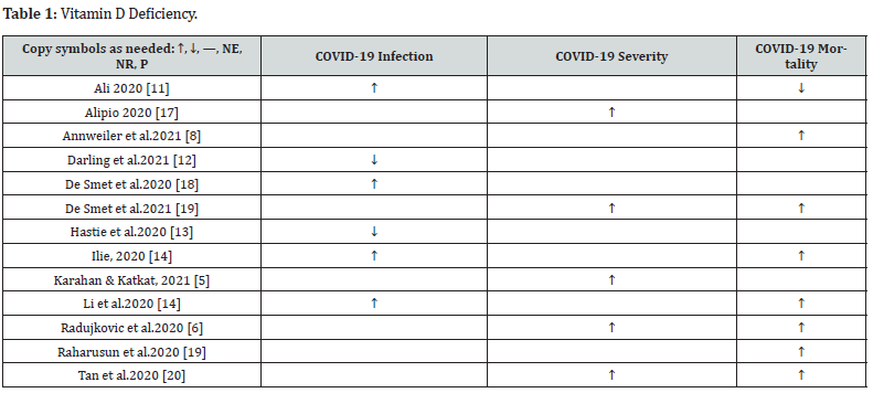
Vitamin D levels have been found to be notably depleted among the aging population, a group that exhibits heightened vulnerability to COVID-19 [15]. Further evidence highlights the prevalence of vitamin D deficiency among hospitalized COVID-19 patients, with 59% of admitted individuals presenting vitamin D insufficiency. Vitamin Ds deficiency upon admission has demonstrated a significant association with COVID-19 severity and mortality, even after adjusting for factors such as age, gender, and comorbidity.

There is possibly a blood level dependent association between vitamin D level and COVID-19 severity. A retrospective multicentric study of 212 patients [7] found that critical COVID-19 cases had the lowest levels of vitamin D, whereas mild cases had the highest levels. Similarly, found similar results when stratifying COVID-19 patients by vitamin D level. There were two additional studies conducted by that reported weaker correlations between vitamin D levels and COVID-19 cases and mortality. Finally, there is a small body of evidence supporting the use of bolus doses of vitamin D3 supplementation administered during or shortly before the onset of COVID-19 (2020) [9] and Karahan and Katkat (2021) [5] both demonstrated a lower incidence of COVID-19 infection and improvement in COVID-19 severity with bolus dosed vitamin D.

There were a few studies that failed to demonstrate a positive effect of vitamin D on COVID-19. These studies were small, completed on a younger and healthier population, and primarily studied the relationship between vitamin D level and COVID-19 infection rates, but did not study the correlation between the vitamin D level and COVID-19 severity or mortality (Table 1).

Discussion

In response to the profound burden imposed by the COVID-19 pandemic and the potential for mitigating severe disease outcomes through the exploration of protective factors, numerous researchers have established a compelling association between vitamin D deficiency and the severity of COVID-19 [18-21]. While research has failed to demonstrate that Vitamin D prevents Covid-19 infection, there is a moderate amount of research establishing that optimal vitamin D levels are associated with less severe cases of COVID-19 and conversely, low vitamin D levels have been associated with more severe cases. Furthermore, bolus dosing Vitamin D3 may provide some protection in the severity of the infection, particularly in populations at risk [22,23].

Implications for Practice

Nurse practitioners manage patients in primary care who are at risk of COVID-19. Staying up to date with the current evidence is crucial in supporting clinical practice. The evidence appraised in this review are all non-experimental research. Observational and cohort studies provide valuable insights into potential associations, and the majority of the reviewed studies indicate a positive correlation between vitamin D levels and COVID-19 outcomes. Moreover, vitamin D supplementation is generally considered safe when administered within the recommended dosage range. Although further experimental research is needed to establish a causal relationship, considering the low risk profile of vitamin D supplementation, it is the recommendation of the authors that nurse practitioners consider prescribing Vitamin D supplementation to improve the severity of COVID-19 infections and that blood levels should be monitored to achieve optimal circulating levels ranging from 75 to 100 nmol/L. As healthcare leaders, nurse practitioners have the responsibility to actively seek opportunities to educate both patients and colleagues. By doing so, we can drive practice change and promote the adoption of evidence-based approaches. Disseminating evidence-based practices is vital in improving patient outcomes and ultimately enhancing the overall quality of care.


To Know more about Journal of Complementary Medicine & Alternative Healthcare

To Know more about our Juniper Publishers

Monday, November 13, 2023

Does the Timing of Intubation and IMV Impact Clinical Outcomes in Adult COVID-19 Patients with ARDS: A Systematic Review and Meta-Analysis - Juniper Publishers

 Pediatrics & Neonatology - Juniper Publishers


Abstract

Background/Aim: COVID-19 can rapidly develop into acute lung injury, and even acute respiratory distress syndrome (ARDS), which has a high risk of death. Patients with ARDS often require intubation. However, the timing of intubation and its effect on clinical outcomes in COVID-19 ARDS (CARDS) patients remains unclear. Thus, the authors explored the impact of intubation time on clinical outcomes in COVID-19 patients with ARDS through a systematic review and meta-analysis.

Materials and Methods: Research articles from PUBMED, CINAHL, MEDLINE, ProQuest Covid database, and Web of Science were searched through December 2021. All patients in the research met the Berlin criteria for ARDS. For the purposes of this review, “Early” intubation was defined as intubation within 24 hours of an ARDS diagnosis, while “Late” was defined as 24 or more hours after diagnosis. The primary outcome was ICU mortality, and secondary measures included length of ICU stay and duration of mechanical ventilation. The meta-analysis was performed using a random-effects model. The quality of cohort studies was assessed using the Newcastle-Ottawa Scale. The methodological quality of the overall evidence in this review was evaluated using the GRADE approach.

Results: After an extensive search, six cohort studies were ultimately included in the systematic review, altogether encompassing 2,739 patients with CARDS. A meta-analysis revealed statistically significant differences in mortality [risk ratio (RR)=0.78; 95% confidence interval (CI),0.69-0.88; Z=3.91, P < 0.0001)]. The mortality rate was 36.2% (817 deaths) in the early group and 48.2% (229 deaths) in the late group, respectively. Results of the narrative analysis showed that early intubation resulted in shorter ICU stays, which was statistically significant. However, no statistical difference was found in the duration of mechanical ventilation.

Conclusions: Early intubation can reduce mortality and length of ICU stay in adult COVID-19 patients with ARDS. However, the timing of intubation did not affect the duration of continuous mechanical ventilation.

Keywords: COVID-19; ARDS; Timing of Intubation; Invasive Mechanical Ventilator; Systematic Review; Mortality

Introduction

In December 2019, COVID-19 was identified as a new clinical syndrome caused by a novel coronavirus. The virus is transmittable through the respiratory tract and is highly contagious. Despite significant efforts to control the spread of COVID-19, it triggered a global pandemic [1-4], an epidemic of scale across international borders [5]. COVID-19 pneumonia may develop rapidly into acute respiratory distress syndrome (ARDS) with a high risk of death [6]. ARDS is an acute respiratory failure caused by increased pulmonary capillary permeability secondary to inflammatory oedema. It leads to alveolar flooding and subsequent deep hypoxemia, in which intrapulmonary shunt is the most important underlying mechanism [7]. However, ARDS caused by COVID-19 is different from ARDS with any other underlying cause. According to Huang et al. (2020) [8], the onset of ARDS associated with COVID-19 is between 8-12 days. There are two distinct phenotypes of COVID-19-associated ARDS (CARDS), L-type and H-type. Type L presents as pneumonia and is limited to mild inflammation of the subpleural interstation.

It is characterized by low elasticity, atelectasis, normal compliance, and low lung weight. On the other hand, patients with Type H meet typical ARDS criteria, including decreased lung compliance, hypoxemia, bilateral lung infiltration, and increased lung weight [9]. Li and Ma [10] have been reporting on respiratory support strategies for patients with CARDS during the past two years, but how exactly the timing of tracheal intubation and use of invasive mechanical ventilation impacts clinical outcomes is still unclear in patients with CARDS. Delayed intubation can cause autologous lung injury (SILI) due to high respiratory drive pressure [11]. However, intubating patients too early can also be associated with some complications, including ventilatorassociated pneumonia, airway injury, ventilator-induced lung injury, and hemodynamic disorders due to positive pressure ventilation [12]. Six primary studies [13-18] have indicated different results regarding the timing of intubation for patients with CARDS, and currently there is no systematic review relevant to this topic. Therefore, a systematic review is necessary to further explore how the timing of intubation impacts outcomes for these patients.

Materials and Methods

The PRISMA statement, which contains a 27-item checklist and four-phase flow chart [19], is used to help authors report systematic reviews and meta-analyses.

Eligibility Criteria

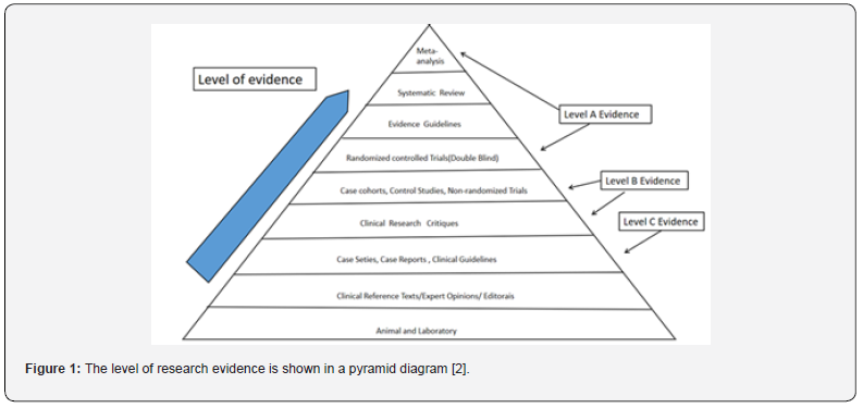
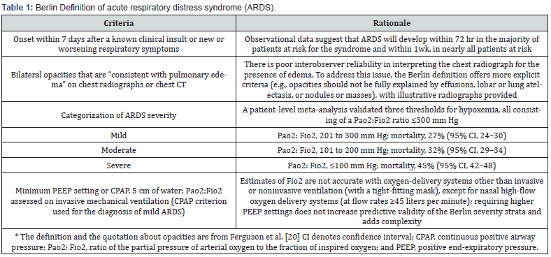
The population included in this systematic review was defined as adult patients (≥18 years old) with PCR-confirmed COVID-19 diagnoses who also had ARDS. ARDS was defined by the Berlin Criteria or American-European Consensus Conference (Table 1) [20]. Early intubation was defined as being intubated within 24 hours of being diagnosed with ARDS; Late intubation was defined as being intubated 24 or more hours after an ARDS diagnosis. The timing of intubation was also defined by authors of four original studies [21]. Systematic reviews and meta-analyses are in the upper echelon of the evidence-based medicine hierarchy of evidence, followed by randomized, controlled, double-blind studies, followed by cohort studies, case-control studies, case series, and case reports [2] (Figure 1). Randomized trial studies were not permitted due to potential ethical issues regarding the timing of intubation of COVID-19 ARDS patients [22]. Therefore, existing cohort studies and case-control studies were sought out to provide high-quality research evidence for this systematic review [23], (Figure 1).

Search Strategy

The authors only searched relevant scientific databases, which included PUBMED, CINAHL, MEDLINE, ProQuest Covid Databases, and Web of Science. Articles were retrieved from December 2019 to December 2021, and the language was restricted to English. In this systematic review, the search strategy developed by the authors consisted of a combination of keywords, medical subject headings (MeSH), free-text words, wildcards, acronyms, synonyms, and transatlantic terms. Boolean operators (“AND” “OR” and “NOT”) were used to combine the terms entered in each search field. Search strategy and keywords are described as follows (Table 2).

Study Selection

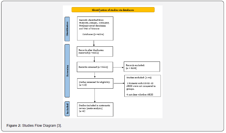
Two authors independently searched for relevant literature by executing the above search strategy and browsing abstracts or full texts to find potential articles. Detailed inclusion and exclusion criteria were used to screen the articles, and six primary research articles were finally selected as suitable for review.

Data Extraction and Risk of Bias Assessment

Two reviewers independently extracted and examined data from each included study. Extracted data included article title, author name(s), the date of publication, language, country, characteristics of participants, type of study, and data pertaining to the study’s outcome. Outcomes included mortality, length of ICU stay, and duration of ventilator use. The Newcastle-Ottawa Scale (NOS), developed by the University of Newcastle in Australia and the University of Ottawa in Canada, is a quality assessment tool for the systematic evaluation of non-randomized studies, especially for cohort and case-control studies [24]. The NOS cohort study version consists of eight multiple-choice questions involving topic selection and comparability, as well as outcome assessment or exposure. A star rating system is used to indicate the quality of the study, up to a maximum rating of nine stars. One star is awarded for each criterion if the reporting methodology is appropriate. Separate scales have been developed for cohort and case-control studies, which can help authors identify low-quality studies and inform sensitivity analyses or meta-regression [25]. NOS developers have examined NOS face and standard validity, reliability among evaluators, and evaluator burden. Surface validity has been assessed as strong by comparing each assessment item with its stem problem [26]. Therefore, NOS can be a helpful tool in assessing the quality of studies included in systematic reviews.

Data Synthesis

Stroup et al. published criteria for conducting and reporting meta-analyses of observational studies to improve the quality of reporting. Dichotomous data and risk ratio (RR) were chosen for data synthesis. The most common, a 95% confidence interval, is used to analysed mortality and favourable outcomes. Narrow confidence intervals are used to indicate that treatment estimates are relatively accurate [27]. In the second stage, the pooled (combined) intervention effect estimates are calculated as a weighted average of the estimated intervention effects in a single study. There are four methods for binary results meta-analysis, including three fixed-effect methods (Mantel-Haenszel, Peto, and inverse variance) and one random-effects method (Der Simonian and Laird inverse variance) [28]. For this systematic review, Rev Man software from the Cochrane Review was used for data analysis. The results were presented using forest maps.

Results

Study Selection.

Assessment Quality

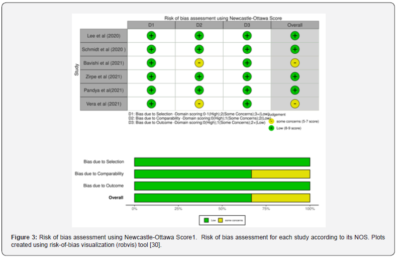
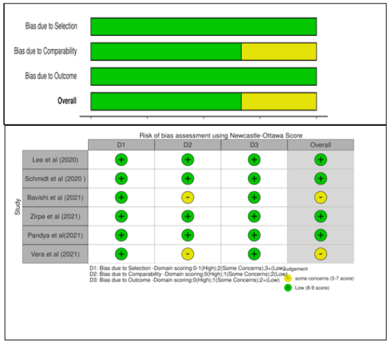
The Newcastle-Ottawa Scale was used to evaluate the quality of the six studies [32] (see Appendix 3). There are detailed evaluation records for each study, as well as summary tables for each of the six studies. The traffic-light plots and summary bar plots were created using the robvis tool (Figure 3), which is a web application for visualizing deviation risk assessment as part of a system assessment [33]. Selection criteria, comparability, and outcome (cohort) or exposure (case-control) were scored on a scale up to 9 (Figure 3).

Main Outcome

The forest plot showed that 2,731 participants across six studies were included in the meta-analysis with a combined RR = 0.78 (95% CI 0.69 to 0.88, Z=3.91, P < 0.0001) (Figure 4). Overall, the results showed that the mortality of the early intubation group was lower than the late intubation group, and the difference was statistically significant. Significant heterogeneity was observed (I2=63%), which indicated a large degree of variation between effect sizes in the included studies (Figure 4).

Length of ICU Stay

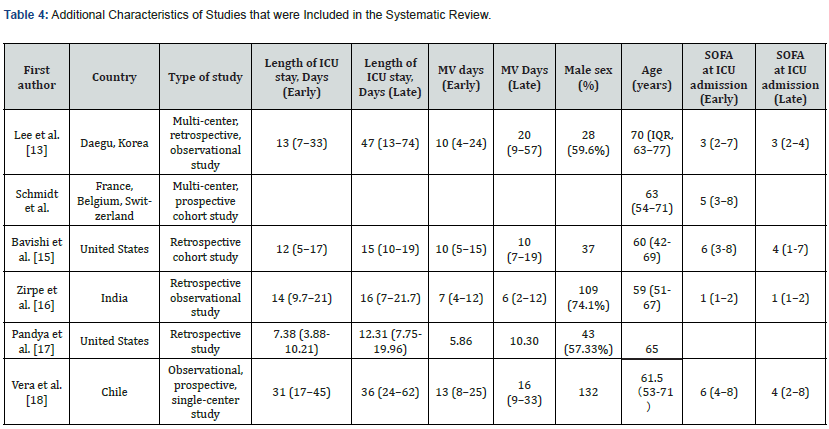
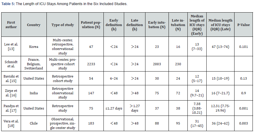
Schmidt et al. failed to extract the length of ICU stay of participants. No statistically significant differences were found between groups regarding the number of days spent in the ICU in three of the other studies (P > 0.05) [34,35]. Two studies indicated that there were significant differences between the groups (P< 0.05). Overall, since analysis found that early intubation results in shorter ICU stays, this was seen as statistically significant (Table 5).

Duration of Ventilation

Ventilation time was not extracted for the participants in one study Schmidt et al. The other five studies reported no significant difference in the duration of mechanical ventilation between the early intubation group and the late intubation group (P>0.05). Overall, the timing of intubation does not appear to impact the duration of mechanical ventilation of CARDS patients in the ICU (Table 5).

Complications

Patient complications were not reported separately in one study. Lee et al. observed that among patients treated with MV, the incidence of ventilator-associated pneumonia (VAP) in the early intubation group was often higher than that in the late intubation group, but no statistical significance was found (30.4%; N = 7 vs 6.2%; N = 1; P = 0.109). In research by the secondary infection rate was 13.3% in the early intubation group, while it was 22.2% in the late intubation group (P = 0.6). Additionally, AKI/RENAL failure was 21.3% in the early group and 18% in the late group (P = 0.1). A total of 16% of patients underwent tracheostomy in the early group, while the percentage rose to 25% in the late group (P = 0.1). Therefore, no statistically significant differences were found regarding secondary infection, Acute kidney injury (AKI), and interventional tracheotomy between intubation within 48 hours (early group) and intubation 48 hours after ICU admission (late group). Two studies did not describe patient complications.

Discussion

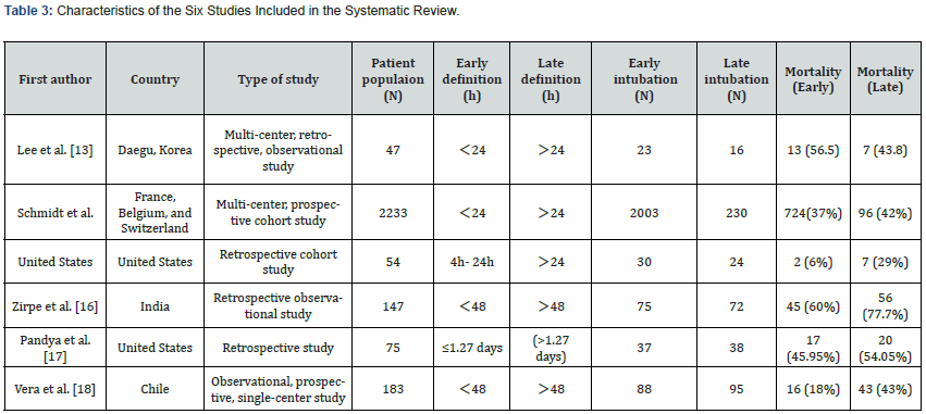
The purpose of this systematic review was to explore the effects of intubation time on clinical outcomes in COVID-19 patients with ARDS. Questions to investigate included whether late intubation increases ICU mortality, length of ICU stay, and duration of ventilator use. Results indicated that early intubation could reduce mortality and length of ICU stay in patients with CARDS. However, intubation time did not affect the duration of continuous mechanical ventilation in patients. There were obvious differences between the definitions of early intubation and late intubation in the six included studies. In three studies early intubation was defined as intubation within 24 hours after diagnosis of ARDS. Two studies defined early intubation as within 48 hours of diagnosis, while defined it as within 1.27 days. Most studies defined the early intubation time as within 24 hours after admission to an ICU [36,37]. However, according to the systematic review reported by the definition of early/late intubation time had no statistical difference in all-cause mortality between the two groups and did not influence the clinical outcomes of COVID-19 patients. Therefore, for the sake of homogeneity, early intubation group and late intubation group data was extracted for analysis according to the respective definitions included in the current study. However, the definition of early/late intubation time directly affected the number of participants between the two groups.

The Effects of Early and Late Intubation

The high mortality associated with late intubation may be related to lung injuries (P-SILI) unintentionally caused by the patients themselves. When COVID-19 patients’ respiratory support was insufficient, their lung function deteriorated in the first week [38]. ARDS is characterized by non-cardiogenic pulmonary oedema, decreased exchange volume of hypoxic blood, and normal gas related to V/Q imbalance, which leads to low respiratory compliance. Hypoxemia may cause patients to inhale spontaneously and violently, leading to lung injury caused by high trans-pulmonary pressure. Early intubation with pulmonary protective ventilation can prevent P-SILI [39]. In a study including 457 ARDS patients, the 60-day mortality rate of the late intubation subgroup (56%) was significantly higher than that of the early intubation group (36%) [40]. The mortality rate of the late intubation group continued to rise during the 2-year follow-up period, which was consistent with the results of the current study.

Chinese critical care experts also suggested that tracheal intubation should be done when critically ill patients are asymptomatic (persistent respiratory distress and/or hypoxemia) after standard oxygen therapy, which was also referred to in the COVID-19 guidelines for patient treatment [41]. Given the high risk of non-invasive respiratory support failure and the risk of virus particle atomization [42], Brown et al. also recommended that early tracheal intubation be performed for patients with respiratory failure who need ventilation support. Other factors influencing death included BMI, age, Sequential Organ Failure Assessment (SOFA) Score, and (Acute Physiology, Age, and Chronic Health Evaluation Ⅱ(APACHEⅡ)); however, no statistical difference was found between the two groups in the early/late stage. In a study by Pandya et al. The included population was characterized by a nasopharyngeal swab-confirmed COVID-19 patient with a mean age of 65 years. A median BMI of 31 was observed in the study, and all patients were more than 50% of the standard BMI and could be categorized as ‘obese’.

These were risk factors associated with mortality. In this study, the mortality rate of patients with mechanical ventilation was as high as 49%. The median age of non-survivors was higher than that of survivors (70 VS 59, p = 0.0006). The median ages of patients in the six included studies were 70, 63, 60, 59, 65, and 61.5, all of which were higher than 59. Therefore, elderly COVID ADRS patients were found to have a higher mortality rate. Compared with the United States, whose patients had a mortality rate of 16.6%, India’s mortality rate was much higher at 68.7%, which may be related to the level of economic development and medical care [43]. Overall, this systematic review found that patients with early intubation were prone to more severe illness, organ dysfunction, and higher SOFA and APACHE scores when diagnosed with ARDS compared to those with late intubation. These results may have great significance in clinical practice, by providing strong evidence for researchers and clinicians to consider when choosing when to intubate COVID-19 ARDS patients. This can assist in rationalizing the allocation of medical resources and reduce the mortality of patients.

Agreements and Disagreements with Other Studies and Reviews

No similar systematic reviews were found pertaining to the topic of this article. Navas-Blanco and Dudaryk (2020) agreed that early intubation can prevent adverse consequences due to delayed intubation in patients with CARDS. Two studies recommend early intubation for COVID-19 ARDS patients. However, a recent review by Papoutsi et al. found no statistically detectable difference in allcause mortality between patients undergoing either early or late intubation (3981 deaths; 45.4% versus 39.1%; RR 1.07,95% CI 0.99-1.15 p = 0.08). The same was true of MV duration (1892; MD- 0.58 days, 95% CI -3.06 to 1.89 days, p = 0.65). Intubation time may have had no effect on the mortality and morbidity of critically ill COVID-19 patients, which was inconsistent with the results summarized in this systematic review. In a study by Papoutsi et al. (2021), participants were critically ill patients with COVID-19. However, the population in this systematic review included ARDS patients with COVID-19.

Critique and Limitation

There are a few notable limitations to this study. For one, the reviewers only searched English-language articles, which can potentially lead to language bias. There may perhaps be articles related to this topic in other languages, but if so, these would have been excluded from the current review. In terms of secondary outcome data extraction, we contacted the authors of the six included studies by email, but failed to obtain specific data on length of ICU stays and MV duration. Therefore, the reliability of secondary measurement results may be reduced.

Conclusion

The findings of this systematic review conclude that early intubation for mechanical ventilation is beneficial to patients with COVID-19 ARDS. However, it appears that early intubation cannot reduce the overall duration of mechanical ventilation. The authors recommend immediate tracheal intubation for patients with moderate to severe COVID-19 ARDS. The treatment and management strategies of ARDS patients have been the continuing focus of researchers. In the face of COVID-19 pandemic, decreasing COVID-19 ARDS patients’ mortality remains an unsolved problem that needs further investigation. Further work is needed to improve research design and solve the problem of high heterogeneity and provide higher quality evidence.

To Know more about Academic Journal of Pediatrics & Neonatology
Click here: https://juniperpublishers.com/ajpn/index.php

To Know more about our Juniper Publishers
Click here: https://juniperpublishers.com/index.php

Friday, December 9, 2022

The Multisystem Disease COVID 19: proBNP at ICU-Admission Might Help in The Prognostication of a Patient’s Hospital-Survival - Juniper Publishers

 Anesthesia & Intensive Care Medicine - Juniper Publishers

Abstract

Background: COVID-19 presents a wide spectrum of clinical manifestations from asymptomatic infection to severe pneumonia accompanied by ARDS and multisystemic failure. N-terminal-pro-B-type natriuretic peptide (NT-proBNP) has been proven to be a good predictor of outcomes in patients with ARDS and might be an indicator of severity for SARS-CoV-2 infection, too.

Methods: We conducted a single-centre, retrospective cohort study of critically ill adult patients at the intensive care unit (ICU) with confirmed COVID-19 infection at the clinical centre of Hanau, Germany. Patients were admitted from March 19th to May 25th and were followed-up until June 25th, 2020.

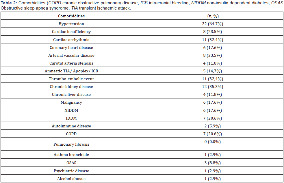
Results: Of 34 patients admitted for COVID-19 to the ICU, 18 (52.9%) survived and 16 (47.1%) died. The majority was male 27 (79.4%). Many patients had cardiovascular diseases 23 (67.6%), chronic kidney diseases 12 (35.3%) and thrombo-embolic events 11 (32.4%). Non-survivors were older than survivors (76.0±7.3 vs. 58.5±14.9 years; p=0.003). In non-survivors we found significant higher levels of d-dimers, creatinine, urea and lactate at ICU-admission compared to survivors. In our population, NT-pro-BNP-Level at ICU-admission could partially help in the prognostication of patients’ hospital survival

Conclusion NT-proBNP may help to categorize the severity of the multisystem COVID-19 disease at admission to the ICU.

Keywords: COVID-19, pro-BNP, ARDS, multi-organ failure, cardiac insufficiency

Abbreviations: COVID-19: Coronavirus disease 2019; pro-BNP: Pro–B-Type Natriuretic Peptide, Acute Respiratory Distress Syndrome: ARDS

Introduction

Coronavirus disease 2019 (COVID-19) has spread rapidly throughout China (Wuhan, Hubei) and almost all countries around the world. Its etiological agent is the coronavirus SARS-CoV-2 [1]. On March 11th, 2020 the World Health Organization (WHO) declared a pandemic. The number of fatalities owing to COVID-19 is escalating and estimated more than 3.1 million patients died world-wide [2].

Patients with COVID-19 present with clinical symptoms of variable severity which might range from detection of virus RNA without symptoms to multiorgan failure and acute respiratory distress syndrome (ARDS) in up to 15% of patients [3,4]. It is difficult to estimate the clinical course in advance and some patients may detoriate rapidly [3].

The immune response to SARS-CoV-2 is known to involve all the components of the immune system that together appear responsible for viral elimination and recovery from the infection [5]. In the late stage of the disease, severe cases suffer from ARDS, metabolic disorders, multiple organ dysfunction (MODS) and coagulation disorders.

Up to now the exact pathophysiological mechanisms responsible for the different clinical courses of COVID-19 patients are not clear. In some patients a severe inflammatory response might lead to a decrease and functional impairment of CD4+ T cells and CD8+ T cells, extraordinarily increased neutrophils, disseminated intravascular coagulation and finally even death [6].

In patients with ARDS of other origin Lai et al. concluded 2017 that N-terminal proB-type natriuretic peptide (NT-proBNP) is a good predictor of outcomes. B-type natriuretic peptide (BNP) was first described in the porcine brain, but BNP in humans originate primarily from the heart’s ventricular myocardium. The secretion of BNP is mediated by the ventricles of the heart in response to excessive stretching of heart muscle cells. Several studies have reported that BNP or NT-proBNP was elevated in patients with ARDS. But only Determann et al. and Park et al. focused on NTproBNP. Accordingly, this peptide may theoretically be used as an indicator of clinical severity for SARS-CoV-2 infection. In a metaanalysis including 13 observational studies and a total of 2248 patients, Sorrentino et al. [4] demonstrated that an elevated NTproBNP level on admission is associated with a worse prognosis in COVID-19 patients [4].

Therefore, we investigated during the first wave of the pandemic in Germany whether the level of NT-proBNP is a possible predictor of mortality in patients with COVID-19.

Materials and Methods

Trial design

We conducted a single-center, retrospective cohort study of consecutive adult patients hospitalized and admitted to ICU with confirmed COVID-19 infection by positive reverse transcription polymerase chain reaction at the clinical center of Hanau, Germany. The hospital is a designated hospital to treat patients with COVID-19 and teaching hospital of the University of Frankfurt, Germany. Patients were admitted from March 19th, 2020 to May 25th, 2020, and they were followed-up until June 25th, 2020. One patient was included despite a negative SARS-CoV-2 PCR due to the typical clinical course and radiological findings in the thorax CT typical for a COVID-19 pneumonia. Clinical information was collected on admission and during ICU stay by attending physicians. This project was performed in accordance with the Declaration of Helsinki and after approval of the local Ethics Committee of the Landesärztekammer Hessen, Frankfurt, Germany (2020-1795-evBO; 28.08.2020).

Data collection

The medical records of the patients were reviewed by a trained team of physicians working in the Hospital of Hanau, Germany, during the epidemic period. Patient data including demographics, medical history, laboratory examinations, comorbidities, complications, procedures, and outcomes (death, need for intensive care unit {ICU}, intubation, mechanical ventilation, renal replacement therapy, ICU- and hospital length of stay {LOS}, and hospital discharge) were collected and analyzed.

Statistical analysis

Continuous variables are presented as mean±SD and median (25%, 75% quartil). Categorical variables are expressed as absolute number of patients and percentage. The mean values for continuous variables were compared using independent group t tests when the data were normally distributed, otherwise, the Mann-Whitney test was used. For pro-BNP we calculated the area under the receiver operating characteristic {ROC} in respect of survival and explored the optimal cutoff value. By means of Kaplan- Meier curves, the survival of patients with pro-BNP below / above the cutoff is illustrated. A p-value less than 0.05 was considered statistically significant. Because of the explorative nature of the study, we did not perform an α-correction for multiple testing, therefore the p-values must be interpreted carefully. All statistical analyses were performed with IBM® SPSS®, version 27 for Windows.

Results

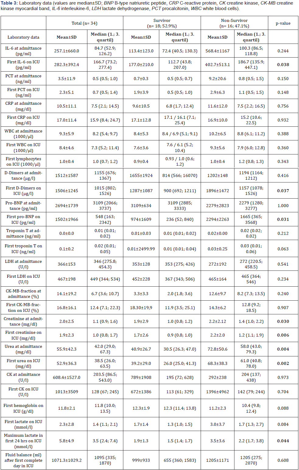
A total of 34 patients were admitted to ICU for COVID-19 during this study period. We had 18 (52.9%) survivors and 16 (47.1%) non-survivors. 27 of them were male patients (79,4%). The age of the total cohort was 67.8±13.9 years. Basic clinical characteristics and respiratory parameters before intubation and extreme values during the first 24 hours of ventilation on the ICU are shown in Table 1. The comorbidities of our patients are demonstrated in Table 2. Most of the patients had cardiovascular diseases 23 (67.6%), chronic kidney diseases 12 (35.3%) and thromboembolic events 11 (32,4%). In the absence of contraindications our patients were anticoagulated slightly elevated to reach a level between prophylactic and therapeutic anticoagulation. Laboratory data are shown at hospital admittance and the first value measured on intensive care unit (ICU) (Table 3).

All 34 patients were admitted to the ICU due to progressive respiratory failure. We treated 28 (82.4%) patients with highflow oxygen therapy, 24 (70.6%) patients were intubated and invasively ventilated, 22 (64.7%) were proned and only 3 patients (8.8%) were non-invasively ventilated. Many patients (28, 82.4%) showed laboratory or clinical signs of kidney injury, 15 (44.1%) patients developed a new AKI, 9 (26.5%) an acute on chronic kidney injury and only 4 (11.8%) patients chronic kidney injury. 19 patients (55.9%) needed renal replacement therapy at any time during their ICU stay whereof only 4 (11.8%) had pre-existing end-stage renal disease.

From our 34 ICU patients, almost half (16, 47.1%) died during their hospital stay. Non-survivors were older than the survivors (75.5±7.3 vs. 60.9±14.9; p=0.003); the youngest non-survivor was 64 years old; the oldest survivor was 88 years old. The laboratory data demonstrated for the non-survivor group at admission on ICU increased levels of IL-6, abnormal levels for d-dimers, pro-BNP, creatinine, urea and lactate compared to survivors. The survivors had a higher paO2 level and Horovitz index before intubation. The total duration of high-flow nasal cannula oxygenation therapy (NFHC) was longer for the survivors.

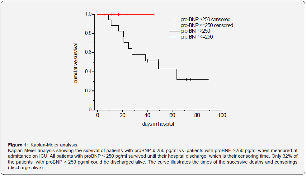
Because of the described elevation of pro-BNP in ARDS and its potential use as a prognostic marker in ARDS we looked in more detail at the pro-BNP level at admittance on ICU and survival. We calculated the receiver operating characteristic (ROC) curve for pro-BNP in respect of survival, which resulted in an AUC of 0.760 (p = 0.007). This is a hint, that pro-BNP is a significant predictor of the survival. We received a pro-BNP value of >244 pg/ml as the optimal cut-off value for our sample. With this cutoff value the sensitivity is 1 and the specifity 0.533. As the adjacent cutoff-value is 252, we determined 250 as optimal cutoff.

The Kaplan-Meier analysis (Figure 1) shows the survival of patients with proBNP ≤ 250 pg/ml vs. patients with proBNP > 250 pg/ml. All patients with proBNP ≤ 250 pg/ml survived until their hospital discharge, which is their censoring time. Only 32% of the patients with proBNP > 250 pg/ml could be discharged alive. The curve illustrates the times of the successive deaths and censorings (discharge alive).

Discussion

In this retrospective analysis of 34 ICU patients with respiratory insufficiency due to a COVID-19 infection during the first part of the pandemic in a municipal hospital in Germany we found clinical courses similar to those in other studies [7,8]. Severe respiratory failure in patients with SARS-CoV-2 infection is only one sign of the multisystem inflammatory syndrome [9-11], which is consistent with the high rate of patients with renal failure and the need for renal replacement therapy in more than half of the patients [12,13]. The high rate of multiorgan failure together with the increased age of our patients may at least partially explain the high mortality of our patients which is comparable to data from other studies with COVID-19 ICU patients [14].

More than two third of our patients were invasively ventilated and only a minority was non-invasively ventilated because at that time - during the “first wave” in Germany - we worried about an increased risk of transmission for the ICU staff with the use of noninvasive ventilation. Accordingly, we suggested a reduced risk of transmission of SARS-CoV-2 when patients were intubated timely. In the meantime, it has been demonstrated that is reasonable, safe and recommended in the guideline to try nasal-high flow oxygen and non-invasive ventilation in patients with respiratory insufficiency due to COVID-19 when patients are closely monitored [2,15-18].

Very soon after the first patients with COVID-19 had been treated it was recognized that there is an increased risk for thrombo-embolic events in these patients due to an inflammatory alteration of the endothelium and an inflammatory pro-coagulatory state [19,20]. It was difficult and potentially misleading to count the number of thrombo-embolic events in our patients because we did not screen systematically for these events as other groups did [20].

The major finding in this - small group - of patients with COVID-19 is that an elevated pro-BNP level on admission to the ICU shows the tendency of a worse prognosis.

Many clinical data described a cardiovascular manifestation induced by this viral infection especially in critical ill patients. Acute myocardial injury manifested mainly by elevated levels of high-sensitive troponin I, and arrhythmias have been detected [9,21]. Guo et al. [3] reported in their study among 187 patients with COVID-19, 52 (27.8%) exhibited myocardial injury as demonstrated by elevation of troponin T (TnT) levels, and the mortality was markedly higher in patients with elevated TnT levels than in patients with normal TnT levels (59.6% vs 8.9%). The authors suggest that myocardial injury due to the inflammation might play a major role in the clinical detoriation of COVID-19 patients and that those patients with elevated troponin T levels (27.8% of patients) had more malignant arrhythmias and a higher mortality [3]. Consistent with our results of increased levels of pro-BNP in non-survivors they found a correlation between elevated pro-BNP and troponin T levels [3].

In our group of non-survivors TnT levels were a bit higher at admission to the ICU (0.03 ± 0.25) and proBNP was significantly higher in the non-survivor group (2294±2263; p =0.031) compared to the survivors. The comorbidities of our patients represented mostly cardiovascular diseases, hypertension on top of all (64.7%). Alvarez-Garcia et al. [5] pointed out that patients with a history of heart failure (HF) hospitalized for COVID-19 face nearly 3 times the risk of mechanical ventilation and twice the risk of mortality compared with patients without HF [5]. Similar to our results Sorrentino et al. demonstrated a correlation between increased pro-BNP levels and severity of COVID-19 disease but in contrast in their study the pro-BNP levels in non-survivors were already increased at admittance to hospital compared to survivors [4]. In our investigation patients with COVID-19 had also elevated levels of proBNP at admission to the hospital. Thus, we learned from several investigations during the pandemic that predictors of a fatal outcome in COVID- 19 cases included age, the presence of underlying diseases, the presence of secondary infection and elevated inflammatory indicators in the blood [5,14,22,23]. Although the high accuracy of NT-proBNP is already established in the diagnosis of acute heart failure, the prognostic value of this marker for patients with COVID-19 remains uncertain [4].

Our study has several limitations. Only 34 patients with COVID-19 were included during the first wave in Germany, and a larger randomized cohort study is needed to verify our conclusions. Unfortunately, we could not provide more information regarding cardiovascular complications as e.g., cardiovascular ultrasound or detailed hemodynamic monitoring. Due to the restricted options and the increased efforts in the isolation ward data was not complete in all patients.

Last but not least, the data of this retrospective study permit a preliminary assessment of the clinical course and outcomes of patients with COVID-19.

The causes of death may involve multiple organ dysfunction in most cases, and it is difficult to differentiate the myocardial injury as the main and direct cause in an individual case. Long-term observation and prospective study design on the effectiveness of treatments are needed. We still have to wait for long-term results after surviving Covid-19 disease [24].

Conclusion

Many critically ill patients with COVID-19 pneumonia suffer from multi-organ dysfunction including cardiac insufficiency. We concluded the measurement of specific laboratory data as NT-proBNP may help to categorize the severity of the COVID-19 disease at admission to the ICU.

Click here: https://juniperpublishers.com/index.php 
Friday, April 29, 2022

Role of Gatekeepers in Suicide Prevention During COVID-19 Pandemic - Juniper Publishers

 Addiction & Rehabilitation Medicine - Juniper Publishers

Abstract

Keywords: COVID-19; Suicide; Mental health; Psychopathology; Gatekeepers

Abbreviations: WHO: World Health Organization; CNS: Central Nervous System; ACE-2 : Angiotensin-Converting Enzyme 2; CRH: Corticotropin Releasing Hormone; mTOR: Mammalian Target of Rapamycin

Introduction

Suicide is defined as death caused by injuring or harming oneself with intent to die and a suicide attempt is an injury or harm caused to self with intent to die but has not resulted in death [1]. Suicide and suicidal behaviors are influenced by multiple psycho-social and biological factors. Factors such as resilience, good social support are protective against suicide while lack of occupation, financial issues, ill health, poor social support are risk factors of suicide. Prevention of suicide is the need of the hour. Amidst the COVID-19 pandemic is a possible suicide and suicidal behavior increase that is being neglected at community level.

Burden of Suicide

World Health Organization (WHO) statistics show that globally more than 800,000 people die by suicide every year, that is, one death by suicide occurs every 40 seconds. For every completed suicide there are 10-20 suicidal attempts that are reported [2]. About 77% of all suicides occur in low and middle income countries [3]. In India, as per National Crime Records Bureau of India 1.53 lakh people have died by suicide in the year 2020 (vs 1.39 lakh in 2019). The most common form of suicide being death by hanging followed by death by consumption of poisons especially insecticides. Total rate of suicides nationwide is 11.3 [4,5]. Though global statistics collected by WHO shows that the rate of suicides have reduced between 2010 and 2019 in low-middle income countries, statistics from India does not reflect this [6].

Influence of COVID -19 pandemic on suicide and suicidal behavior

Suicide and self-harm behavior are influenced by multiple psychological, social and illness aspects and reflects the severity of the mental health crisis and illness in a given community. COVID-19 infection has impacted the psychological, social and physical health globally and thus self-harm and suicide behavior as well. A study in Nepal has shown that suicide rates were higher during lockdown than immediate post lockdown period and same time period of pre-COVID years. Delay in arrival to hospital, admissions and in hospital deaths due to self-harm were also higher during lockdown [7]. Indian study compiled from news reports and social media showed that more than half of them were tested positive for COVID-19, about one-third was in institute setting and that the most common mode of suicide was by hanging followed by fall from height.

Nearly two-thirds of the patients had contacted a physician within 2 weeks prior to their self-harm. More than 80% of the people who completed suicide did not have pre-existing psychiatric or physical co-morbidities [8]. This may indicate that COVID-19 pandemic and its effect on psychological health and social factors may be an independent risk factors for suicide. Comparing pre-COVID data with the current data shows that depressive illness has increased two to three fold than before. Anxiety and stress levels in the general population have also increased significantly [9-11]. As 90% of people who have completed suicide have a pre-existing psychiatric diagnosis [12], increase in psychiatric illness can lead to increase in suicidal behavior.

Psychosocial factors influencing suicidal behavior during COVID-19 pandemic

Studies have shown that stressors related to COVID-19 infections such as fear of infection, increased vigilance about changes in body and wrongly attributing it to COVID-19 infection, deterioration of physical health due to COVID-19 infection, reduced social support and caregiver ability have impacted the mental health of the people [13]. People who follow COVID-19 related news closely are more prone to develop anxiety as most of the news is distressing and emphasizes the infectivity of the virus and its associated morbidity and mortality. Rumors, fabrications and misinformation on social media also contribute to the rising anxiety levels and exacerbate depressive symptoms [14-16]. Isolation due to lockdowns, work from home and infection can cause loneliness, helplessness and stress. Physical isolation with social and emotional closeness by using social networks and virtual platforms can be a protective factor [17].

Pathogenesis of psychopathology associated with COVID-19 infection

COVID-19 or SARS CoV 2 2019 is well known for its entry through mucosal surfaces and its effect on respiratory system, morbidity and mortality associated with the same. However, lesser-known fact is that SARS Cov-2 infection and associated immune response affects multiple organs and organ systems of the body; one such system being the Central Nervous System (CNS). SARS CoV-2 virus enters the epithelial cells via angiotensin-converting enzyme 2 (ACE-2) receptors. This in turn leads to down-regulation of ACE-2 receptors expression. In animal studies down-regulation of ACE-2 expression has resulted in increased sympathetic activity, reduced tryptophan uptake and production of serotonin; thus, compromising the body’s ability to respond to stress and hence increasing the individual’s susceptibility to depression and anxiety.

ACE-2 receptors in hypothalamus suppress fear responses, anxiety and its related behavior as well as Corticotropin releasing hormone (CRH) which plays an important role in response to physiological stress. ACE-2 receptor down-regulation hampers negative feedback mechanism of glucocorticoids in reducing excessive inflammation. Therefore, in SARS CoV 2 infection an excessive and dynamic inflammation is observed. SARS CoV 2 virus has the ability of infecting all tissue having the ACE-2 receptor including the brain tissue. Hence, direct infection and increased immune response both play a role in pathogenesis of psychiatric illnesses associated with COVID-19 illness. This can be prevented by preformed antibodies occupying the ACE-2 receptors in the brain thus preventing entry of the virus into the neuronal cells. Hence, ACE-2 receptor modulator drugs may have a role in treatment of COVID-19 infection and prevention of psychiatric complications [13].

Though rennin angiotensin aldosterone system plays a role in pathogenesis of stress related depression and anxiety, it is minimal. Bradykinin and mammalian target of rapamycin (mTOR) play a major role in depression and effect of COVID-19 infection on them is not known [13]. Inflammatory mechanisms activated by SARS CoV 2 virus increase inflammatory cytokines such as TNF-A, Interleukin 6 and Interferons in both peripheral and central tissues leading to cell apoptosis. This leads to increased risk of developing mood disorders, anxiety and psychosis. Hence, augmenting anti-depressant or anti-psychotic drugs with immunomodulatory drugs or anti-inflammatory drugs are more beneficial than anti-depressant or anti-psychotic drug treatment alone [13]. Effect of COVID-19 pandemic on pre-existing psychiatric illness and access to mental health care services.

The COVID-19 pandemic, precautionary measures implemented to limit its spread and burden on the health care services due to infection had a major impact on non-pandemic related illnesses and treatment for the same. Restrictions in movement, transport, burden on health care infrastructure made accessing health care services in timely manner difficult globally [18,19]. This has also had an effect on the help seeking attitude and availability of services for people with pre-existing mental illnesses. Re-allocation of all available services in health care services including mental health care professionals to tackle the burden of COVID pandemic may have resulted in lack of timely professional help for people with mental illnesses [20]. As psychiatric facilities were found to be a high risk area of spread of COVID-19 infection, community based or home based treatment through telepsychiatry, psychological support and home delivery of the medications would have been recommended and ideal.

However, due to sudden and unexpected impact of the pandemic mental health services were not equipped to handle this crisis [20]. Study comparing people with pre-existing psychiatric illnesses and those without showed that people with psychiatric illnesses had significantly higher depression, anxiety, stress, PTSD like symptoms, anger, impulsivity and suicidal ideation [21]. Lack of access to mental health care services and medications could have resulted in untreated mental illnesses and above findings. This results in impaired ability of the individuals to cope with stress and in turn cause increase in suicidal ideation and attempts.

Bridging the gap…Gatekeeper?

In India, there exists a huge gap between the requirement of mental health services and the personnel who are trained to provide adequate services. The National Mental Health Survey of 2015-2016 shows that lifetime risk of psychiatric illness is 13.67% and point prevalence was 11.56%. But the available mental health professionals were 0.3 per 100,000 population for psychiatrists, 0.07 per 100,000 population for clinical psychologists and 0.12 for mental health nursing staff [22]. To meet the minimum mental health needs of the population of India there should be 3 psychiatrists per 100,000 populations and for optimum care or in an ideal situation 6 psychiatrists per 100,000 populations. Keeping attrition rate of psychiatrists to 0%, approximately 2700 new psychiatrists need to be trained annually the next 10 years to meet the demands of our country [23].

However, as of 2021-2022 academic year approximately 1200 Post-graduate students are being trained as psychiatrists throughout the country, which is less than half the required numbers [24]. COVID pandemic has also increased the risk factors for suicide thus increasing the need for mental health services in prevention of suicide. Gatekeepers can help in bridging this gap. Gatekeeper refers to people who have primary contact with individuals at risk for suicide, and who can identify such individuals by recognizing suicidal risk factors and refer to a professional. Gatekeeper training refers to training individuals to identify risk factors for suicide among people and refer for appropriate help [25]. Gatekeeper could be teachers, family members, supervisors, religious leaders, faith healers, physicians, police, colleagues and friends. Most of them are however not aware of the signs that people who have risk of suicide exhibit and hence are unable to identify and provide timely health.

Most of the people in the community, especially the youth are hesitant approaching mental health professionals for help for suicide due to stigma attached with suicide and lack of awareness of avenues of help available and fear of being judged. Often times when they approach lay people for help with suicidal ideation, their concerns are trivialized or ignored (probably due to ignorance and their own discomfort regarding suicide) making them hesitant to reach out for help again. Gatekeeper training is a strategy designed to improve early identification of individuals at high risk for suicide and to facilitate timely mental health referrals, responding to the fact that suicidal youth are under-identified and few are using these services.

Identifying people who have contact with population at large and adequate training to recognize and provide first aid for people with risk of suicide before professional help is available becomes important. Educational institutes (like schools, colleges and universities) and workplaces should have a crisis management team who have had gatekeeper training from professionals. Crisis team should include people from the administration and employees who are willing to undergo gatekeeper training and help in suicide prevention.

Click here: https://juniperpublishers.com/index.php  

Artificial Intelligence System for Value Added Tax Collection via Self Organizing Map (SOM)- Juniper Publishers

  Forensic Sciences & Criminal Investigation - Juniper Publishers Abstract Findings:  Based on our experiments, our approach is an effec...