Thursday, July 16, 2020

Risk Factors, Epidemiology and Common Presentations and Complications of Slipped Capital Femoral Epiphysis - Juniper Publishers

Arthritis & Bone Research - Juniper Publishers

Mini Review

Slipped capital femoral epiphysis (SCFE) occurs when the metaphysis of the neck of femur is displaced antero-superiorly while the epiphysis remains in the acetabulum, most common presentations are hip, groin or knee pain, a limp and fixed external rotation [1]. SCFE is considered one of the most frequent hip disorders with an incidence of 0.33 up to 24.58 per 100,000 children from ages 8 to 15, with an average of 12.0 years old for boys and 11.2 years old for females, it varies with different racial and ethnic frequency with 1.0 for Caucasians, blacks at 3.9, and 2.5 for Hispanics , the average onset of symptoms is 4 to 5 months [2], Latency in the diagnosis of SCFE is found to be related to poor prognosis, it also increases degree of slippage as the disease is not detected in the pre-slip phase, patients with referred pain and patients with stable slips are more prone to be misdiagnosed, hence the delay of the diagnosis [3]. As the international epidemic of childhood obesity continues to grow, an increasing number of children are developing SCFE [4]. According to a study conducted over the last 20 years in Scotland, a close association was observed between rising childhood obesity and an increase in the incidence of SCFE [5]. Moreover, an epidemiological study of SCFE in Sweden showed a mild increase for girls over the years 2007-2013, However, it also showed that overweight or obesity was one major characteristic for boys with SCFE but to a lesser extent for girls [6].

Although the etiology of SCFE is not yet clear, it’s known to be correlated with endocrine disorders [7]. It was found that the prevalence of SCFE is increased in children who have hypothyroidism, who are receiving growth hormone supplementation, or who have hypogonadism [8-9]. Additionally, it is found that patients with SCFE associated with hypothyroidism are commonly obese or overweighed, but a persistent hypothyroidism may be a risk factor itself for SCFE, even without obesity [10]. The management of SCFE is still controversial and the management differs in case of stable or unstable slips, a survey was sent to 287 members of the European Pediatric Orthopedic Society (EPOS) ,where only 72 participated , 90 percent of the respondents agreed upon not performing a reduction in case of a stable slip, however there was a controversy in the way of managing unstable slips ,(46% by positioning, while 35% would manage by manipulation ,and only 11% went with open reduction [11]. Moreover, to understand how obesity increases the risk of SCFE, we have to fully comprehend the mechanism in which the injury occurs. It is well known that this type of deformity is caused by an increase in the force applied through the epiphysis, or a decrease in the resistance of the physis itself to shearing. This type of deforming mechanics occurs mainly in the hypertrophic zone of the growth plate. For that reason, other risk factors such as coxa profunda, which is a deeply seated acetabular socket, and femoral or acetabular retroversion can lean to an increase incidence of SCFE [12]. The most common complain associated with SCFE is hip pain. This pain is usually aggravated by physical activities such as running, jumping and pivoting motion of the hip joint. Chronologically, slipped capital femoral epiphysis can be subdivided into three main types: acute, acute on top of chronic, and chronic. The most common presentation of SCFE in the acute phase is severe hip pain that can radiate to the groin area or around the ipsilateral knee. In addition, limited range of motion mainly in hip abduction and internal rotation. While in the acute on top of chronic, patients tend to have an altered gait and moderate pain.

In the chronic SCFE, patients are able to walk with slight pain on top of mild to moderate shortening of the affected leg and atrophy of the thigh muscles. Other classifications were introduced to further highlight certain types of SCFE, the most well-known was proposed by Loder et al. [13] and it is based on the physeal stability [13]. This stability is judged based on the ability to walk on the affected leg with or without crutches. Loder et al emphasized that this distinction is very important as the prognosis and treatment will vary based on it. Other literature has further elaborated on the stability in relation to radiographic signs including clear separation between the head and metaphysis, absence of the metaphyseal remodeling and incidental reduction of the slip angle by more than 10 degrees during surgery [14]. Complications of SCFE can vary from persistent hip pain, limp and impingement to osteonecrosis of the femoral head. By far, osteonecrosis is considered the most devastating complication to such patients. In a study conducted by Larson et al, Osteonecrosis was found to be the most common reason for hip arthroplasty in patient with SCFE [15]. Although, long-term studies have shown that excellent functional outcome can be expected until fifth decade if the hip can be stabilized without the occurrence of osteonecrosis [16].


Tuesday, July 14, 2020

Polycythemia Vera: Current Therapeutic Approaches - Juniper Publishers

Tumor Medicine & Prevention - Juniper Publishers

Abstract

The hemoglobin level threshold required to diagnose PV is now established at 16.5 g/dL for men and 16 g/dL for women by the 2016 WHO classification for MPNs. The clinical course of PV might be interrupted by thrombohemorrhagic complications and disease transformation to MF or acute myeloid leukemia. JAK2 oncogenes homozygosity may result in distinct tumorigenic consequences. Different therapeutic strategies and good candidates’ patients for such therapies are illustrated.

Keywords: Polycythemia vera; Thrombohemorrhagic; Tumorigenic; Myeloid leukemia

Abbrevations: PV: Polycythemia Vera; STAT: Signal Transducer and Activator of Transcription; AML: Acute myeloid leukemia; MF: Myelofibrosis; MPNs: Myeloproliferative neoplasms

Introduction

PV is one of the lower-risk subtypes of MPNs [1] that is characterized primarily by clonal erythrocytosis [2]. It has an annual incidence of 0.21-2.27 per 100,000. Median age at diagnosis is estimated at 71 years. A male preponderance is noted. JAK2 V617F is found in >95% of PV patients and JAK2 exon 12 mutations in ~4% [3]. JAK2 exon 12 mutations such as insertions or deletions are relatively specific to JAK2V617F-negative PV and not found in ET and PMF [4]. JAK2 homozygosity is neither necessary nor sufficient for a PV phenotype as indicated by presence of small or undetectable homozygous clones in some PV patients [5]. Heterozygous mutant erythroblasts of PV patients have distinct transcriptional profiles that precede acquisition of homozygosity and reflect differential activation of phosphorylated STAT1, which modulate erythropoiesis [5]. CALR and MPL have distribution frequency of 0 and 0% [6]. Other mutations (e.g., LNK) have been reported [2]. Co-occurrence of CALR mutation exon 9 with JAK2V617F, usually occurs in less than 1% of PV [4].

JAK2, MPL, and calreticulin mutations are driver mutations that activate the JAK2 pathway, but additional recurrent somatic mutations in several genes (TET2, ASXL1, DNMT3A, CBL, LNK, IDH1/2, IKF1, EZH2, TP53, and SRSF2), encoding transcriptional and epigenetic regulators and signaling proteins, modulate disease progression and can also occur as a primary mutation [3]. Clinical features of PV include mild-to moderate degree of splenomegaly, mild-to-moderate degree of constitutional symptoms, including fatigue and pruritus [6] (usually after bathing) [2], symptoms of hyperviscosity, leukocytosis, thrombocytosis, microvascular symptoms (e.g., headaches, lightheadedness, visual disturbances [6] e.g., blurry vision [2], atypical chest pain, acral paresthesia and erythromelalgia [6] i.e., erythema, warmth, and pain in distal extremities) [2], and thrombotic and bleeding complications [6].

The risk of thrombosis exceeds 20% [7]. History of hypertension predicted arterial thrombosis and advanced age venous thrombosis [7]. Hepatic and portal vein thrombosis is a well-recognized phenotypic association with JAK2 V617F and is often observed in younger women, with either a masked phenotype, or lower leukocyte counts and lower allelic burdens. The affected patients present a unique exception with regard to demographics, clinical phenotype, and allelic burden [8]. Some patients may develop AvWS, especially in the presence of extreme thrombocytosis (platelets >1000 x 109/L) and are at risk for aspirin associated bleeding [7]. JAK2 oncogenes homozygosity may result in distinct tumorigenic consequences, but direct evidence has been elusive (5). JAK2V617F homozygosity was associated with reduced platelet survival that is likely to reflect increased platelet apoptosis and/or clearance. Moreover, homozygosity results in reduced platelets numbers, consistent with the lower platelet levels seen in PV compared with ET [5]. Increased JAK2V617F signaling may enhance (or reduce) platelet reactivity, a concept that may be relevant to thrombotic (or hemorrhagic) complications in PV patients [5]. JAK2 V617F homozygous allele burden has been associated with older age, male sex, pruritus, and splenomegaly; associations between homozygous or increasing allelic burdens and thrombosis (arterial and venous), as well as MF transformation have also been suggested [8]. JAK2 exon 12e mutated PV, is more likely to be associated with younger age and [2] may have higher hemoglobin, and lower platelet and leukocyte counts compared with those with JAK2 V617F mutated PV, clinical outcomes do not differ, with similar incidences of thrombosis, MF, AML, and death [8] PV may present with thrombocytosis and because hydroxyurea suppresses erythropoiesis, misdiagnosis of PV patients with prominent thrombocytosis as having ET may have occurred. Iron deficiency may mask the polycythemia [9].

Estimated median survivals for PV are 14 years; the corresponding median survivals in younger patients were 33 and 24 years [2]. In WHO-defined PV, the 10- year projected rates for survival, leukemic transformation and fibrotic progression were >75%, <5%, and <10%, respectively [7]. Patients who developed MF (post-PV MF) had a higher rate of leukemic transformation [9]. The goal of therapy in PV is primarily to prevent thrombohemorrhagic complications, without increasing bleeding risk, and secondarily to control the microcirculatory symptoms. Treatment is tailored to individual patients according to their risk for thrombosis or bleeding [7]. The presence of hypertension and leukocytosis is taken into consideration, when deciding treatment in certain circumstances [6].

Low-risk patients (i.e., those <60 years and without a history of thrombosis) are commonly treated with phlebotomy [10] (to a hematocrit target of 45%) [7] and antiplatelet therapy [10]. Hydroxyurea or interferon may be indicated in low-risk patients in quite rare instances such as those who need, and poorly tolerate, frequent phlebotomies, unmanageable disease-related symptoms, extensive thrombocytosis or progressively increasing leukocyte count, or symptomatic splenomegaly [11].

High-risk patients (i.e., presence of any thrombosis history or advanced age with JAK2 mutation) receive cytoreductive treatment in addition to low-dose aspirin (depending on the type and date of the previous thrombotic event, oral anticoagulation may be indicated instead of low-dose aspirin) [10]. Twice-daily aspirin is used in arterial thrombosis in older patients or harbor JAK2 mutations or in the presence of CV risk factors. In patients with venous thrombosis, systemic anticoagulation is advised and the addition of once-daily low dose aspirin, in the presence of JAK2 mutation or CV risk factors, is reasonable [7]. High-risk patients should receive hydroxyurea (starting dose 500 mg BID) as first-line drug to minimize their risk of thrombosis. The dose of hydroxyurea is titrated to keep the platelet count in the normal range [7]. Complete response was defined as: hematocrit <45% without phlebotomy, platelet count <400 x 109/L, white blood cell count <10 x 109/L, and no disease-related symptoms. Partial response was defined as: hematocrit <45% without phlebotomy, or response in 3 or more of the other criteria [11].
Hydroxyurea resistance and intolerance in PV patients is defined by ELN consensus as
1. Need for phlebotomy to keep hematocrit <45% or
2. Uncontrolled myeloproliferation, i.e., platelet count >400 x 109/L AND white blood cell count >10 x 109/L, or
3. Failure to reduce massive splenomegaly (i.e., extending >10 cm from the left costal margin) by >50% as measured by palpation, OR failure to completely relieve symptoms related to splenomegaly, or
4. All these first 3 criteria after 3 mo of at least 2 g per day of hydroxycarbamide, or
5. Absolute neutrophil count <1.0 x 10⁹/L OR platelet count <100 x 109/L or hemoglobin <100 g/L at the lowest dose of hydroxycarbamide required to achieve a complete or partial clinicohematological response, or
6. Presence of leg ulcers or other unacceptable hydroxycarbamide-related non-hematological toxicities, such as mucocutaneous manifestations, gastrointestinal symptoms, pneumonitis, or fever at any dose of hydroxycarbamide [11]. The three drugs, pegylated IFN-α, busulfan and ruxolutinib are currently considered as second-line therapy for PV [6]. PV patients who are either intolerant or resistant to hydroxyurea are effectively managed by INF- α (pegylated preparations preferred) or busulfan [7]. The use of INF-α is preferred for patients younger than age 65 years and busulfan in the older age group. There is no controlled evidence to support or refute such a strategy. Busulfan is started at 4 mg/day, withheld in the presence of platelets <200 x 109/L or WBC <3 x 10⁹/L, and the dose is reduced to 2 mg/day when treatment is resumed after withholding. Pegylated IFN-α is usually started at 45 mcg once-a- week and titrate up to 180 mcg once-a-week if tolerated [7]. Long-term safety data are considered acceptable for pegylated IFN-α and busulfan. Both drugs display broader activity against clonal myeloproliferation and display better quality of response, including the ability to induce molecular remission compared to ruxolutinib [6]. There are no controlled studies that implicate either hydroxyurea or busulfan as being leukemogenic in PV [7]. The risk of acute leukemia was not significantly increased until after 8 years of hydroxyurea exposure [9]. Ruxolutinib is recommended only in PV if there is severe protracted pruritis [12], severe constitutional symptoms [7] or marked splenomegaly that is not responding to interferon alpha or busulfan [12]. Advise patients about possible herpes zoster reactivation and skin tumors [11].

Conclusion

High-risk patients should receive hydroxyurea. The risk of acute leukemia was not significantly increased after hydroxyurea exposure Long-term safety data are considered acceptable for pegylated IFN-α and busulfan as second line. Use of ruxolutinib is limited to certain indications.

Monday, July 13, 2020

Omniverse: The Final Existence - Juniper Publishers

Trends in Technical & Scientific Research - Juniper Publishers  

Opinion

As in my last communication in the field of Space Science I claimed and agreed with the postulate, phenomenon and hypothesis of existence of “Parallel Universes” called in lucid and brief word “Multiverse” can be defined in another word is “ Set/Collection of ALL Universes” with some references, concepts and Déjà vu examples. But again we have question mark on the basis of some expert observations and purposed space theories which claims Not a single Multiverse or Multiverse is not a final existence of ALL and again researches in space science open new door to explore more where Astrophysicist, learners, practitioners and researchers not only purposed Multiverse is not final existence of ALL space elements and we have instead of single Multiverse several “Multiverses or several sets of parallel universes” but also forms, laws, forces, formation, reactions, forces and nature would be different from one another and Nine new types of Multiverse defined as “Quilted Multiverse, Inflationary Multiverse, Brane Multiverse, Cyclic Multiverse, Landscape Multiverse, Quantum Multiverse, Holographic Multiverse, Simulated Multiverse and Ultimate Multiverse”. Hence with further collecting all these types of Multiverses what form final called “Omniverse”. 

In other word several supposed co-existing multiverses. Where I would like to define as “Omniverse is a collection or super set or ultimate set of every single Universe, Multiverse, Megaverse, Dimension and Realm with all Omniversal substance’s like matters, anti-matters, black holes, worm holes, white holes and dark energy in singleconfined group”. Now if after defined someone raised question to me is there further possibility? And the answer is Big No, because this is the “Point of No Self-creation” at this stage Omniverse means in contrast up to the this final expansion of space called “Omniverse” is the “Point of Self-creation” with ALL possibilities and forms asteroids, planets, stars, energy stars, solar systems, galaxies, milky ways, black holes, nova, supernova, interstellar space, clusters, super clusters, universes, multiverses, megaverses to finally last at point “Omniverse: The Final Existence”. To know after Omniverse just like to try to find someone who death analogically, because no one knows what after death and if knew unable to share with us. If we are looking in hierarchical order of all these elements our solar system and again in our solar system planet earth just like a tiny particle and this is the only reality. Here from lowest like stars, planets, solar systems, superclusters to biggest elements like universe, multiverse, Megaverse and finally Omniverse just based on frequencies, lights, matters, anti-matters, dark energy, forces, laws and vibrations. Hence from existence small entity like us Humankind to final set “Omniverse” we all made-up of same building blocks and in motion with same frequencies, laws, forces and vibrations and answer of them lies in theory of relativity, space and time, space and light-years, string theory, quantum mechanics and finally in “Theory of Everything”. Hence explore space to explore our existence to answer unsolved question of life and space.


Friday, July 10, 2020

Morphological Changes in Diabetic Foot - Juniper Publishers

Pulmonary & Respiratory Sciences - Juniper Publishers  

Abstract

Objective: Diabetic feet and toe deformities is commonly associated with diabetes mellitus and neuropathy is the causative factor combined with vasculopathy. In this study we list out various morphological changes which occur in Diabetic foot lesions, the pathophysiology and the management of the deformed toes and feet.
Materials and Methods:A prospective study was carried out in patients presenting to the Diabetic Clinic at a tertiary care hospital in Delhi. 70 patients who were labelled as diabetics formed the study group.
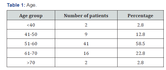
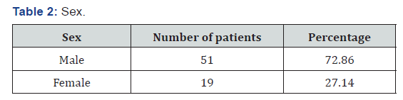
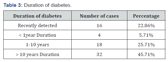
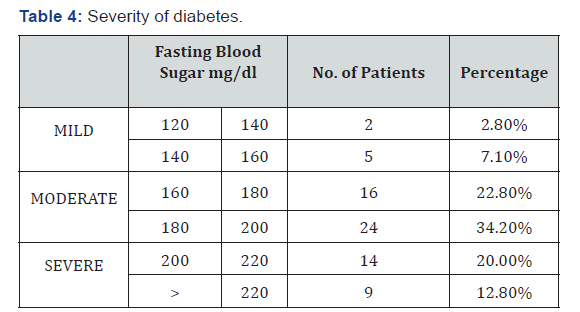
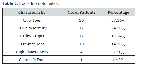
Results: The most common age group affected belonged to 51-60 age bracket which included 41 patients (58.5%). The male to female ratio was approximately 3:1. Majority of patients 32 (45.71%) had diabetes of >10 years duration. Foot and toe deformities affected majorly diabetics with moderately controlled diabetes mellitus and the study revealed that 40 patients (57.2%) had moderately controlled diabetes with blood sugar levels ranging from 160-200 mg/dl. The highest number of patients i.e. 29 patients (41.4%) who had foot/ toe deformities were on oral hypoglycaemic agents. Clawing of the toes (26 patients, 37.14% of patients), was the commonest morphological change in the architecture of the foot followed by Varus deformities (17 patients, 24.28% of patients). Nail changes in the form of hypertrophied and brittle nails affected 37 patients (52.85%).
Conclusion: We concluded that team approach, adequate diabetic control combined with patient education about foot care and footwear will help prevent complications and improved quality of life of the patients suffering from diabetic foot and toe deformities.

Keywords: Diabetic foot; Toe deformities; Hammer toes; Hallux valgus; Bunion; Claw toes; Charcot’s foot

Introduction

Foot problems are common in patients with diabetes, often requiring prolonged and costly hospital stay and eventually leading to lower extremity amputation [1]. Motor neuropathy affects the function of the intrinsic and extrinsic musculature of the foot, thus upsetting the delicate balance between flexors and extensors of the toes [2]. Atrophy of the small muscles responsible for metatarsophalangeal plantar flexion is thought to lead to the development of hammertoes, claw toes and prominent metatarsal heads [3]. The prevalence of all diabetic foot complications increased clearly with age and diabetes duration [4]. Clinical recommendations for people with diabetes include provision of special footwear to individuals with foot risk factors [5].

Aim

To study the morphological changes in the form of foot and toe deformities occurring in diabetic patients presenting to a tertiary care centre in New Delhi, India.

Materials and Methods

A prospective study was carried out in patients presenting to the Diabetic Clinic at a tertiary care hospital in Delhi. The patient profile comprised of patients who were known Diabetics or recently detected diabetics under treatment and follow up and had obvious foot and/or toe deformities. A detailed history regarding foot and toe deformities was followed by a thorough physical examination and the following were noted: -
1) Age and sex of the patient
2) Duration and severity of Diabetes.
3) Treatment by Oral Hypoglycaemic agents and / or Insulin/p>
4) Evidence of Neuropathy -loss of touch, vibration, temperature sensation, using a 10gm monofilament, 128 Hz tuning fork, deep tendon reflexes were examined using percussion hammer.
5) Evidence of ischaemia – thinning of skin, loss of subcutaneous fat, hair loss, brittle hypertrophied nails, callous formation, ulceration, peripheral pulses and venous filling time were noted.
6) Foot and/or toe deformities
7) Nail changes

Results

International Journal of Pulmonary & Respiratory Sciences
Out of the 70 patients, 2.8% were below 40 years of age, 9 patients (12.8%) were in the group between 41-50. 41 patients (58.5%) belonged to the 51-60 age group, 16 patients (22.8%) were in the 61-70 age group and only 2 patients were in the age group above 70 years (Table 1). Out of 70 patients, 51 patients (72.86%) were males and 19 patients (27.14%) were females who presented with various foot and toe deformities (Table 2). Of the 70 patient, 16 patients (22.86%) were detected to have diabetes during their first presentation to the hospital. 4 patients (5.71%) had foot lesion with diabetes of less than 1year duration, while 18 patients (25.71%) had diabetes of <10 years duration (Table 2). Majority of patients 32 (45.71%) had diabetes of >10 years duration (Table 3). Only seven patients (10%) had well controlled blood sugar levels, 40 patients (57.2%) had moderately controlled diabetes with blood sugar levels ranging from 160-200mg/dl.23 patients (32.8%) had poorly controlled diabetes (Table 4). 16 patients (22.8%) were unaware about their disease hence were taking no treatment (Table 3). 29 patients (41.4%) were only on oral hypoglycaemic agents, 14 patients (20%) were being managed with insulin, 11 patients (15.7%) were being treated with both oral hypoglycaemic agent and insulin (Table 5). Clawing of the toes (26 patients, 37.14% of patients), was the commonest morphological change in the architecture of the foot followed by Varus deformities (17 patients, 24.28% of patients) . Hallux valgus (17.14%), Hammer toes (14.28%), high plantar arch (5.71%) and Charcot’s foot was seen only in two (1.42%) patients (Table 6).
International Journal of Pulmonary & Respiratory Sciences

Nail Changes

Hypertrophy with brittle nails (52.85%) were seen in 37 patients.

Follow Up

All patients were followed up at monthly intervals whenever they used to come to collect their medicines for diabetes. They were given specific advice about foot care and footwear.

Discussion

Normal foot

The skin of the healthy foot is elastic but resilient. It should not show signs of scaliness, dryness or brittleness, nor should it be thin and shiny. Plantar skin is about twice as thick as that on the dorsum, is tougher and moves less readily over the deeper structures.

Gait cycle

During walking the weight is taken first on the point of the heel (“heel strike”). It is then transferred smoothly to the outer aspect of the sole and the metatarsal heads of the second to fifth toes. It then shifts medially to the foot (first metatarsal head) and the body is propelled forward by flexion of all the metatarsals, and of the big toe (“toe off”). This smooth transfer of forces is lost when the foot loses its normal plantar arch, or the ligaments and soft tissues become less elastic. The result is that some parts of the sole take increased pressure and therefore become liable to neuropathic ulceration. This is most likely to occur over the second and third metatarsal heads, and the big toe.

Diabetic foot

Diabetic foot

Peripheral neuropathy

Sensory Neuropathy: Diabetes leads to abnormal function of peripheral nerves due to Metabolic causes –due to accumulation of sorbitol. Ischaemia - the nerves dependent on the vasa nervous for their nutrient supply which suffer ischaemic damage from microvascular disease.

Motor neuropathyy

The integrity of the arch of the foot is preserved by muscles, ligaments and connective tissue. The muscles become weak and atrophic when affected by motor neuropathy. This causes loss of normal balance between the toe flexors and extensors; hence the persons may develop either a flat foot or one which is excessively clawed.

Autonomic neuropathy

Vasomotor: The fine control of distribution of blood is dependent on the action of Vasomotor nerves. These are responsible for opening and closing of arterioles and venules such that blood is shunted to the areas where it is most needed. The foot affected by autonomic neuropathy may have abnormal distribution of blood even though the macrovascular supply is good. The altered blood flow due to arterio-venous shunting leads to increased bone turnover, resorption and destruction of bones and joints called the Charcot’s joint. Charcot joint was considered when bones, joints, and soft tissues of the foot and ankle are inflamed in the presence of neuropathy with or without history of trauma leading to variable degrees of bone destruction, subluxation, dislocation, and deformity [8].

Sweating

Autonomic neuropathy may result in sudomotor dysfunction leading to abnormal sweating and dry skin with cracking and fissuring facilitating the bacterial infection of the foot [9]. The risk for diabetic foot ulceration increases by sevenfold in patients with peripheral diabetic neuropathy [10]. 45% to 60% of all ulcerations in patients with diabetes are mainly due to neuropathy, while 45% of the ulcers are due to combined neuropathic and ischemic factors [10]. Distal bilateral symmetrical neuropathy is the commonest presentation and usually starts in the lower limbs, has a progressive course. It usually presents in a glove and stocking pattern of abnormal sensations [11]. “Burning feet syndrome.” is a form of neuropathy which arises at night and is accompanied by high sensation of pain [12]. Peripheral neuropathy is accompanied by autonomic neuropathy in 30% to 50% of cases [13]. The foot skin affected by autonomic neuropathy is dry and fissured with the consequence of finding a reduced protective skin function and thus increased risk of injury.

Lesions of neuropathic foot

a) The foot is typically pale and puffy, with clawing of toes. The skin is dry and fissured and the planter arch may be exaggerated. Clawed toes are defined as toes with extension of the metatarsophalangeal joint and flexion of the proximal and distal interphalangeal joints. Claw toes is caused by an imbalance between the extrinsic and intrinsic foot muscles [14].
b) Hammer toes: The metatarsophalangeal joint of the first digit is extended, and the proximal interphalangeal joint is flexed. Atrophy of the small muscles responsible for metatarsophalangeal plantar flexion is thought to lead to the development of hammer toes [15].
c) Varus deformities: The third, fourth and fifth toes drift medically due to muscle imbalance, to gouge adjacent toes producing ulcers.
d) Hallux Valgus: Hallux valgus is defined as a lateral angulation of the first MTP joint of the great toe [16].
e) Tailor’s Bunion: It is an exostosis of the lateral part of the fifth metatarsal head which is associated with Varus deformity.
f) Charcot’s foot: It is a rare but serious complication of the diabetic foot. It occurs in less than 1% of people with diabetes, most commonly during the fifth and sixth decade in insulin dependent patients.
The duration of diabetes is usually greater than 12 years. Normally it develops unilaterally, but in 20% of patients it develops bilaterally. It is defined as a chronic, painless degenerative process affecting the weight bearing joints of the foot. The ethology is thought to be repeated minor trauma in a neuropathic foot and due to arterio- venous shunting. Charcot’s foot develops through 3 stages, described by Eichholtz in 1966. Eichholtz stage 1 is characterized by acute inflammation associated with hyperaemia and erythema. During this stage, the bone softens and fragments, fracture dislocations occur, with pure dislocations more common. Stage 2 is characterized by bony coalescence, reduction in swelling of the foot, and periosteal new bone formation. In stage 3 bony consolidation and healing occurs. Normally, this whole process lasts about 2 to 3 years, although progress through the stages can be quite rapid.

Limited joint mobility

Diabetes causes abnormalities of the connective tissue due to glycosylation of proteins like collagen which renders it stiff and inflexible. These changes restrict foot mobility, which while walking leads to trauma and ulceration.

Management of Foot and toe deformities in Diabetes Mellitus

Multidisciplinary foot care team consisting of diabetologist, surgeon, diabetic nurse, chiropodist and orthotist forms the foot care team.

Skin care

The feet are often dry and scaly as a result of anhidrosis secondary to autonomic neuropathy. Regular applications of moisturizing creams will help soften the skin and prevent skin fissures which might get infected.

Nail care

International Journal of Pulmonary & Respiratory Sciences
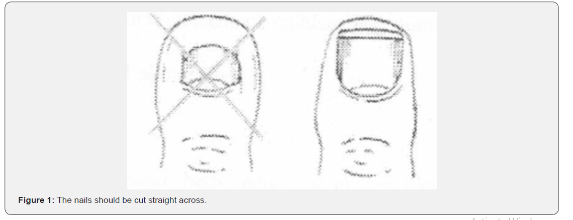
Obesity and poor eyesight prevent elderly patients to cut their nails properly which may lead to damage of the adjacent skin or may leave a portion of nail uncut, resulting in an ingrowing toenail (Figure 1). The toenails should be cut short with no remaining sharp edges. Mycological infections should be diagnosed early and be treated. A grossly deformed mycotic nail should be treated by nail excision.

Use of proper footwear

International Journal of Pulmonary & Respiratory Sciences
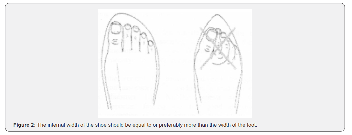
Proper footwear help reduce points of high pressures. In most of the cases, the problem is caused by arching of the foot resulting in high pressure at the metatarsal heads. Custom Fitted Shoes should have generous instep support to re-distribute the weight more evenly (Figure 2). The problem of clawing is that there is an increased requirement of a deep shoe. The principle of good footwear is to provide a shoe which is deep, broad but not floppy. The material should be soft with good in step support. Bespoke footwear may be needed for feet which are grossly deformed following Charcot’s arthropathy.

Conclusion

Diabetes affects the foot in various ways including lesions affecting the skin, nails, bones and connective tissues. Team approach, adequate diabetic control combined with patient education about foot care and footwear will help prevent complications and improved quality of life of the patients suffering from diabetic foot and toe deformities. Studies have confirmed that inappropriate footwear is the most common source of trauma and cause of ulceration in patients with diabetes [17] and hence patients with diabetes should be educated regarding use of proper footwear.


To Know more about our Juniper Publishers


Thursday, July 9, 2020

Mammosomatotropic Adenoma and Acromegaly; What Particularities? - Juniper Publishers

Brain, Spine & Neural Disorders - Juniper Publishers  

Abstract

Acromegaly is a rare condition, usually secondary to excessive production of somatotropic hormone (GH) by a pituitary adenoma, which is clinically expressed either by acromegaly or by gigantism depending on the age of onset. Several histological types are involved. We usually distinguish somato-prolactin adenoma. The mammosomatotropic adenoma is a particular entity, characterized by an intense anti-GH immunohistochemistry and a lower anti-prolactin immunopositivity, but within the same cell. We report 4 observations of a histological type: mammosomatotropic adenoma.

Keywords: Acromegaly Mammosomatotropic adenoma GH prolactin IGF1 Immunohistochemistry

Introduction

Acromegaly is a disease related to hypersecretion of growth hormone (GH), a pituitary adenoma somatotropic in more than 90% of cases [1,2]. She is responsible for an acquired dysmorphic syndrome, and rheumatological, cardiovascular, respiratory, metabolic, etc. consequences. Which condition the prognosis: they are indeed all the more severe as the excess of GH has been prolonged and important [3,4]. The severity of acromegaly can also, of course, stem from the pituitary tumor that is the cause, and which can be the cause of a tumor syndrome, marked by headaches and / or visual disturbances (by chiasmatic compression). The diagnosis of acromegaly is based on the demonstration of a high plasma concentration of GH and especially non-breakable by oral hyperglycemia (greater than 0.4 μg l-1). Once the diagnosis of acromegaly has been made, it is necessary to evaluate, by magnetic resonance imaging [MRI], the volume and possible expansions of the pituitary tumor [5-10]. Several histological types are involved. Immunohistochemistry provides conclusive evidence that significant diversity exists between tumors secreting excess growth hormone (GH). We report 4 observations of a particular histological type: mammosomatotropic adenoma.

Observation

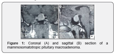
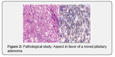
It is about four patients consulting at the endocrinology department of Med VI University Hospital of Marrakech for an acromegaloid syndrome (in 3 cases) with a case of acrogigantism. The average age was 42.7 years, with a sex ratio H / F of 0.25. The biology has shown a high level of IGF1 in all cases, with a highprolactin level in a single patient. Magnetic resonance imaging of the hypothalamic-pituitary region has demonstrated the presence of a pituitary macroadenoma in all these patients. (Figure 1) The latter benefited from first line transsphenoidal surgery, whose anatomopathological and immunohistochemical study objectified an aspect in favor of a mamo somatotropic adenoma (Figure 2), for which our patients were all placed under cabergoline with a somatostatin analogue.
Theranostics of Brain, Spine & Neural Disorders

Discussion

Acromegaly is a rare condition, usually secondary to excessive production of somatotropic hormone (GH) by a pituitary adenoma, either alone or in combination with another hormone, including prolactin. We usually distinguish somato-prolactin adenoma. The mamo somatotropic adenoma is characterized by an intense anti-GH immunohistochemistry and a lower antiprolactin immunopositivity, but within the same cell. Electron microscopy confirms this granular colocalization of the two hormones. The diagnosis of pluro hormonal somatotropic adenomas requires the routine practice of immunohistochemical tests because there is no specific clinical presentation. Even if they remain rare, their treatment does not differ from the other types described in acromegaly [11-19].

Conclusion

The mamo somatotropic adenoma is very rare, characterized by an intense anti-GH immunohistochemistry and a lower antiprolactin immunopositivity, but within the same cell. Electron microscopy confirms this granular colocalization of the two hormones.


To Know more about our Juniper Publishers

Wednesday, July 8, 2020

Periorbital Necrotizing Fasciitis in a Young Woman with Anorexia Nervosa-the Management and Considerations of Cosmetic and Functional Outcomes of Fulminant Disease in a Critically Unwell Patient - Juniper Publishers

Head Neck & Spine Surgery - Juniper Publishers  


Abstract

Necrotizing fasciitis is a rapidly progressive and potentially deadly bacterial infection of the superficial fascia. The horrific and grossly debilitating nature of necrotizing fasciitis has led to an increased prominence of its reporting in mainstream media however the disease entity has not been historically well understood, classified or reported in medical literature. While capable of manifesting in numerous bodily locations following inoculation during trauma, the periorbital region is the most commonly involved facial site and presents unique challenges in treatment of the disease due to the unique anatomical and functional attributes of this area. This case report describes the diagnosis, surgical and medical management in a young woman with severe malnutrition due to anorexia nervosa.

Introduction

Necrotizing Fasciitis (NF) is a devastating rapidly progressive bacterial infection involving the superficial fascia leading to necrosis of overlying skin and systemic toxicity [1,2]. We present a case of periorbital NF in a chronically malnourished and unwell patient following facial trauma.

Case Report

A 36-year-old female presented to the emergency department following a mechanical fall with extensive left facial bruising extending to the neck and chest wall. The patient featured a complex medical history including severe anorexia nervosa. Pathology revealed significant electrolyte derangement, hypothermia, osteoporosis, bone marrow suppression and synthetic hepatic impairment. A clinical diagnosis of NF was made 6 days into her ICU admission following the development of anaesthetic, necrotic-appearing skin associated with purulent exudates from the palpebral fissure and a corresponding rise in the patient’s White Cell Count (WCC) and C-Reactive Protein (CRP). Examination of the eye was unremarkable, with the exception of a subconjunctival hemorrhage and nasally oriented corneal dellen. Initial emergency debridement was undertaken as a joint operation between ophthalmology and ENT teams, stripping back necrotic periorbital tissues down to the lower cheek and performing a lateral tarsorrhaphy to prevent exposure keratopathy. ENT undertook further exploration and debridement down to the neck and submental region, leaving a VAC dressing in-situ and harvesting multiple tissue samples for histopathological evaluation. Histology revealed marked suppurative inflammatory changes in the reticular dermis, skeletal muscle and subcutaneous fat in association with venous congestive changes and large regions of muscular and dermal necrosis (Figure 1).

Pan-sensitive Streptococcus pyogenes and penicillinresistant Staphylococcus aureus (MSSA) were isolated from the excised tissues, while Pseudomonas species were isolated from the superficial left facial and eye swabs. Pending sensitivities, targeted antimicrobial therapy was instituted including the use of intravenous flucloxacillin, vancomycin, piperacillin-tazobactam and meropenem. Subsequent operative debridement and exploration was undertaken two days later, excising further nonviable periorbital tissues down to the buccal fat pad. Inspection of oral tissues was performed under direct laryngoscopy along with extension of tissue debridement from the neck area, noting that progression did not appear to track along fascial planes. Two months later, initial reconstruction surgery was performed. After washout of the left cheek and periorbital wounds, a Split Skin Graft (SSG) was applied to the left cheek (from a left thigh donor site) and Full-Thickness Skin Graft (FTSG) was applied to the superior and inferior left eyelids (abdominal donor site). The left tarsorrhaphy was also revised. Six months following her initial presentation, horizontal lateral canthotomy and division of tarsorrhaphy was performed to allow for a wider palpebral fissure. Finally, nine months after the initial diagnosis of NF, debulking of the upper lid was performed in conjunction with suturing of trace remaining elevator fibers to the tarsal plate. Reconstruction of the lower eyelid with FTSG (donor site medial upper arm) was also performed to relax the cicatricial nature of the earlier procedures (Figure 2).

Post-operative best corrected visual acuity was 6/9+2 in the affected eye with 5-6mm of lagophthalmos and complete pupillary exposure, allowing for good binocular vision without altered head posture. The patient remains able to use frontalis and orbicularis action to consciously moisten her cornea. The patient continued to use regular preservative-free lubricants to prevent exposure keratopathy. Periorbital NF is a rare rapidly progressing ophthalmic emergency that requires early recognition and operative debridement to prevent a fulminant course [1,3-5]. First described by Hippocrates in the fifth century B.C. as a complication of erysipelas [5], NF was then first properly described in modern literature by Confederate army surgeon Joseph Jones in 1871. Inconsistent nomenclature and inadequate linkage to sufficient bacteriological data led to confusion in the literature and a delay in the understanding of NF as a single disease entity [6]. Infection of the head and neck is uncommon in NF with only asmall number of cases being described in literature since 1960. In the post-antibiotic era, the overall mortality of head and neck NF has been estimated at between 9 and 31%, but its relatively frequent association with multi-system shock and permanent disfigurement render NF a particularly devastating and terrifying illness [3,7] (Figure 3).

The most common trigger for periorbital NF is an antecedent injury leading to the breakdown of the skin barrier and concomitant inoculation of the causative organism(s) [6-8]. Swelling, pain, erythema, fever, haemorrhagic bullae, skin necrosis and crepitus are the most common clinical signs relevant to diagnosis [2,4]. Diabetes mellitus, immunosuppression, chronic renal impairment, pulmonary disease, recent surgery and traumatic injury have been identified as predisposing factors for NF; however, it is worthwhile noting that a large number of published cases remain idiopathic in nature [1,9-10]. Immunosuppression, age greater than 50 years, cirrhosis, congestive cardiac failure, gout and development of toxic shock syndrome (a late sign of infection) have been identified as independent risk factors most associated with increased mortality from NF [1,2,11]. Relative risk indicators have been identified, utilizing biochemical indicators such as white cell count, haemoglobin, CRP, serum creatinine, sodium and blood glucose to stratify NF severity in the acute setting [12].

Periorbital NF is divided into 4 classifications on the basis of microbiological culture. Type 1 (polymicrobial) infections are related to both anaerobic and aerobic (including facultative anaerobes such as Enterobacteriaciae and non-typable Streptococci) organisms most commonly manifesting on the abdomen, trunk and perineum. Type 1 NF is the most common type to develop in patients with diabetes. Type 2 (monomicrobial) NF involves the extremities, cutaneous and muscular tissues; and features no clear underlying risk factors. Group-A betahaemolytic Stretococcus (S. pyogenes) and/or S. aureus are the most commonly implicated organisms in Type 2 disease, characterizing its strong association with toxic-shock syndrome. Type 3 infections are caused by Clostridium, Vibrio spp. and other gram-negative organisms, typically involving the abdominal wall and/or perineum. They are often rapidly progressive infectionsfollowing salt-water injuries, seafood ingestion and penetrating traumas. Type 4 NF describes fungal infections involving the extremities, most frequently occurring in immunosuppressed individuals [1,9,10,13].

Our patient was a young, immunosuppressed, malnourished female. On hospital presentation she had severe electrolyte derangement (hypomagnesaemia, hypokalaemia and hyponatreamia), hypothermia, impaired hepatic synthetic function and coagulopathy (INR 2.0 with hypovitaminosis-K) and ischaemic digits. She developed severe facial Type 2 NF (S. aureus and S. pyogenes) following a break in the periorbital skin overlying a large haematoma, initially caused by blunt mechanical trauma. No underlying fractures were identified on computed tomography imaging. Anaesthetic necrosis of the thin eyelid skin allowed the early clinical recognition and diagnosis of necrotizing fasciitis, nevertheless the infection was rapidly progressive in nature, resulting in severe permanent disfigurement despite prompt operative debridement and broad-spectrum antimicrobial cover. This case affords some insights into the pathophysiology of NF and also presented challenging aspects in the consideration of reconstructive techniques, attempting to maximize both long-term functional stability of the eye in limiting exposure keratopathy whilst optimizing the patient’s cosmesis Operative exploration and debridement in this patient demonstrated that this infection did not respect fascial planes, considered anomalous in the typical subcutaneous spread pattern of NF [14]. It is possible that traumatic disruption of subcutaneous compartments involving all skin and muscular layers, in conjunction with a large haematoma facilitated alternative pathways for the dispersion of infection in this patient. This is of particular importance to keep in mind in such cases wherethe musculoaponeurotic structures of the face and neck may be breached, allowing the spread of infection into the superior mediastinum [11].

Discussion

Periorbital NF is the most commonly involved facial site and behaves differently from NF elsewhere in that the highly vascularized orbicularis oculi acts as an effective barrier to prevent the spread of infection from the skin to the underlying periorbita, thereby retarding the spread of infection into the orbit. As in this case, necrosis of the thin eyelid skin occurs rapidly, facilitating early detection of the condition due to the obvious cosmetic abnormality on examination [1]. Given the fragile general health and constitution of the patient, decisions regarding the donor sites for reconstructive skin grafting were based purely on pragmatism, with cosmesis playing a clearly secondary consideration to what were emergent and life-saving surgeries. Gross malnutrition and immunosuppression meant that donor tissue was chosen to maximize graft viability, to facilitate adequate coverage of the relatively large areas left exposed following extensive surgical debridement and to maximize the chances of adequate postoperative healing in this setting. Subsequent procedures were required in this case to balance the cosmesis and function of the patients eyelids, her vision (and the initial requirement for a chinup posture to peer through her tight palpebral fissure,) and the necessity to prevent an exposure keratopathy in a patient with significant lagophthalmos secondary to an almost obliterated orbicularis.

Conclusion

The rapidly destructive nature of NF necessitates early recognition and diagnosis to minimize the risk of a fulminant course with high morbidity and mortality. Physicians must remain vigilant and mount a thorough clinical suspicion for NF in suitably vulnerable patients with clinical features suggestive of the disease such as severe pain, swelling, erythema, bullae and tactile anaesthesia. Urgent resuscitation, emergency surgical debridement, broad-spectrum IV antibiotics and other novel therapies such as hyperbaric oxygen must be implemented without delay to prevent the frequently debilitating, disfiguring and life-threatening sequelae of NF [1,8,11,15]. This case report demonstrated some of the complexities in managing periorbitalNF in the context of pre-existing medical comorbidities and highlighted the challenges in optimizing the functional and cosmetic outcomes of reconstructive surgery following extensive emergency debridements of neck and facial tissues.


To Know more about our Juniper Publishers


Tuesday, July 7, 2020

Transdermal Methimazole for Feline Hyperthyroidism - Juniper Publishers

Journal of Toxicology - Juniper Publishers


Abstract

Hyperthyroidism is a common disorder in older cats causing detrimental adverse effects if left untreated. The three most recommended treatment options include thyroidectomy, radioiodine treatment, and antithyroid medication therapy. Oral methimazole has been the most widely used option due to low cost and accessibility. The topical application of transdermal methimazole is an ideal route of administration for cat owners. The purpose of this review article is to give insight into the efficacy and recommended indication for use of the pluronic lecithin organogel (PLO) formulated transdermal delivery system of methimazole, in the treatment of feline hyperthyroidism. PLO compounded methimazole is uniquely transported through the skin, and chronic use has been shown effective in treating feline hyperthyroidism. In many cases, once daily application of the gel has provided enough methimazole activity for lowering hormone levels. The compounded formulation also allows for more individualized dosing than the oral tablets. There is limited information regarding long-term treatment of PLO methimazole, however, the formulation continues to satisfy both veterinarians and owners, and effectively lower serum thyroxine (T4) concentrations.

Keywords: Feline; Hyperthyroidism; Transdermal; Methimazole

Abbrevations: PLO: Pluronic Lecithin Organogel

Introduction

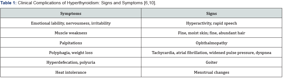
Over the past 20 years, the prevalence of feline hyperthyroidism has increased astoundingly [1-3]. It has become the most common endocrine disorder in cats, and the risk worsens with each year of increasing age, being most common in middle to older-aged felines [1]. The disease is primarily characterized by an excessive production and release of the thyroid hormones thyroxine (T4) and triiodothyronine (T3) most commonly due to a functional, benign adenomatous hyperplasia of the thyroid gland. At present, there is not a feline specific thyroid stimulating hormone (TSH) assay test available, therefore unlike human hyperthyroid diagnosis, veterinarians do not commonly depend on a low TSH value for primary hyperthyroid diagnosis. Hyperthyroidism diagnosis in cats is generally based on a high free T4 level and the presence of clinical signs and symptoms. Some of the clinical complications of hyperthyroidism that may be present include emotional lability, hyperactivity, palpitations, tachycardia, and a plethora of other manifestations of the disease (Table 1). Although the exact etiology is unknown, many nutritional and environmental causes are suspected, including canned cat food products containing iodine, soybean, phthalates, polyphenols, and polychlorinated biphenyls [2,4,5].
Regardless of the etiologic origin, medical management of prolonged thyroid hormone elevation is very important. Untreated hyperthyroidism can have many consequences on the cat. Many cats initially present with a change in personality or behavior, often being more easily agitated and mean, as well as with unexplained weight loss, changes in eating habits, accelerated heart rates, and a goiter. Hyperthyroidism, if left untreated, can also have life threatening adverse effects, such as causing hypertension, cardiac tachyarrhythmia, atrial fibrillation, and even death [6,7]. These result from elevated thyroid hormone levels and cause up-regulation of various gene expressions involved in the body’s metabolism, thermogenesis for heat regulation, nerve function, and muscle and bone function [7]. They also function to increase activation of the sympathetic nervous system, which elevates the heart rate, the heart’s force of contraction, and increases cardiac output overall [8,9]. Clearly, both the symptoms of the disease, as well as the enhancement of these biochemical pathways, can pose serious health risks to the feline patient. The longer a cat goes without treatment, the worse their complications become [6,10,11].

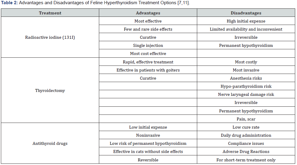
Like the management of hyperthyroidism in humans, there are several different treatment options available for cats. The top three recommended therapies include surgical thyroidectomy, radioiodine therapy, and medication treatment. Thyroidectomy and radioiodine treatment can be permanent solutions to the disease. However, limitations such as expense and permanent hypothyroidism prevent these from being practical options for most feline patients [7,11] (Table 2). Medication therapy is often the most practical and accessible way to manage hyperthyroid cats. Methimazole (Tapazole, Felimazole) is the most common and favored agent in the United States [12]. Other alternatives include carbimazole (a prodrug of methimazole marketed only in the UK), iodine-containing agents, iodine dietary restricted food, and homeopathic regimens [6,12-14]. Dietary iodine restriction is another option, however, there is limited supporting data to determine a true benefit.
Although several treatment options are available for hyperthyroid cats, each therapy option has considerable drawbacks to both the client and the feline patient. Oral methimazole has historically been the most accessible and affordable choice. However, gastrointestinal side effects and an unfavorable twice-a-day oral administration schedule often limit its ultimate therapeutic outcomes in the cat. Both negative attributes are avoided with use of the transdermal methimazole gel compound. Due to the limited amount of data available on transdermal methimazole, this review aims to evaluate whether the pluronic lecithin organogel (PLO) compound of methimazole is effective in treating hyperthyroid cats. In addition, it also serves to provide insight on the recommendations for its use.

Methods

A PubMed search was conducted to identify articles in which the safety or efficacy of transdermal methimazole for treatment of feline hyperthyroidism was assessed. Key MeSH search terms included feline hyperthyroidism with a subheading for treatment. In addition, feline hyperthyroidism plus one of the following search terms were searched: treatment, drug-related side effects and adverse drug reactions. A free-text search was also conducted to identify articles not included in the MeSH term search. Metaanalyses, randomized controlled clinical trials, and case reports were included in the review if the primary focus of the article related to the use of oral or transdermal methimazole for feline hyperthyroidism. Studies were excluded if published in languages other than English. In addition, studies highlighting mechanisms of action, studies of pharmacodynamics or pharmacokinetic effects were excluded.

Results

Clinical data on the topic of feline hyperthyroidism treatment is limited. A PubMed search revealed 14 articles with transdermal methimazole and feline hyperthyroidism as a subheading. Of the articles used in this review, there were six that directly assessed the use and efficacy of transdermal methimazole in the treatment of feline hyperthyroidism. Of those six, five were small clinical studies and one was a case report/series.

Evaluation of oral methimazole

Oral methimazole has remained the mainstay of feline hyperthyroidism treatment since the early 1980’s. It reversibly suppresses thyroid hormone levels by inhibiting thyroid peroxidase. It does not inactivate circulating T4 and T3, resulting in a 2 to 4-week delay before serum T4 concentrations begin to normalize [8]. While it accumulates in the thyroid gland, it does not block the release of preformed hormone, nor does it help reduce goiters [8,15]. Oral methimazole has variable bioavailability ranging from 27 to 100% so its efficacy varies from patient to patient [6]. The recommended dose for maximum efficacy is 2.5mg administered twice daily.

In a randomized, unblinded, clinical trial by Trepanier et al. [11], forty methimazole naive cats with newly diagnosed hyperthyroidism were studied to compare the efficacy of one daily dosing of oral methimazole to twice daily dosing. Owners completed a questionnaire of their cat’s baseline behavior status and reported any changes that occurred during the study. The overall efficacy of once daily methimazole was found to be less effective than twice daily dosing. Serum T4 concentrations were considerably higher in cats receiving once daily dosing, and only 54% (13/24) were found to be euthyroid at two weeks, compared to 87% (13/15) euthyroid in the twice daily group [16]. Both treatment groups showed considerable clinical improvement of many complications caused by hyperthyroidism. However, among the initial 40 cats studied, one cat in the once daily dosing group was removed prior to the 2-week point due to considerable gastrointestinal (GI) upset. Of the remaining 38 feline patients, 17 (44%) developed some type of adverse event throughout the four-week duration. Throughout the remainder of the study, 23% (9 cats) reported similar GI upset. Among the 24 cats treated once daily, 42% (10/24) required discontinuation of therapy, in order to resolve oral methimazole induced adverse events. Facial excoriation was reported in six patients, five reported from the once daily dosed group alone. Five of the six total facial excoriation cases reported were from the once daily dosed group. Manifestations of blood dyscrasias and hepatopathy were not significantly reported in either group [16].

Not only were adverse events such as GI upset and facial excoriations, found to be less prevalent in cats dosed twice a day, but also these cats were also more likely to obtain the goal euthyroid state. Cats also show rebound increases in serum T4 concentrations and a return to hyperthyroid state within 24 to 48 hours of methimazole discontinuation [3,16,17]. This likely correlates with the need for twice daily dosing in cats, and further research should be performed to help determine methimazole’s true intrathyroidal residence time in cats. Oral methimazole is not a cure for feline hyperthyroidism, and treatment must be continued indefinitely. With the intolerable GI upset from the oral tablets and the difficulty many owners face administering the medication twice daily to uncooperative cats, the alternative transdermal route of administration poses significant benefits [16].
 

Transdermal methimazole formulation

Despite the limited clinical studies on transdermal methimazole, some clinicians have achieved a good therapeutic benefit to using this dosage form in cats. Pluronic lecithin organogel is a microemulsion-based gel containing lecithin, isopropyl palmitate, and pluronic acid to effectively deliver both hydrophilic and lipophilic drugs topically across the stratum corneum and may aid in the administration of methimazole [18- 22]. PLO is composed of both an oil phase (lecithin phase) and an aqueous phase (pluronic phase). It includes isopropyl palmitate acts as a solvent and permeation enhancer while lecithin also serves as a permeation enhancer by increasing the fluidity of the stratum corneum, and slightly disorganizing the skin structure to permit substance permeation [23-25]. PLO reversibly turns into a thick gel at body temperature, leading to an increase in dehydration of the aqueous solution, forming a shell-like structure of aggregated micelles [7,24-28]. Methimazole is an ideal drug for transdermal delivery due to its low molecular weight, high lipid solubility, water solubility, low daily dose, and is non-irritating and non-sensitizing to the skin [20,24].

Efficacy of the PLO methimazole

In a small retrospective study examining dispensing records for 16 hyperthyroid cats undergoing transdermal methimazole treatment, the transdermal formulation was effective at reducing serum T4 concentrations in 15 of the 16 cats studied. One cat showed an increase in serum T4 level, but there is no mention or clarification of appropriate application or other possible contributing factors. The only adverse event reported was a single case of increased blood urea nitrogen level, thought to be the unmasking of prior renal disease. This study also demonstrates variability in dosing and administration frequency of the topical, ranging between 5 mg once a day to a twice daily dose of 7.5mg every morning and 5 mg every night. This wide variation between each feline patient, limits our ability to recommend a standard dose or administration frequency, but does indicate the need for patient-specific doses and frequencies in order to effectively reach the euthyroid goal [29].

In a randomized clinical trial conducted by Sartor et al, 47 newly diagnosed hyperthyroid cats were used to investigate whether PLO formulated transdermal methimazole was safe and efficacious in controlling feline hyperthyroidism. At two weeks of treatment, more cats in the oral methimazole group had serum T4 concentrations within the reference range (14 of 16 [88%], p=0.035). By week four, there was no difference between the oral and transdermal methimazole. The PLO transdermal methimazole group took longer to reduce serum T4 concentrations to the acceptable reference range, however, it was as effective as oral administration in producing euthyroidism by the fourth week of treatment [30]. Fewer GI adverse events were reported with the transdermal formulation (1/27 vs 4/17 in the oral group). The reduction of GI upset deems consideration as it is often the cause of discontinuation of oral methimazole [30,31].

Lecuyer et al evaluated the efficacy of transdermal methimazole in 13 newly diagnosed hyperthyroid cats. The feline patients received 5mg methimazole concentrated in PLO, applied to the inner ear twice daily. In addition to reaching the euthyroid state, all 10 cats that completed the study also showed improved clinical signs related to hyperthyroidism consistent with other previously reported studies [16,32-33]. No GI adverse events were reported, and investigators concluded that PLO transdermal methimazole is a safe and effective alternative to oral methimazole [6].

Duration of t4 suppression

A study by Boretti et al. [33] evaluated the duration of serum T4 suppression among newly diagnosed hyperthyroid cats treated with once daily transdermal methimazole versus twice daily dosing. Twenty cats were treated with the PLO-based methimazole formulation dosed either 2.5mg every 12 hours (10 cats, group 1) or 5mg every 24 hours (10 cats, group 2). Serum T4 concentrations were measured one and three weeks after initiation of therapy, immediately before and every two hours after gel application for up to 10 hours. Cats were limited to a maximum of five blood samplings in one day [33]. A sustained suppression of T4 concentration for at least 24 hours was seen following gel application and there was no significant difference in change in serum T4 concentration immediately before or any time after gel administration in either group. As also discussed in Lecuyer’s study [6], further research is needed concerning the duration of intra thyroid methimazole accumulation [6,33,34]. Among the twice daily dosing group, reductions were required in three cats, and a dose increase was required in one patient. Of the once daily dosing group, two cats required a decrease in dose, and one cat required an increased dose, after three weeks of treatment as a result of sustained hyperthyroid levels [33]. Investigators concluded that once daily application of the PLO methimazole compound can effectively reduce serum T4 concentrations in most hyperthyroid cats. Once a day dosing is most convenient for the owner, and thus promotes better compliance [33]. The compounding of this preparation allows for changes in dose or frequency and allows for the individualization of therapy.

PLO vs. novel lipophilic base

In a 12-week prospective study by Hill et al, a novel lipophilic formulation of methimazole was investigated. The study included 45 cats newly diagnosed with untreated, naturally occurring hyperthyroidism [12]. The study used a novel lipophilic formulation prepared with methimazole, “carrier compounds” (propylene glycol, polyethylene glycol 4000, dimethyl formamide, and cyclodextrin), and several penetration enhancers, chosen from fatty acids, terpenes, pyrrolidones, a short chain alcohol, glycol ethers, acetins, and triglycerides. The formulation was determined to be stable for 12 months after preparation, by the International Cooperation on Harmonization of Technical Requirements for Registration of Veterinary Products. Cats were treated with a starting dose of either oral carbimazole (5mg twice a day) or the novel transdermal methimazole formulation (10mg, or 0.1mL applied to the inner ear once a day). Both the once daily novel transdermal methimazole and twice daily oral carbimazole were effective in the treatment of feline hyperthyroidism in cats with compliant owners. All owners were satisfied with the improved clinical symptoms.

The novel lipophilic transdermal formulation had several advantages over the oral carbimazole, as the transdermal medication was tolerated better, and caused no gastrointestinal side effects in the cats. Owners reported that administering tablets to their cats was a challenge, and 35% admitted to missing doses or cats spitting out the medication [12]. Unlike the rare occurrences of pruritus reported with the PLO formulation of methimazole, no adverse events of pruritus or erythema of the inner ear were reported [6,12]. The study suggests that since methimazole is a lipophilic drug, a lipophilic vehicle might more suitable than the PLO base. Although this study clearly highlights the effectiveness of once a day use of this novel lipophilic formulation, it would have been more appropriate to study it in comparison with the PLO methimazole formulated topical. The novel lipophilic formulation appears to be less irritating to the skin among cats than the PLO. However, this has not been shown clinically significant in any study, and thus does not provide enough evidence to recommend one transdermal formulation over the other [6,12,33]. Further evaluation and study are needed to compare the costs, efficacy, stability, accessibility, and adverse event rates between the PLO and novel lipophilic formulations of methimazole.

Discussion

Transdermal drug delivery is an appealing route of administration for veterinary medicine, especially for clients with uncooperative pets. PLO used for methimazole is recognized as a viable transdermal delivery tool because of its enhanced drug transport capabilities. It can effectively deliver both hydrophilic and lipophilic drugs. Transdermal methimazole circumvents the liver’s first pass metabolism, potentially allowing a lower drug dose for an equal effect while also avoiding the intolerable GI upset often caused by oral drugs leading to discontinuation. Following chronic daily application of PLO formulated methimazole to the inner ear of cats with hyperthyroidism, successful resolution of clinical signs and lower T4 levels have been noted [6,18,30,31,33].

Although ultimately effective, delayed onset of action was noted and transdermal methimazole takes longer to achieve therapeutic serum T4 concentrations compared to oral methimazole activity. Oral administration may be more suitable in cats with very severe hyperthyroidism, requiring rapid reduction of thyroid hormone levels. Repeated dosing with the PLO formulation can lead to exfoliation of the inner ear, mild inflammation, and may cause a depot of drug in the skin [30,35]. As the PLO works to compromise the skin barrier over time, more drug is absorbed. Therefore, maximum effectiveness is not seen immediately, but most feline patients will reach a euthyroid level by week 4 of treatment. Transdermal methimazole can be deemed noninferior to the widely approved oral formulation.

Oral methimazole has only been proven effective if dosed twice a day in cats [16]. Once daily dosing of transdermal methimazole was successful, however, the need for twice daily dosing was recognized early in treatment. Once daily dosing presents an obvious advantage as it is most convenient for the owner and aids in promoting good compliance. Near perfect compliance is imperative when treating hyperthyroidism, because serum T4 concentrations can return to their hyperthyroid level within 48 hours after the last dose. Another unique advantage of the transdermal formulation is that it can be compounded into any dosage concentration needed.

In the past, transdermal methimazole was recommended only for short-term use in cases of oral methimazole induced GI upset or an uncooperative cat. Oral methimazole was indirectly favored due to the cost, variable stability, and unknown pharmacokinetic information of the transdermal form. However, more recent studies have suggested extended effectiveness with long-term use of the transdermal methimazole. Also, upon diagnosis of hyperthyroidism, most cats are near the end of the life and shortterm treatment is usually enough in resolving the hyperthyroid illness until the cat expires due to other unrelated diseases. Although the transdermal formulation is more expensive, it is still a more reasonable cost compared to the expense of thyroidectomy and radioactive therapy. Cat owners reported missing oral doses or cats spitting tablets, thus the transdermal gel may be worth the extra cost in order to manage the disease. Clients at large reported satisfaction with the compounded medicine, with only a few reports of precipitation of the gel [6].

Conclusion

Transdermal use of PLO compounded methimazole is an effective therapy for lowering serum T4 concentrations in cats. It is safe, posing fewer adverse effects than the oral formulation. It can be effectively used to treat feline hyperthyroidism through individualized dosing and frequency of administration. Owners should rotate ears each application and remove any residue with a damp cotton ball prior to the next application. Cats tolerate it very well, and it is favored by owners for its convenience and resolved GI upset events. Frequent monitoring of the cat’s liver function tests, BUN, creatinine, CBC, platelet count, and serum T4 concentration is recommended. Very little data exists regarding its pharmacokinetic properties and formulation stability, and the significance of the information available is limited by the small sample sizes studied.


To Know more about our Juniper Publishers

How Does the Multi-Disciplinary Team Impact Chronic Kidney Disease Management? - Juniper Publishers

  Urology & Nephrology - Juniper Publishers Abstract Chronic kidney disease (CKD) is a condition where a gradual kidney function loss le...