Endocrinology and Thyroid Research - Juniper Publishers
The control of muscle mass depends mainly on the
action of two signaling pathways: IGF-1 axis and Myostatin. While one
(IGF-1) signals increases in protein synthesis, the other (myostatin)
acts by inhibiting muscle growth. The interaction of these signaling
pathways with other mechanisms determines muscle mass. Furthermore, an
important player in the process are the satellite cells in a dependente
or non-dependent way to hypertrophy process.
Keywords: Skeletal muscle; Muscle mass control; Muscle protein syntesis
Its described that the size of skeletal muscle is
dependent upon the kinetic processes of muscle protein synthesis (MPS)
and muscle protein breakdown (MPB), the difference between which (MPS
minus MPB) dictates net protein balance [1,2]. A positive protein
balance (when MPS is greater than MPB) determines the increase in
skeletal muscle fiber volume called hypertrophy. In recent decades,
studies in molecular biology and biochemistry have shown two
intracellular pathways to control the size of muscle fibers [3-5]. The
first of them positively regulates muscle growth, comprising a cascade
that includes the growth factor IGF-1. The second pathway is an
inhibitory mechanism. It down regulate muscle growth and includes
myostatin [3-7]. The purpose of this work is to show that the signaling
for muscle hypertrophy is done via activation or not of satellite cells.
It has been showed that the inactivation of IGF-1 in
the skeletal muscle of animals impairs growth and reduces the number and
size of muscle fibers. On the other side, an overexpression of IGF-1
promotes muscle hypertrophy, in a process dependent on the activation of
the PI3K-Akt-mTOR cascade [8-10]. Akt phosphorylation is able to
increase protein synthesis, through the activation of mTOR, which
interacts with other proteins, to form complexes mTORC1 with a raptor
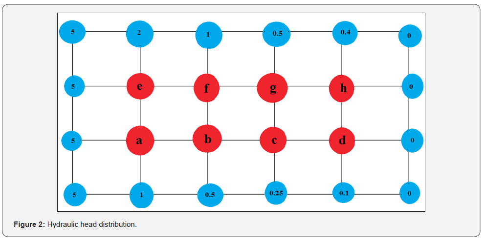
subunit and mTORC2 with a rictor subunit [10,11](Figure 1).
Amino acids are capable of activating mTOR [4,12].
The effect of amino acids in the regulation of protein synthesis seems
to be mediated mainly by leucine, through a mechanism that can be via
insulin or not. It is important to highlight that although amino acids
are fundamental as a substrate for protein synthesis, the simple
increase in intake does not induce hypertrophy per se [13]. Based upon
animal and cell culture studies, only leucine is required to induce
protein synthesis and mTOR signaling, however, in vivo human studies are needed to validate this hypothesis [14].
Knock-out animals for mTOR, present reduced postnatal
muscle growth and expressive reduction in size only of the fast-twitch
fibers with preservation of the slow ones despite severe myopathy [15].
Despite the activation of two complexes associated with mTOR, complex 1
has a more important role in protein synthesis, being also the target of
the inhibitory potential of rapamycin, whose treatment inhibits
development, muscle regeneration and compensatory hypertrophy caused in
experimental models by eliminating synergists [11,15].
As showed, although mTORC1 is capable of influencing
the 4E-BP1 (eukaryotic translation initiation factor) and S6K1 (S-6
Kinase 1) factors, only the latter is essential for muscle hypertrophy
and, in fact, its deletion it results in atrophy and prevents the
anabolic effect of the constitutive activation of Akt [5,15]. It has
also been shown that the protein AMP kinase (AMPK), activated when
intracellular energy levels are decreased, is able to block the inducing
effect of mTORC1 on muscle hypertrophy and AMPK-deficient mice
demonstrate hypertrophy [16,17]. Despite this, other factors must be
involved in the activation of hypertrophy independently of mTOR
activation, as rapamycin is able to only partially inhibit the
accumulation of ribosomal RNA necessary for protein synthesis and which
normally occurs after activation of mTORC1 in conjunction with the
synthesis of tRNA and nuclear RNA polymerases I, II and III [7].
Interestingly, it was affected that the transcription
factor Yin-Yang (YY1) is able to physically interact with mTORC1 and
mediate the expression of mTOR-dependent mitochondrial gene expression
[7]. Apparently, mTORC1 seems to be able to induce YY1 phosphorylation,
inducing the displacement of a transcription repressor complex of
several genes that directly contributes to the expression of numerous
genes involved in the insulin/IGF-1 signaling pathway, including IGF1,
IRS1, IRS2, Akt1 and Akt2 [7,18]. In this context, the administration of
rapamycin, by inhibiting mTORC1 and dephosphorylating YY1, recruits
again the repressor complex of the promoter region of these genes and
blocks its transcription [7].
A second muscle hypertrophy control pathway involves a
myostatin, a member of the transforming growth factor superfamily
(TGF-β). Myostatin is produced by skeletal muscle itself and negatively
regulates muscle growth [16]. The exposure of purified myostatin
inhibits the protein synthesis of cultured myotube cells and its
systemic administration promotes muscle atrophy in mice [16]. The
participation of myostatin in this phenomenon has been classically
represented by the high levels of hypertrophy observed in several
species of mammals that have mutations in the myostatin gene [18]
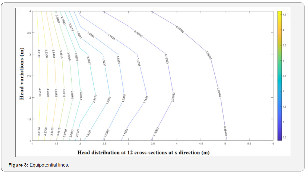
(Figure 2).
A protein known as follistatin has also been
identified, which is produced by skeletal muscle and capable of
inhibiting the action of myostatin, in addition to binding and
neutralizing the activity of activin-A, another member of the TGF-β
family that it also acts as a negative regulator of muscle growth [19].
Follistatin has a more profound impact on hypertrophy than the myostatin
gene deletion itself [20]. Myostatin and activin-A interact with each
other and activate heterodimeric receptor in a process that antagonizes
protein synthesis and muscle hypertrophy, and which can be inhibited by
administering the soluble form of activin-A type II receptor (ACVR2B)
[19]. The signal mediated by the myostatin/activin-A complex is believed
to occur through phosphorylation and nuclear translocation of
transcription factors SMAD2 and SMAD3 and formation of heterodimers with
SMAD4. Although the targets of these factors are not yet known, it is
speculated that they interfere negatively in the Akt-mTOR pathway
[21,22].
In fact, muscle hypertrophy produced by ACVR2B
transfection can be inhibited by rapamycin and that induced by
follistatin is blocked by inactivation in different segments of the
IGF-1/Akt/mTOR pathway [9,21]. Likewise, follistatin overexpression,
induced by viral injection in skeletal muscle, increases Akt
phosphorylation and signals the activation of protein synthesis, in a
process dependent on mTOR activation and which is suppressed by exposure
to rapamycin [7]. Apparently, inhibiting the interaction of SMAD3 with
follistatin is critical for Akt-mTOR signaling even though, as
demonstrated in other cell systems, SMAD3 is able to directly interact
with Akt and suppress protein synthesis in that environment [21,23].
Such evidence demonstrates that there is
communication between the myostatin/activin-A complex and the IGF-1
signaling pathway in order to control the degree of muscle fiber
hypertrophy [24]. However, it has been shown that other pathways are
also involved in the skeletal muscle hypertrophy process. These include
serum responsive factors (SRFs), which are required to induce
hypertrophy in synergists' ablation models, in a role mediated by
interleukins 6 and 4 (IL-6 and IL-4) which act in a paracrine manner to
induce proliferation and satellite cell fusion [25,26]. It has also been
shown that SRFs activate Akt through the microRNA, miR-486, which is
shown to be able to inhibit PTEN phosphatase, which negatively affects
PI3K-Akt signaling [27].
Androgens and beta-2 adrenergic agonists are equally
capable of activating the PI3K-Akt-mTOR pathway and contribute to
increasing the protein synthesis process. In this sense, the
administration of testosterone or mimetics that prioritize the
development of male secondary sexual characteristics are capable of
producing hypertrophy and animal models where there is a lack of
testosterone, it reduces Igf-1 mRNA, Akt phosphorylation and rate of
protein synthesis in processes that are reversed with nandrolone [19].
Similarly, clenbuterol or formoterol, beta-2 adrenergic agonists, are
able to activate, through the increase of cyclic AMP, Akt
phosphorylation, in a process that is blocked by rapamycin [28].
Another extracellular protein known as Wnt7a is
capable of contributing to hypertrophy through activation of satellite
cells and binding to the Fzd7 receptor, which is also involved in the
activation of the PI3K-Akt-mTOR pathway [29]. This process seems to be
enhanced by the positive impact of tensional changes in the sarcolemma
and membrane caveoli, which contain an abundance of the nitric oxide
synthetase enzyme in its neuronal isoform, which is abundant in skeletal
muscle. It is believed that the released nitric oxide would be able to
interact with superoxide anions produced by the contractile fiber
mitochondria during exercise, and promote the synthesis of
peroxynitrite, an aggressive reactive oxygen species that, however, has
already been involved in the activation of vanilloid receptors Trpv1
present in the sarcoplasmic reticulum, and subsequent release of calcium
ions. Thus, the moderate increase in the concentration of calcium ions
in the cytosol would be able to positively influence numerous metabolic
aspects and also activate mTOR to promote alterations in the protein
synthesis process [14,19].
Mechanic stimuli in the membrane would also be able
to activate mTOR through an Akt-independent pathway that involves the
participation of phosphatidic acid and activated phospholipase D [7,30].
On the other hand, the fourth isoform of the transcription factor
PGC-alpha was recently identified, whose isoform 1 is traditionally
involved in mitochondrial biogenesis and is targeted by AMPK.
Differently, PGCα-4 would be involved in increasing muscle mass and
strength, being able to repress the action of myostatin and, in fact, in
clenbuterol-induced hypertrophy it is abolished by deletion of the
PGCα-4 gene [16].
Finally, although we have previously discussed the
role of satellite cells in the hypertrophy process, it should be noted
that muscle regeneration recaptures embryonic and neonatal myogenesis in
a process that involves the activation, self-renewal, proliferation and
fusion of myogenic and near stem cells always includes immune
activation and inflammation [31]. Anyway, it is important to emphasize
that muscle hypertrophy may exist even in the absence of significant
activation of satellite cells and in fact, when this phenomenon is
stimulated by clenbuterol, the muscle growth process does not involve
satellite cell activation, as occurs in reintroduction of gravitational
load, after a period of intentional removal to induce muscle atrophy
[26,32].
Even in follistatin-induced hypertrophy signaling,
satellite cells do not appear to play a role [15]. However, in the
aforementioned experimental models, where synergists are intentionally
removed from the limbs of animals to produce compensatory hypertrophy of
the agonists, there is immediate proliferation and fusion of satellite
cells, with well-evidenced increases in the number of myonuclei in a
process that is also verified after eccentric contraction performed with
high load and for a long time and capable of producing micro-lesions
and localized inflammation [31]. Such experimental models are
traditionally performed in rats and involve the removal of the
gastrocnemius or tibialis anterior to observe, respectively, the
hypertrophy of the soleus or extensor digitorum longus [33,34].
Likewise, it is well demonstrated that IGF-1-mediated
hypertrophy involves the activation of satellite cells, and that the
cytokines IL-6 and IL-4 are directly involved in muscle growth in
experimental models involving overload [24]. Anyway, it is important to
remember that even with the elimination of 90% of satellite cells
through the use of toxins for PAX7, it is still possible to verify some
muscular hypertrophy. It seems evident that, in mammals, there may be
hypertrophy with or without the involvement of satellite cells [15,18].
In this context, acute and aggressive stimuli such as the elimination of
synergists or intense exercise with a high eccentric component and
presence of muscle damage, seem to necessarily involve the participation
of satellite cells for regeneration and compensatory adaptive
hypertrophy[35]. On the other hand, more gradual physical exercise
stimuli and experimental models that replace the load on one or both
limbs after intentional removal (functional overload model), the
verified hypertrophy, does not seem to involve the activation of
satellite cells, being exclusively associated to the increase of the
protein synthesis process [36,37].
Based on the above, we can verify the central, but
antagonistic, roles of IGF-1 and myostatin in controlling muscle
hypertrophy. The factors shown effect their actions through complex
intracellular signaling mechanisms. In addition, other mechanisms
contribute to the control of hypertrophy, such as amino acids, androgens
and inflammatory molecules. Satellite cells are not always involved in
hypertrophy, and there is an activation of them mediated or not by IGF-1
and other factors.