Wednesday, May 25, 2022

Conservative Versus Surgical Management for Non-Traumatic Subarachnoid Hemorrhage: A Mini Review - Juniper Publishers

 Head Neck & Spine Surgery - Juniper Publishers

Abstract

Subarachnoid hemorrhage diagnosis is always a challenge for patients and physicians because it has different presentations. There are two main causes of subarachnoid hemorrhage: traumatic and no traumatic subarachnoid hemorrhage. Both groups share clinical characteristics, but not treatment. Therefore, it is essential to recognize the signs, symptoms, and types of presentation for proper management. The objective of this article is to inform our audience about the significant difference between conservative and surgical treatment for non-traumatic subarachnoid hemorrhage because it carries a high risk of morbidity and mortality, requiring emergency management and well-trained physicians to evaluate patients suspicious for the diagnosis. This article is a compilation of several articles that have been selected from different databases, International Journal of Emergency Medicine, and Journal of Neurosurgery. Traumatic subarachnoid-related articles were excluded from our search. There is no consensus yet about the approach of patients with non-traumatic Subarachnoid Hemorrhage(ntSAH) among experts worldwide, so SAH diagnosis is often overlooked due to the clinical manifestations and inconsistencies in individual findings, especially atypical presentation arrives at the ER. The most significant limitations of timely and aggressive management of SAH are the lack of clinical suspicion and the delay from the CT scan order until the CT scan report is ready. The most repeated cause of subarachnoid hemorrhage is aneurysm rupture. A timely aneurysm repair is considered the most critical strategy to reduce the risk of aneurysm re-rupture. Therefore, detection of the cause of bleeding and prompt management can make the difference between life and death.

Keywords: Subarachnoid hemorrhage; Surgical Management of SAH; Conservative Management of SAH; Medical management of SAH; Nontraumatic Subarachnoid hemorrhage

Abbreviations: SAH: Subarachnoid Hemorrhage; NTSAH: Non-Traumatic Subarachnoid Hemorrhage; GCS: Glasgow Coma Scale; DCI: Delayed Cerebral Ischemia

Introduction

The most common cause of patients going to the emergency room is headache [1]. As physicians, we must be able to recognize headaches that could be life-threatening. The medical history and the physical findings will allow us to differentiate a simple headache from those that can be deadly. "The worst headache of my life," this is the way how medical students and physicians can start thinking about subarachnoid hemorrhage, but not all the patients can recognize the worst headache in their life; some of them die before they arrive at the emergency room. There are many tools and strategies to approach and treat patients with severe headaches, and we must understand the strengths and limitations of each strategy.

The clinical presentation should be considered before proceeding with the different diagnostic modalities for subarachnoid hemorrhage. Once a subarachnoid hemorrhage is suspected, a CT scan must be requested. Subarachnoid hemorrhage diagnosis is always a challenge for the physician. Every second and minute will determine a poor or great outcome in each patient. There are multiple causes of subarachnoid hemorrhage (SAH), but we can differentiate two big groups: Traumatic and no traumatic subarachnoid hemorrhage. Both groups share some similar clinical characteristics, but not the treatment.

There are several scales to categorize SAH. The systems used to predict the patient outcome are the Hunt and Hess score and World Federation of Neurological Surgeons grading, and the Fisher grade helps predict vasospasm. In terms of patient-centered results and prognosis, specific scores were not performed better than the Glasgow Coma Scale (GCS). As we search into the diagnosis of SAH, it is essential to note that some patients with SAH, for example, Hunt and Hess lower grades are more commonly failed to see because the clinical presentation is mild, and they may have smaller aneurysms with scant subarachnoid blood. These patients do not necessarily recover or have less morbidity with rupture or re-rupture [1].

Accurate data is not available about the management of subarachnoid hemorrhage, but some of the Egyptian, Greek, and Arabic literature report some clues of earliest management in 1800. Unfortunately, the management is still controversial, especially when the discussion is between surgical and medical management of SAH. That is why reviewing many studies worldwide will allow colleagues to understand how to face this particular situation.

The most crucial strategy to reduce the risk of aneurysm re-rupture is timely aneurysm repair is generally considered. However, evidence for the ideal timing of treatment is limited, and it is undefined if ultra-timely treatment (within 24 hours) is superior to timely aneurysm repair (within 72 hours) [2]. A recently published retrospective data analysis that compares ultra-early treatment with repair performed within 24-72 hours after hemorrhage suggests that aneurysm occlusion can be performed safely within 72 hours after aneurysm rupture [2]. The American Heart Association/American Stroke Association suggests as a Class IB guidance that surgical clipping or endovascular coiling of the ruptured aneurysm should be implemented as early as achievable in most patients to decrease the risk of re-bleeding after SAH [2]. The treatment modality option between surgical clipping and endovascular coiling is a complex endeavor that requires an interdisciplinary team's expertise, including neuro-intensivists, interventional neuroradiologists, and neurovascular surgeons. The endovascular approach is superior for aneurysms to be considered equally treatable by both modalities, associated with better long-term outcomes.

Retrospective data on clipping and coiling in poor-grade patients suggests that surgical clipping and endovascular are equally effective. An early and short course of an antifibrinolytic drug, including tranexamic acid, started as soon as the radiological diagnosis of SAH is made and stopped within 24-72 hours, has been associated with a decreased rate of ultra-early re-bleeding and a non-significant improvement in long-term functional outcome. This approach remains controversial, and short-term administration of tranexamic acid to prevent re-bleeding is being studied in a multicenter randomized trial (Dutch Trial Registry number NTR3272) [3]. The avoidance of extreme levels of blood pressure is another medical intervention applied to prevent aneurysm re-rupture. The American Heart Association/American Stroke Association and the Neurocritical Care guidelines advise keeping the mean arterial blood pressure below 110mm Hg or systolic blood pressure below 160mm Hg (or both) in the presence of a ruptured unsecured aneurysm.

Serum biomarkers to detect the risk of delayed cerebral ischemia (DCI) are showing promising results [3]. Changes in serum protein S100B levels interacted with DCI status (presence vs. absence): F= 3.84, p= 0.016. Patients with DCI had higher S100B concentration level on day 3 than those without DCI (3.54±0.50ng/ml vs. 0.58±0.43ng/ml, p= 0.001). S100B concentration on day 3 following a SAH predicted DCI (p= 0.006). The multivariate logistic regression analysis has shown that impaired cerebral autoregulation and elevated S100B concentration on day three increase the likelihood of DCI [3]. Subarachnoid hemorrhage (SAH) is a medical emergency that requires urgent management. Around Eighty-five percent of cases of atraumatic SAH result from a ruptured aneurysm. Other factors such as arteriovenous malformation, Ehlers-Danlos disease can also be the cause [4].

The diagnosis of SAH ought to be considered in any patient with a severe and sudden onset or rapidly escalating headache. With many such patients presenting to the ED with a chief complaint of headache, differentiating those with a benign cause from an emergent etiology such as SAH can be difficult. Establishing the diagnosis of SAH, the most critical time-sensitive goals include confirmation of airway security and stabilization of hemodynamics. In the setting of a low Glasgow Coma Scale Score or the lack of ability to protect the airway, intubation should be undertaken, but care should be taken to mitigate increases in mean arterial pressure during the intubation process [5]. These therapeutic modalities should be addressed with the admitting neuro-intensivist or neurosurgery team. In addition, continuous electroencephalogram monitoring may be started in the intensive care unit.

It is essential to determine adequate management in every case, as this can be the difference between life and death. According to preoperative neurologic function, location, size of the aneurysm, the timing of the operation, severe initial bleeding, re-bleeding (usually within two weeks), and delayed ischemia were the major preoperative problems; ten percent died, and 13 percent deteriorated before surgery. Operative mortality was 5 percent, ranging from 1.6 percent of patients with normal preoperative neurologic function to 35 percent of severely disabled patients. Intraoperative complications (5 percent of cases) related primarily to the size and location of the aneurysm, postoperative delayed ischemia (minor and reversible in 10 percent and severe in 5 percent) related to operation timing and occurred primarily in patients afflicted within the previous ten days [4]. The outcomes of surgical treatment, including preoperative deaths, were better than the natural history of the illness. The difference became apparent after one month of observation.

Once a bleeding aneurysm is identified, the ultimate therapeutic goal is to secure it surgically by coiling or clipping. While coiling is the preferred method since it is less invasive than open surgical clipping, data is indeterminate as to whether long-term outcomes are better with either procedure, but protocols propose that coiling should be performed if both are possible [6]. In some cases, tortuous vascular anatomy or other contraindications to coiling make open surgery necessary. Timely treatment and securing the aneurysm are associated with a lower risk of re-bleeding. If surgical treatment is delayed, antifibrinolytics such as aminocaproic acid may be used for a short time to mitigate the risk of re-rupture [6].

Nine articles have been selected from Pubmed, Google Scholar, International Journal of Emergency Medicine, Journal of Neurosurgery, International Journal of Emergency Medicine, and other Databases. The articles were published within the previous ten years and written in the English language. The studies reviewed include review articles, clinical articles, systematic reviews, single-center, retrospective studies, prospective, multicenter cohort studies, cross-sectional studies, observational studies, and clinical trials. Traumatic subarachnoid-related articles were excluded from our search. The objective of this article is to inform our audience about the significant difference between conservative and surgical treatment for non-traumatic subarachnoid hemorrhage.

Discussion

There is no consensus about treating patients with hemorrhage (no traumatic Subarachnoid Hemorrhage) among expert clinicians within the United States and worldwide. Many concerns arise from an attempt to establish a protocol for the individual patient. However, at least in some areas, the wide variety of management practice testifies to a lack of agreement in the medical community. Therefore, we sought to design a survey that would highlight areas of controversy in the modern management of ntSAH and identify specific areas of interest for further research. Additionally, we performed a comprehensive review of the existing literature on several of these controversial subtopics in the management of ntSAH [7].

Although the timing of surgical intervention after SAH is controversial, it should be based on the clinical-grade, site of the aneurysm, and patient's medical condition. There are many factors to consider when treating patients with SAH, such as patient neurological condition and aneurysm location (Ex. Basilar aneurysms) aneurysms, unusually large or irregular aneurysms [8]. Patients with a non-peri mesencephalic SAH have an increased risk of a worse neurological outcome. Therefore, these patients should be monitored attentively. When an aneurysm breaks down, patients require a calcium channel blocker to reduce vasospasm risk due to ischemia. For example, The Mayo Clinic experience of 1,947 patients who underwent surgical treatment because of aneurysmal SAH or aneurysmal repair for about 20 years shows the results after a follow-up that 1,445 had an excellent outcome, 231 had an acceptable outcome, 171 had a poor outcome, and 100 died. Aggressive management can benefit many patients with severe neurologic injury after SAH by preventing rupture of the aneurysm, attenuating the severity and sequelae of vasospasm, and decreasing the surgical complications [8].

Clinically, subarachnoid hemorrhage diagnosis is often missed due to the various clinical manifestations and inconsistencies in individual findings, especially when atypical presentation arrives at the ER. In addition, there are several etiologies of non-traumatic SAH, such as perimesencephalic SAH, intracranial arterial dissection, pituitary apoplexy, mycotic aneurysms, reversible cerebral vasoconstriction syndrome, cerebral venous sinus thrombosis, moyamoya, vasculitis, and even cocaine use [9]. When SAH is suspected, the best initial step would be a CT scan of the head or LP. Once the diagnosis of SAH hemorrhage has been made, it is essential to classify and grade the patient's risk to lead to the urgency of further management and prevent neurological consequences [9].

Subarachnoid hemorrhage carries a high risk of morbidity and mortality, requiring emergency medicine physicians to evaluate patients suspicious for the diagnosis cautiously. It is crucial to consider the restrictions of diagnostic modalities and early implementation of grading/scoring systems even in a nontraditional presentation. Giving the SAH complications, making a timely diagnosis, initiating management in the ED, and employing suitable consultations or admission for possible early intervention is crucial for care [9].

The two most significant limitations of timely and aggressive management of SAH are the lack of clinical suspicion from physicians and the delay from the CT scan order until the CT scan report is ready [8]. We suggest starting a SAH standardized protocol that includes the high priority of imaging studies (CT scan) to reduce the time from diagnosis and management. Performing a prospective cohort study using the protocol could lead us to better conclude aggressive and early management in non-traumatic SAH.

Limitation

This systematic review uses data collected in nine articles that included cohort studies, a cross-sectional study, and several observational studies and clinical trials. Given the nature of this investigation (secondary data review), the main limitation of this study is the lack of control over the desired study population, variables of interest, and the study design. Problems with secondary data could be that bias may have crept meanwhile obtaining the data; this bias will go unnoticed and may inadvertently affect the results.

Furthermore, the primary data may not include certain demographic information (e.g., respondent zip codes, race, ethnicity, and specific age) relevant to the study. For example, in the specific case of this investigation, age, availability of conditions to perform endovascular procedures, the severity of the SAH, and other variables could be ignored. In such cases, the data would create an aggregate pooled effect that may be misleading if there are important reasons to explain variable treatment effects across different types of patients.

In addition, secondary data analysis research cannot establish causality. This kind of investigation is limited to descriptive, exploratory, and correlational designs and nonparametric statistical tests. By their nature, they are retrospective, and the investigator cannot examine causal relationships (by a randomized, controlled design).

These significant limitations were addressed and minimized by:

1. Assuring that the correct type of studies was eligible for the review and guaranteeing that identifying all relevant information was comprehensive.

2. Considering publication bias.

3. Confirming that the methods used in each study were appraised and had an appropriate data abstraction.

Conclusion

Non-traumatic subarachnoid hemorrhage is a medical emergency. Early diagnosis and adequate management are crucial for a patient's survival. Therefore, conservative or surgical management should be promptly established. Intense headache is one of the most common alarm symptoms of non-traumatic subarachnoid hemorrhage that bring a patient to the emergency room; frequently described as “the worst headache of my life”. There are many tools and strategies to approach and treat our patients with severe headaches, and we must understand the strengths and limitations of each strategy.

One of the most frequent causes of subarachnoid hemorrhage is aneurysm rupture. This can be caused by certain conditions such as arteriovenous malformation, Ehlers-Danlos disease, collagen deficiencies, uncontrolled high blood pressure, uncontrolled Diabetes Mellitus. A timely aneurysm repair is considered the most vital strategy to reduce the risk of aneurysm re-rupture. However, evidence for optimum timing of management is insubstantial, and it is unclear whether ultra-early actions to resolve the subarachnoid hemorrhage (less than 24 hours) are superior to early aneurysm repair (within 72 hours) [2].

Retrospective data on clipping and coiling in low-grade patients suggests that surgical clipping and endovascular are equally effective. Early detection of the cause of bleeding and prompt determination of management can make the difference between life and death, as it requires prompt and adequate management.

Click here: https://juniperpublishers.com/index.php  

Tuesday, May 24, 2022

Clinical And Evolutive Characteristics in Acute Toxic Methemoglobinemia in Children - Juniper Publishers

 Open Access Journal of Toxicology - Juniper Publishers

Abstract

Introduction: Acquired or toxic methemoglobinemia is the result of exposure by swallowing, inhalation or dermal absorbtion of medicines or non-drug chemicals with oxidizing properties. The main objectives of the study were to highlight the main causes of methemoglobinemia in children, to identify the associated factors and their influence on the clinical form of the disease as well as the response to antidote treatment with methylene blue.

Methods: All patients with acute toxic methemoglobinemia hospitalized over a period of six years were included in the study, the inclusion criteria being: age < 18 years, suggestive symptomatology (generalized cyanosis, asthenia, dyspnea) and methemoglobin values >3%.

Results: 82 patients with acute toxic methemoglobinemia were identified of which the majority (94%) were between 0-5 years. 69 cases were secondary to exposure to nitrates from water used to prepare food. The severity of methemoglobinemia was significantly higher among anemic and dehydrated patients. All patients received antidote treatment with methylene blue one or more administrations, and all had a favorable outcome.

Conclusions: Acute toxic methemoglobinemia is mainly observed in children up to 5 years of age, the majority being infants. The main cause of it in children is exposure to exogenous substances among which nitrates that contaminate well water occupy the first place. Age under 1 year, hemoglobin values lower than 11g/dl, and acute dehydration syndrome represent risk factors for severe forms. Methylene blue is the treatment of choice for acute toxic methemoglobinemia in children and its administration is effective in the majority of cases.

Keywords: Acute toxic, Methemoglobinemia, Children, Methylene Blue

Introduction

Acquired or toxic methemoglobinemia is defined as an increase in methemoglobin blood levels above 1%, as a result of exposure to medicines or chemicals with oxidizing properties. The medicinal etiologies of toxic methemoglobinemia include, acetanilide, phenacetin; local anesthetics (benzocaine, lidocaine, prilocaine, and articaine), chloroquine, dapsone, flutamide, metoclopramide, nitric oxide, nitroglycerin, nitroprusside of sodium, antibiotics of the class of nitrofurans and sulfonamides, sodium nitrate, silver nitrate, silver sulfadiazines, antiepileptics (sodium valproate, phenytoin), phenazopyridine, sulfasalazine, and zopiclone [1]. Among non-drug chemicals that can produce methemoglobinemia, the most common include aniline-based paints, silver nitrate, naphthalene, trinitrotoluene, nitrobenzene, Vicia faba, nitrates and nitrites from well water or contaminated vegetables, paraquat, phenol, or fires [1].

Acute toxic methemoglobinemia in children can be triggered by any of the above-mentioned xenobiotics It has the potential to cause severe injury or death but if recognized early, patients can significantly benefit from antidote treatment. The antidote of methemoglobinemia is methylene blue a thiazine coloring agent, which in the presence of NADPH-dependent methemoglobin reductase and nicotinamide adenine dinucleotide phosphate causes the reduction of methemoglobin and its subsequent transformation into hemoglobin. Antidote treatment with methylene blue is commonly indicated in all symptomatic patients or cases where methemoglobin concentration is above 30% [1].

In Romania acute toxic methemoglobinemia in children was recognized as a public health problem for many years [2]. A national report of the Ministry of Health published in 2012 revealed that Romania is characterized by the existence of numerous rural territories with frequent and important nitrogenous substances contamination [2]. Nitrates contaminate the soil from where water infiltrates into artisanal wells existing in rural areas. The toxicity of these salts is due to their transformation into nitrites under the action of bacteria at the level of the soil or gastrointestinal tract of the child. Nitrites exert a direct oxidant effect on hemoglobin, which subsequently loses its physiological capacity for carrying oxygen. The level of nitrates in the water depends on several elements, and hence it is difficult to quantify soil permeability, well depth < 15 m, agricultural activities, and inadequate measures for eliminating organic waste, making it difficult to identify populations at risk based only on geographic origin [3]. The World Health Organization has regulated the maximum permissible concentration of nitrates (NO₃) in drinking water to <50 mg/L [4]. Various local research has been proven that in countries like Romania, a country with poor infrastructure in rural areas, exposure to nitrates remains the main cause of acute toxic methemoglobinemia in children [5].

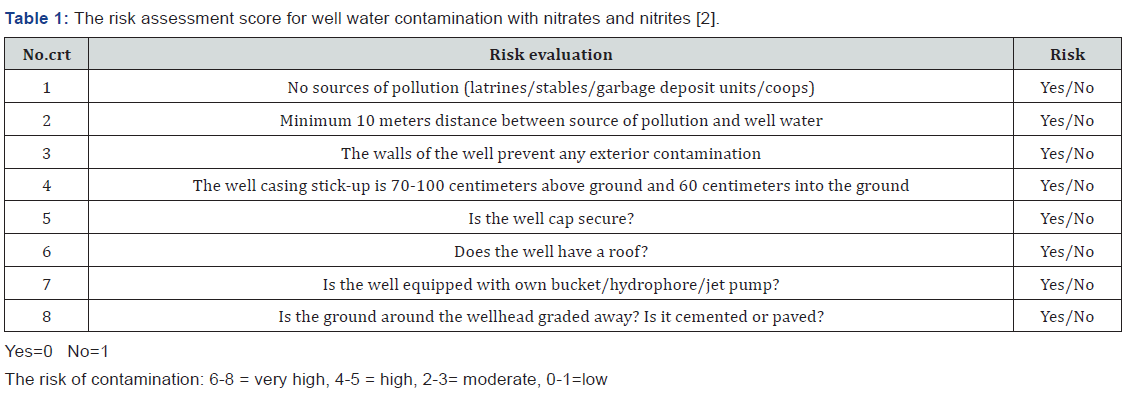
To come to health care specialists’ aid, the Ministry of Health of Romania has developed a score for assessing the risk for well water contamination with nitrates and nitrites (Table 1) [2].

The research presented in the study details a retrospective study including cases of acute methemoglobinemia that were hospitalized and treated at the Pediatric Poisoning Center of the Emergency Clinical Hospital for Children”Grigore Alexandrescu” in Bucharest Romania and analyzed , for a period of 6 years. The main objectives of the study were to highlight the main causes of acute methemoglobinemia in children, to identify the associated factors (age and associated pathology), and their influence on the clinical form of the disease as well as the response to antidote treatment with methylene blue. The study design will be of significant help to global researchers and pharmaceutical firms looking forward to analyze methemoglobinemia in study populations in their respective countries.

Methods

All patients with acute toxic methemoglobinemia hospitalized at the Pediatric Poisoning Center of the Emergency Clinical Hospital for Children “Grigore Alexandrescu” in Bucharest, Romania between January 01, 2015, and December 31, 2020, were included in the study. The inclusion criteria were age < 18 years, patients with suggestive symptomatology (generalized cyanosis, asthenia, dyspnea, and altered neurological status), and methemoglobin values > 3%. For this purpose, we used the computer data base of clinical consultation sheets and medical reports upon discharge of patients using for searching the ICD 10 code of diagnosis for acute toxic methemoglobinemia. The following data were analyzed: medical history data, demographic characteristics, data provided by clinical examinations from patient hospitalization and daily evaluations during hospitalization, the presence of methemoglobinemia (measured by spectrophotometric method), and associated conditions. The therapy regimens provided to the patients and the outcome of each patient were also analyzed, including the duration of hospitalization, the need to repeat the dose of the antidote, and admission to the intensive care department. The data obtained were statistically processed using the IBM® SPSS® Statistics 20program, considering significant results of statistical probability (p) less than 0.05.

Results

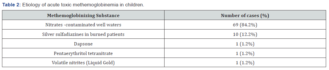
During the mentioned period, 82 patients with acute toxic methemoglobinemia were identified representing a percentage of 1.58% from the total of 5122 patients diagnosed with acute intoxication in the Pediatric Poisoning Center in Bucharest. Etiology Regarding etiology: clinicians ascribed 69 cases as secondary to exposure to nitrates from well water used to prepare food. The risk assessment score for well water contamination with nitrates and nitrites (Table 1) was used, all the 69 children having a risk score between 6 to 8. Ten cases were secondary to topical exposure to silver sulfadiazine used to treat severe burns, two cases were instances of medicines poisoning: one patient had accidental acute poisoning with pentaerythritol tetranitrate, (a coronary vasodilator) and another patient was undergoing chronic treatment with dapsone. In the study group, a case of acute toxic methemoglobinemia was identified, wherein the patient was a chronic consumer of drugs of abuse and was hospitalized following an overdose of volatile nitrites by inhalation (cigarettes soaked with a street substance known as “Liquid Gold”) (Table 2).

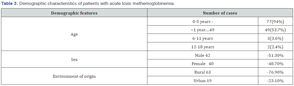
The average age of the patients was 23.11 months, and the median age was 6 months. The majority of patients were under 5 years old: 77(94%), The infants subcategory (children aged under 1 year) within this age group was most represented with 49 patients (53.7% out of the total of cases). At the same time the number of cases was much lower for other age groups (Table 3). Cases among patients of rural origin were significantly higher than those from urban areas (63 versus 19 cases, p<0.05). In addition, rural origin was significantly associated with exposure to nitrates contaminated well water (binomial test, p<0.0001). The study group included 42 boys and 40 girls, the difference in patient sex being insignificant (Table 3). The most common route of exposure was the oral route (87%), followed by cutaneous (12.2%), and inhalation (0.8%). In the majority of cases (85.3%), toxic exposure was accidental.

The mean methemoglobin concentration in the study group was 41.53%, with values ranging from 6.6% to 87% (Table 4). The mean value of methemoglobin for cases of exposure to nitrates -contaminated well water was 41.5%, and it was 39.2% for cases involving topical exposure to silver sulfadiazine, used in patients with severe burns. The methemoglobin values were: 41.9% for case of accidental acute poisoning with pentaerythritol tetranitrate, 44.2% for voluntary inhalation, with new substance of abuse “Liquid Gold “and 6.6% for a patient undergoing dapsone chronic treatment. Since the number of cases caused by exposure to dapsone, pentaerythritol tetranitrate, and new substance of abuse “Liquid Gold “(volatile nitrites) was small (one per etiology), we could not perform a statistical analysis of methemoglobin concentrations in these etiologies. Regarding the mean concentrations of methemoglobin secondary to exposure to nitrates contaminated well water and silver sulfadiazine, the difference was not statistically significant.

Clinical Manifestations:

Upon admission to the toxicology department, all patients showed clinical manifestations with different grades of severity. Clinical manifestations are summarized in Table 4.

The lowest concentration of methemoglobin recorded in this study was 6.6%. Regardless of the methemoglobin concentration, all patients experienced altered general conditions and cyanosis. For values between 6.6% and 20%, other clinical manifestations besides cyanosis, including dyspnea and agitation, were observed in 2 of the 10 identified cases (20%). The majority of patients with methemoglobin values between 20% and 50%, had dyspnea (24.4%), tachycardia (19.5%), psychomotor agitation (31.7%), and drowsiness (12.2%). Those with methemoglobinemia between 51% and 70% showed clinical manifestations, with a higher frequency of dyspnea (37%), tachycardia (29.6%), and altered sensory function (25.9%). Two patients in this group (7.4%) experienced generalized tonic-clonic seizures. These two patients had not preexisting neurologic disease and were 3 months respectively 7 months old. Seizures were ceased after 5 mg diazepam intrarectal administration and required one dose of 1 mg/kg of methylene blue.

All patients with a methemoglobin value above 70% experienced tachyarrhythmia, dyspnea, and impaired sensory function (Table 4). Evolution was favorable in all cases, and all patients were declared cured upon discharge.

Associated pathology We analyzed the associated pathology presented by patients, as shown in medical documents (Table 5). Of the 82 patients, 58 (70.7%) had a blood count at the time of admission, which revealed hemoglobin levels below 11g/dL. We found that the severity of methemoglobinemia was significantly higher among patients with hemoglobin levels below 11g/dl., (p=0.03). The concomitant presence of respiratory tract infections, cardiac pathology, or malnutrition was not significantly associated with a greater severity of methemoglobinemia. Eight patients (9.76%) experienced manifestations suggestive of an acute enterocolitis (fever, diarrhea, and vomiting). Of these in three cases, clinical and paraclinical changes of acute dehydration syndrome were obvious. The presence of dehydration syndrome was associated with a greater severity of methemoglobinemia (Mann-Whitney test, p<0.05).

A greater severity of methemoglobinemia was correlated with certain characteristics, including patients from rural areas, use of nitrates contaminated well water, age <1 year, presence of dehydration and hemoglobin values bellow 11g/dl, in a statistically significant manner. We analyzed the direct relationship between the severity of methemoglobinemia and the common presence of these parameters using multilinear regression. The results proved that the relationship between co-factors is complex and does not allow the establishment of a linear relationship between the severity of methemoglobinemia and a certain factor.

All patients received antidote treatment with methylene blue 5mg/mL solution intravenously, at a dose of 1mg/kg. The clinical status and methemoglobin value were analyzed during the first 24 hours post administration. In 60 cases (73.2%) cyanosis and the other symptoms disappeared in the first 60 minutes and no further administrations were required Of these 60 patients who received a single dose of antidote, 9 had methemoglobin values <20%, 31 had values ranging between 20 and 50%, and 20 had concentrations above 50% at the moment of diagnosis. Eighteen patients (21.9%) required two doses of methylene blue (1mg/kg), among whom 1 patient had an initial methemoglobin concentration <20%, 6 patients had values between 20% and 50%, and 11 patients had values >50%. Among the 4 patients (4.9%) requiring three doses of antidote, one patient had a methemoglobin concentration <20%, while the rest had concentrations above 50% before starting treatment.

Of the 22 patients who required additional doses of the antidote 20 (91%) had hemoglobin values bellow 11g/dl at the time of diagnosis. However, no significant association was reported between the need for additional doses of antidotes and the presence of hemoglobin values bellow 11g/dl. (Mann Whitney test, p=0.36). It was necessary to examine whether the lack of response to methylene blue administration is secondary to other causes. However, since this was a retrospective study, we did not have the values of glucose-6-phosphate dehydrogenase (G6PD) activity or Sul hemoglobin concentration. For all patients with acute dehydration syndrome, additional doses of antidote were required; in this situation, a statistically significant correlation (Mann-Whitney test p=0.009) was observed. All patients had a favorable outcome. In the analyzed group, there were no deaths and two patients required admission to the intensive care unit. The average duration of hospitalization was 3days; however, the majority of patients (51.2%) were discharged after 24h of staying.

Discussion

Acute toxic methemoglobinemia is a rare condition in pediatric toxicology, as evident in the present study, which revealed a prevalence of 1.58% of all intoxication cases in children. However, it can lead to severe functional anemia, especially in infants or in cases of associated pathologies. Early identification allows for quick initiation of antidote treatment with methylene blue, ensuring an excellent prognosis for patients.

Based on the data detailed above, the present study proposed a detailed analysis of cases of acute toxic methemoglobinemia from the point of view of etiology, clinical manifestations, associated factors (age, associated pathology), and their influence on the clinical form of the disease, as well as the outcome under treatment. The limitations of this analysis include difficulty in quantifying the extent to which each factor is independent of the severity of methemoglobinemia. the small sample size that hindered the predictive model, the difficulty associated with the evaluation nitrate concentrations in the wells of the patients and bias created by the type of admissions in an emergency hospital like the one we were referring to a large number of chemicals or drugs have been reported in literature as capable of producing oxidation of hemoglobin with the accumulation of methemoglobin. However, in this study, we identified only five categories of toxic substances inducing acute methemoglobinemia in children. Most of the cases in this study - 69 (84.1%), were secondary to exposure to nitrates contaminated well water used to prepare food in rural areas.

In a report published in 2009 by the National Institute of Public Health of Romania within the National Programme for Monitoring the Triggering Factors in the Life and Work Environment, 89 cases of toxic methemoglobinemia have been reported as a source of food contamination [5]. In the group of patients analyzed in the present study consisting exclusively of children, this source was not identified. In all cases, anamnesis referring the living conditions , using the risk assessment score for well water contamination with nitrates and nitrites [2]. indicated that the water used in the preparation of tea or milk was the source of the disease.

In 12 patients, the drug-induced cause of acute methemoglobinemia was identified. Of these, 10 patients (12.2%) were children with severe burns of at least II degree on extended body surfaces, who experienced episodes of acute methemoglobinemia during hospitalization. Analyzing the substances with the oxidant potential to which the children were exposed, we found that the common element was the repeated topical application of silver sulfadiazine on extended skin surfaces.

Silver sulfadiazine is an antibiotic of the sulfonamide class and exerts antibacterial and antifungal effects through the simultaneous action of silver ions and sulfadiazine. Its application on extensive skin areas and/or with significant destruction of superficial layers, such as burns, favors systemic action, leading to adverse effects similar to those of sulfonamides, including oxidation of hemoglobin with the formation of methemoglobin.

Another drug identified in our study as an inducer of methemoglobinemia was dapsone (dianinodiphenyl sulfone), a sulfone-class compound, considered in literature as one of the most common causes of toxic methemoglobinemia [6,7]. Dapsone is used in immuno depressed patients for the treatment of infection with Pneumocystis jiroveci or Toxoplasma gondii, in the treatment of leprosy, malaria, or some dermatological diseases, such as bullous pemphigoid and dermatitis herpetiformis.

In our study, dapsone was identified in a 4-year-old boy who was undergoing treatment for bullous pemphigoid. Clinical manifestations occurred at a methemoglobin concentration of 6.9%, which is consistent with the findings of other studies [8,9], confirming that therapeutic doses may trigger suggestive symptomatology at serum methemoglobin levels < 10%. Combination with other drugs with oxidant effects or concomitant presence of other diseases, such as heart, respiratory disease, and anemia, are considered to be promoting factors [9,10]. In the analyzed case, we only identified anemia with a hemoglobin level of 9.7 g/dL.

Another drug-induced cause of acute toxic methemoglobinemia was pentaerythritol tetranitrate.an organic nitrate that can induce methemoglobinemia in case of accidental or voluntary overdose. Methomoglobinemia occurs especially in cases of accidental or voluntary overdoses, with limited cases of methemoglobinemia occurring because of therapeutic doses.

In the analyzed group, we identified a female patient aged 2 years who presented with acute toxic methemoglobinemia due to accidental acute poisoning with pentaerythritol tetranitrate, with a methemoglobin concentration of 41.9%.

In one case, volatile substance were identified as etiological agents of methemoglobinemia. It was about a teenager aged 16 years, institutionalized, chronic consumer of substances of abuse, who presented with suggestive clinical manifestations: generalized cyanosis not responsive to oxygen administration, dyspnea, and tachycardia about an hour after smoking cigarettes soaked in a liquid street substance named “Liquid Gold”

Volatile abuse substances are another category with a well-documented role among the causes of toxic methemoglobinemia [11]. Volatile nitrites (amyl nitrite or isobutyl nitrite) are esters of nitric acid, which cause an intense vasodilating effect, with a rapid but short onset at the level of the central nervous system [12]. Although there are legislative regulations regarding the possession or sale of volatile nitrites in Romania as in the other European countries, these substances are available under various names: “Liquid Gold”, “Rush”, “Pig Black”, and are used both for inhalation and application in classical cigarettes. Most often, users of volatile substances are male adolescents, with poor socio-economic backgrounds, from disorganized families, and have low educational qualifications [13].

Although exposure to substances with oxidant potential may cause the accumulation of toxic concentrations of methemoglobin regardless of age, in the pediatric population, cases involving infants present the highest risk [14,15] due to functional particularities. The main peculiarity is the predominance of fetal hemoglobin, especially up to the age of 3 months, which can be oxidized much more easily than in adult form, which becomes predominant after 18 months of age. At this age, cytochrome b5 methemoglobin reductase activity is reduced [16], and gastric pH is less acidic than in older children or adults, thus facilitating bacterial proliferation, which plays an important role in converting nitrates in food into nitrites [17,18]. The analysis of the group of patients provided consistent results; the age subcategory of under 1 year being the best represented with 49 infants (59,7%) ,and the median age in the studied group was of 6 months.

Clinical manifestations are similar regardless of the incriminated oxidant substance and depend primarily on the concentrations of methemoglobin. In the vast majority of cases, levels below 10% are well tolerated, and patients are asymptomatic [15,19]. Cyanosis is the main clinical manifestation and is an indispensable clinical element for positive diagnosis along with an increase in methemoglobin values, as evidenced by literature. Most previous studies have shown that cyanosis becomes obvious at methemoglobin concentrations of more than 10%. However, in our study, the lowest value at which cyanosis was noted was 6.6%. As methemoglobin concentrations increase, the clinical picture is supplemented with changes secondary to hypoxia: cerebral symptoms (agitation, restlessness, irritability, headache and subsequently drowsiness, obnubilation, seizures, or coma), cardiac symptoms (tachyarrhythmias, circulatory failure), or respiratory symptoms (dyspnea, tachypnea, respiratory failure). Values above 70% are generally associated with an increased risk of death. In our study, the severity of clinical manifestations varied with the concentration of methemoglobin, but cases with values above 70% (72%–86% in our study) had a favorable outcome and no death was reported. There were 4 cases with methemoglobin values above 70%. All of them were infants and in addition to cyanosis presented tachycardia dyspnea and neurologic symptoms (drowsiness or agitation) Of these, 3 required three administrations of methylene blue and 1 required two doses.

In case of association with other pathologies that cause cell hypoxia (respiratory, cardiac, hematological conditions), the symptomatology is more severe at reduced concentrations of methemoglobin [15,20]. The analyzed group included a large number of patients with such associated pathologies, identifying a significant correlation between the presence of hemoglobin levels below 11g/dl, and the severity of clinical manifestations in patients with methemoglobinemia. Another situation encountered in the study, which has been significantly associated with a higher severity of the clinical picture of methemoglobinemia, was acute dehydration syndrome in patients with acute enterocolitis, most likely secondary to associated metabolic acidosis.

Exposure to methemoglobin zing substances can be life-threatening, so that the antidote treatment with methylene blue can be administered as soon as possible. Methylene blue is a thiazine coloring agent, which in the presence of NADPH methemoglobin reductase and NADPH is reduced to an intermediate metabolite, methylene blue leuco, capable of donating an electron to methemoglobin to reduce it to oxyhemoglobin.

The indication to initiate antidote treatment is symptomatic methemoglobinemia, since there are situations where the concentration of methemoglobin does not correlate with the severity of clinical manifestations [21]. The dose is 1-2mg/kg, administered intravenously over 3-5min, and repeated, if necessary, when the symptomatology does not cease after 60 min, up to a maximum dose of 7 mg/kg [9,15].

Although extremely effective in a vast majority of situations, the administration of methylene blue is not without risk. Digestive side effects (nausea and vomiting), electrocardiographic abnormalities (inversion of T wave, flattened R waves), profuse sweating, dyspnea, retrosternal pain, and oral dysesthesia usually occur at doses above 7 mg/kg [9]. It can also cause a paradoxical increase in methemoglobin through its oxidant effect, especially in patients with glucose-6 phosphate dehydrogenase deficiency. In the study group, methylene blue administration was limited to a single dose in most situations, 73.2% (60 patients), and the majority (51 of 60) had methemoglobin concentrations over 20%. In 22 cases, it was necessary to administer one or two additional doses, most of them [16] having hemoglobin levels lower than 11g/dl. Even that there were not possible to establish a significant association between the need to supplement the initial dose and the presence of hemoglobin levels lower than 11g/dl. The total dose of methylene blue used in the treatment of patients in this group did not exceed 3 mg/kg, and no adverse effects of the treatment were reported.

Conclusion

Acute toxic methemoglobinemia is mainly observed in children up to 5 years of age. This age group represents 94% of all cases in our study, of which the majority belong to the subcategory of 0-1 year. In countries as Romania where the rural infrastructure is less developed the main cause of acute toxic methemoglobinemia in children is the exposure to nitrates contaminated well water used for food preparation. Silver sulfadiazine cause acute toxic methemoglobinemia when applied to extensive areas, repeatedly and/or under the condition of significant destruction of the surface layers of the skin. Methemoglobinemia secondary to exposure to volatile substances, such as amyl nitrile or isobutyl nitrile, used as new psychoactive substances, is a newly described condition in recent years and must remain under the attention of specialists.

Although the relationship of co-factors is complex and is difficult to quantify to what extent the severity of the clinical picture is in linear correlation to a certain factor. We believe that the age under one year, hemoglobin values lower than 11g/dl, and acute dehydration syndrome are factors that can aggravate clinical symptomatology and outcome in acute toxic methemoglobinemia.

Methylene blue is the treatment of choice for acute toxic methemoglobinemia in children, and its administration is effective in a single dose of 1mg/kg in 73.2% of cases without adverse reactions.

Click here: https://juniperpublishers.com/index.php  

Monday, May 23, 2022

Knowledge and Utilization of Iodized Salt and Associated Factors among Households of Fitche Town, Central Ethiopia, 2020: A Community based Cross-Sectional Study - Juniper Publishers

 Public Health - Juniper Publishers

Abstract

Adequate knowledge and proper utilization of iodized salt in the household is a significant factor in the prevention of iodine deficiency disorders. This study was aimed at assessing knowledge and utilization of iodized salt and associated factors among the Fitche town community. A total of 470 individuals in the household were participated. About 58.6% of the respondents had good knowledge of iodized salt use and 52.6% use adequately iodized salt. Having higher monthly income [adjusted odds ratio (AOR)=2.97, 95%confidence interval (CI):1.05-8.42], having family health insurance [AOR=2.57, 95%CI:1.15-5.74] and being aware of iodine deficiency consequences [AOR=1.88, 95%CI:1.03-3.40)] were significantly associated with proper utilization of iodized salt. Besides, acquiring health advice on iodized salt [AOR=2.20, 95%CI:1.10-4.40] and educational status [AOR=0.14, 95%CI:0.03-0.56] were shown significant association with the knowledge of respondents on iodized salt use. Even though knowledge of iodized salt use in the study area is higher as compared to other findings, the coverage of proper iodized salt utilization is less than the world health organization recommendation of iodized salt use in households. Therefore, promoting the knowledge and utilization of iodized salt should be strengthened. Additionally, routine testing and monitoring for iodine levels in salt sold in the markets and used by households have enormous benefits.

Keywords: Knowledge; Utilization; Iodine; Salt; Fitche; Ethiopia

Abbreviations: AOR=Adjusted Odds Ratio; COR=Crude Odds Ratio; KM=Kilometer; PPM=Parts Per Million

Introduction

Iodine is an essential micro-nutrient and dietary mineral which is required for human growth, development, and maintenance of normal levels of thyroid hormone synthesis [1]. For the past six decades, iodine deficiency has been recognized as a major public health problem, posing huge risks to the population's health and growth. According to a new global report on iodine status, the number of countries where iodine deficiency is a public health problem has halved over the past decade. However, 54 countries are still iodine-deficient [2]. World health organization (WHO) recommends all food-grade salt used in household and food processing should be fortified with iodine as a safe and effective prevention and control strategy for iodine deficiency disorders [3]. Salt is the most preferable and effective vehicle for distributing iodine to the public because it does not spoil and is consumed in a more predictable amount than most other commodities [4]. According to WHO, the recommended daily allowances of iodine intake is 90mcg for preschool children (0-59 months), 120mcg for schoolchildren (6-12 years), 150mcg for adolescents (>12 years) and adults, and 250µg for pregnant and lactating women [5]. Globally, the problem of iodine deficiency was recognized as a major public health problem affecting billions of people with less developed countries affected more than the rest of the world [6]. In 2020, 28 countries have insufficient iodine in their diets worldwide [7]. Likewise, nearly 1 billion people did not consume iodized salt in 2018 [8]. Insufficient daily consumption of iodine causes a range of functional and developmental abnormalities collectively known as Iodine Deficiency Disorders (IDD) [1]. This Disorder refers to all the ill-effects of iodine deficiency including mental retardation, goiter, reproductive failures such as abortions, congenital abnormalities and stillbirths, mental retardation, impaired mental function [9] hypothyroidism, intellectual disability, psychomotor defects, hearing, and speech impairment [3]. Universal Salt Iodization (USI) is a key strategy proposed by WHO, UNICEF, and ICCIDD to eliminate IDD at the population level [3]. In reality, even though salt iodization programs with high coverage exist, the programs remain vulnerable to changes in the knowledge and practice of the community.

In Ethiopia, the practice of adequate iodized salt use showed a noticeable growth from 15% in 2011 to 89% in 2016 [10,11]. However, inconsistencies of the practice are detected among residences and economic standing [12]. As an illustration, iodized salt utilization is highest in Addis Ababa City [12] and lowest in northern Ethiopia [13]. Concerning the knowledge of iodized salt utilization, the highest prevalence was observed in Addis Ababa City [12] and the lowest was observed in the southern part of the country [14].

On top of the observed discrepancy of iodized salt practice and knowledge at household in the country, further studies to identify the knowledge and awareness of the general population about the use of iodized salt are indicated essential to address barriers in general [4] and particularly in Ethiopia where there are needs to improve coverage of adequately iodized salt [15]. Additionally, the knowledge and practice of iodized salt use appear unknown in Fitche town households. So, the current study aimed at filling this evidence gap by providing significant information on the knowledge and utilization of iodized salt and the reason behind it.

Materials and Methods

Study design and setting

A community-based cross-sectional study was conducted in Fitche town, Oromia regional state from May 01 to June 30/2020. Fitche is the capital town of the North Shewa zone, Oromia regional state, and found to the North of Addis Ababa, the capital city of Ethiopia. According to data obtained from the town municipality, the town's total population in 2019 is estimated to be 44,265, of which 21,000 were males. There are 11,020 households in the town, which is divided into 4 kebeles (the smallest administrative unit).

Study participants and sampling procedures

The source populations were all households in Fitche town. The member of a selected household who is responsible for food preparation and aged ≥18 was considered as a study unit. Individuals (study units) who resided in the study area for at least 6 months were included in the study. The sample size was determined by using single proportion formula, n=((Z1-α/2)2*pq/d2), considering the following assumptions: 52.8% proportion of good knowledge of iodized salt utilization [14], 95% level of confidence, 5% marginal error, and 15% non-response rate. Thus, the final sample size was 440. All four administrative kebele in the town were included in the study. Preliminary household enumeration (census) was done to identify eligible households. Then, the total sample size was allocated to each kebele using proportion allocation to size based on the total number of eligible households in each kebele. Finally, the required sample from each kebele was selected by a simple random sampling technique using a computer-generated number in SPSS.

Data collection technique and instruments

Data was collected using a pretested structured, interview administered questionnaire, and a rapid field iodine test kit was used to collect data from the study participants. Two BSc nurses who have experience in supervision and four data collectors female BSc nurses participated in the study. Rapid test kits were used as semi-quantitative estimations of iodine content. Rapid field iodine test kits manufactured by MINI KITS INTERNATIONAL, India were used to test the iodine content of household salt. A small amount of household salt was obtained from each respondent and Rapid Field Iodine Test Kits were used to test the iodine content. The test was done by adding two drops of the test solution to each salt sample and this was expected to produce light or deep violet color within one minute depending on the iodine content of the salt. The color of the salt was compared to the color chart provided to determine the iodine content. On samples where no color appeared after one-minute, fresh salt samples were obtained and about five drops of the test solution were added. The color was again compared to the color chart to determine the iodine content. Depending on this test, if the iodine content in the salt is ≥15ppm then the salt was taken as adequately iodized. But, if the iodine content in the salt is <15ppm then salt is inadequately iodized [16,17]. After validation of whether the salt is iodized or not, the practice of iodized salt at that specific house was determined and classified as practicing and not. Utilization status was classified as proper and improper after determining whether individuals in the household were using adequately iodized salt (≥15ppm) iodine and use salt after cooking finished. Additionally, knowledge status was determined by using a nine-item question regarding the benefits and risk iodine deficiencies for participants to choose. Those who were able to answer above 50% were categorized as having good knowledge while those who answered less than five correct responses were categorized as having poor knowledge. Additionally, the questionaries consist of different socio-demographic, health system-related, and individual-level factors.

Operational Definition

Knowledge of iodized salt

Respondents who answer half and more than half of the knowledge-related questions were considered as [13,16,17].

Utilization of iodized salt

Refers to a respondent who used adequately iodized salt (≥15ppm) by using a rapid test kit [3,17].

Data analysis procedures

After data collection, each questionnaire was checked for completeness and consistency of the information obtained from the respondent. Then, the data were entered into EpiData manager version 4.4.6 and exported to SPSS version 24 for analysis. Descriptive analysis was done to describe the variable based on their nature. Bivariable analysis was conducted to see the association between dependent and different explanatory variables. Then, all variables with p-value ≤0.25 were considered as a candidate for the multivariable logistic regression model. Backward stepwise multivariable logistic regression was used to identify the predictor variables. Adjusted Odds Ratio (AOR) with the corresponding 95% Confidence Interval were estimated to show the strength of association. Finally, variables with a p-value <0.05 were considered statistically significant.

Data quality management

The questionnaires were initially designed in English and translated into the local language (Afan Oromo) by experts, and then translated back to English by a third person to check for consistency. Data collection instruments were pretested on 10% of the total sample size in Hambiso town which is 12.2 km far from the study area. Accordingly, necessary measures were taken to correct the observed error before entering the actual data collection process. Also, data collectors and supervisors were trained for two days on the techniques of data collection before starting data collection. The functionality of the iodine field test kit was checked during the entire process and proper handling of the kit was also maintained.

Results

Socio-demographic characteristics of the respondents

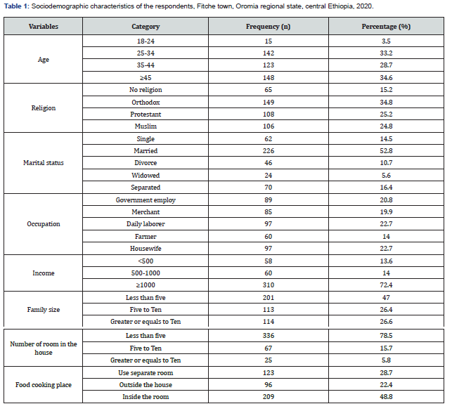
A total of 430 respondents were participated in the study yielding a 97.3% response rate. The mean age of the respondents was 40.48 years with (SD=10.88). One-third of the respondents belong to age 25-34 years. The majority of the respondents, 149(34.8%) were orthodox religion followers and 264(61.5%) attend formal education with 98(22.7%) of them reported not having a job outside the house. About 303(70.8%) households mentioned husband as a head of a family with nearly one third 145(33.9%), and 101(23.6%) of the participant stating husband and wife were the last decision-makers respectively. In about 134(31.3%) and 48(11.2%) of the household joint husband and wife and relatives were mentioned as the last decision-maker at the household level respectively. Almost half, 213(49.6%) of the household had using iodized salt for less than five years and 195(50.8%) were using it for more than ten years. About 242(62.4%) of the household store iodized salt at home for a period lasting less than two months and the rest 146(37.4%) store for more than two months (Table 1).

Health system-related characteristics

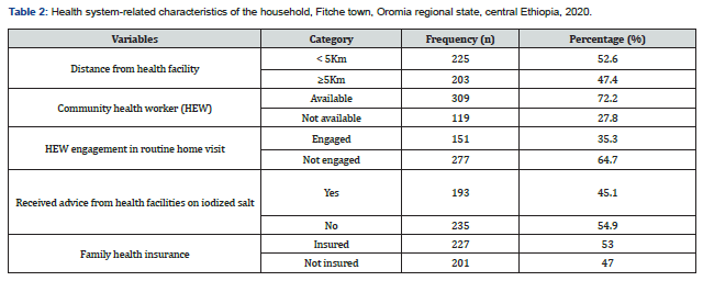
In this study, about 225(52.6%) of the respondents reside within a five-kilometer diameter from a health facility with slightly more than half, 228(53.0%) of the respondent accessed family health insurance service coverage. Out of the total respondents, 193(45.1%) stated as they have been received advice from health facilities on iodized salt (Table 2).

Knowledge of the respondents on the iodized salt use

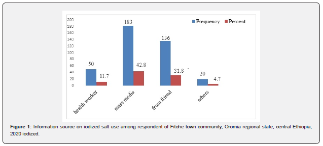
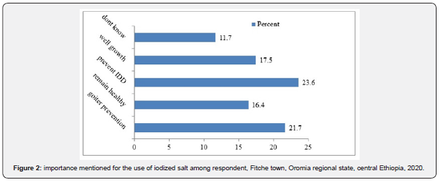
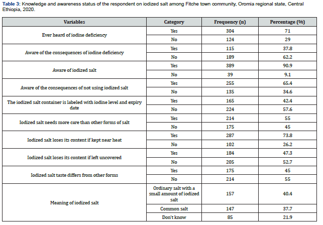
This study demonstrated that more than half, 58.6% of the respondents have good knowledge of the benefit of iodized salt use. About 304(71.0%) of the participants had heard iodine deficiency with only 115(37.8%) of the participants stating some form of health defect/consequences resulting from iodine deficiency. Related to the care of iodized salt more than half (55.0%) of respondents revealed that iodized salt needs more care than other commonly used types of salt. On the other hand, about 82(32.8%), 84(33.6%), and 78(31.2%) of the respondents stated that iodized salt must be stored far from heat, moisture, and sunlight respectively (Table 3). The study also revealed that various sources of awareness related to the importance of iodized salt use among the respondents who have awareness. Accordingly, the majority, 183(42.8%) of the respondents mentioned mass media as their major source of information (Figure 1). On the other hand, a series of questions have been asked the respondents on the importance of iodized salt who were initially aware of iodized salt. Accordingly, the majority 101(26.0%) respond as it prevents iodine deficiency disorder (IDD) followed by goiter prevention 93(23.9%) (Figure 2).

Utilization practice of iodized salt in fitche town community

The practice/utilization of adequately iodized salt i.e., iodine (≥15ppm) in Fitche town household was 52.6%. out of the total participants, 10.3% was not having any iodine content (0ppm) and 37.1% having less amount of iodine (<15ppm) (Table 4).

Reasons for non-using of iodized salt

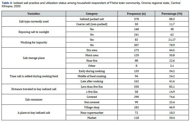
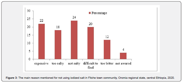
From the total respondents, 71% of the respondent claimed that they heard about iodized salt with around 88.3% revealed as they are using iodized packed salt. But the other, 11.7% reported that they are using coarse salt. Being expensive (22%) and not being salty (24%) were the main reason mentioned for not using iodized salt (Figure 3).

Factors associated with knowledge and utilization of iodized salt

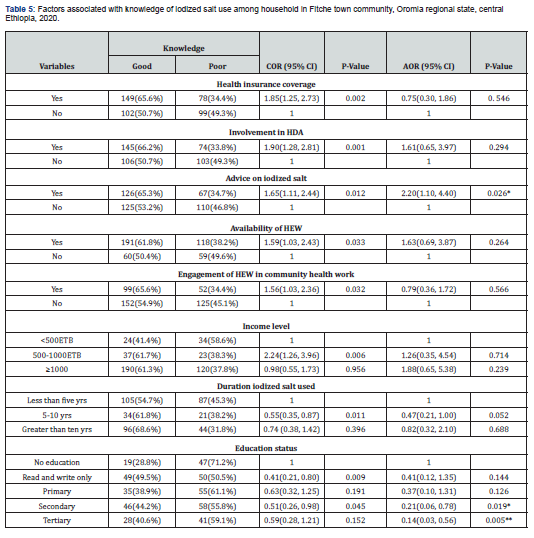
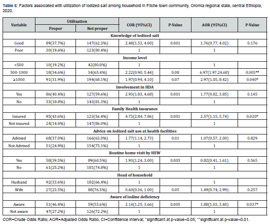
On multivariable logistic regression analysis covariates such as educational status and receive advice on iodized salt at health institution, were significantly associated with knowledge of individuals. Accordingly, individuals who did not attend any formal education were 79% less likely knowledgeable on iodized salt use as compared to those who did attend secondary education (AOR=0.21, 95%CI:0.06, 0.78). Individual who received health advice on iodized salt was more than two times more knowledgeable compared to those who did not receive (AOR=2.20, 95%CI:1.10, 4.40) (Table 5). Furthermore, income level and being aware of the consequences of iodine deficiency were significantly associated with proper utilization of iodized salt. Accordingly, the odds of proper iodized salt utilization were about three times higher in those individuals earning greater than 1000 birr as compared to those individuals earning less than 500 birrs (AOR=2.97, 95%CI:1.05, 8.42). The odds of proper iodized salt utilization were almost two times higher among individuals who have been aware of the consequences of iodine deficiency compared to those who have not been aware of iodine deficiency consequences (AOR=1.88, 95%CI:1.03, 3.40) (Table 5).

Discussion

This study was aimed to assess the knowledge and utilization of iodized salt in Fitche town community. In this study, 58.6% of the respondents have good knowledge of the use of iodized salt. This finding is in line with studies conducted in Ethiopia (63.8%) [18], Sudan (56%) [19] and India (56.2%) [20]. However, it is higher than the study conducted in different parts of Ethiopia including Mecha district (28.5%) [21], Wolaita Sodo (44.7%) [22], Arba Minch (52.8%) [14] and Debreberhan (53%) [23]. Demographical proximity of Fitche town to the center of the country may increase the chance of accessing information helping in knowledge improvement. Besides, the current study has enrolled only urban dwellers which was found to increase the knowledge level [22]. In contrast, this finding is lower than the study conducted in Addis Ababa (78%) [12], Axum (80%) [24], Ghana (72%) [25], and India (64.6%) [26]. The variation may also be due to differences in awareness creation and educational activities [14]. The differences might be due to iodized salt availability and accessibility in the local market and monitoring concerning the use of iodized salt in those areas [13]. About 52.6% of households in the study area were practicing adequately iodized salt for food preparation. This finding is comparable with the study done in Kore town (56.6%) [13]. On the other hand, this finding is higher than the studies conducted in Wolaita (37.7%) [27], Dabat district (33.2%) (16), Gondar (28.9%) [28], Benishangul Gumuz (26.1%) [29], Laelay Maychew District (33%) [30], and zuway (30.7%) [31]. This discrepancy could be correlated to a change in the time when the studies were done [32]. As evidence from the Ethiopian demographic and health survey, the coverage of iodized salt increased from 28.4% in 2000 to 89% in 2016 [11]. In opposite to the above, this finding is lower than the study done in Saudi Arabia (95.2%) [33] and India (83.1%) [34]. The difference could be due to iodine deficiency has been recognized as a major public health problem in Ethiopia as compared to other countries where the studies have been conducted. Beyond assessing knowledge and utilization of iodized salt, this study also points out predictors of knowledge and utilization of iodized salt. Accordingly, receiving health counsel on iodized salt use was identified as a predictor of knowledge in this population. This might be because health counsel/education has a positive impact on knowledge [35].

The educational status of the respondents was also found to be an independent predictor of knowledge on iodized salt use. This finding was in line with the studies conducted in Arba Minch [14] and Laelay Maychew district [30]. This could be because educated respondents have learned and could read about the importance of iodized salt. This study also identifies independent predictors of proper utilization of iodized salt. So, earning high monthly income was identified as an independent predictor of proper utilization of iodized salt. This finding is in line with the findings from Arba Minch town [14] and Addis Ababa [17]. This might be due to the reasons that families with high income can buy packed salts and have better information about iodized salt [17]. Moreover, family insurance coverage was another factor identified as a predictor of proper utilization of iodized salt. This is possibly due to the insured family have a higher chance of obtaining health-related information which might help them to practice iodized salt utilization properly. In this study, higher odds of proper utilization of salt were observed among households who have awareness about iodine deficiency disorder. This finding is supported by the study Laelay Maychew District [30]. This might be due to the fact that having awareness on how iodine deficiency affects health help to give due emphasis on its proper utilization.

Conclusion

Even though knowledge of iodized salt use in this community is higher as compared to other findings, the coverage of proper iodized salt utilization is less than 90% world health organization's recommendation of iodized salt use in the household [36]. Advice on iodized salt and education status were found to affect the knowledge of iodized salt use at the household level, while income level, family health insurance, and iodine deficiency awareness were independently associated with iodized salt use. Therefore, the town health facilities should provide health education programs to promote the knowledge and consumption of iodized salt. Additionally, routine testing and monitoring for iodine levels in salt sold in the markets and used by households are essential.

Click here: https://juniperpublishers.com/index.php  
Friday, May 20, 2022

A Possible Cluster of AEFI (Adverse Event Following Vaccination) Following AZD1222 (Covishield) Vaccination in India? - Juniper Publishers

 International Journal of Pulmonary & Respiratory Sciences 

Abstract

In order to mitigate the spread of the COVID-19 infection in India, the Indian government granted approval for the emergency use of Oxford-AstraZeneca’s COVID-19 (coronavirus disease 2019) vaccine AZD1222 (Covishield). The biggest large-scale COVID-19 vaccination drive in the world is being carried out continuously till date in India. In this research article, a possible cluster of AEFI (Adverse Event Following Vaccination) following AZD1222 (Covishield) vaccination in India has been briefly presented.

Keywords: AZD1222 (Covishield); Covishield vaccine; COVID-19 vaccination; AEFI (Adverse Event Following Vaccination); Acute myocardial infraction

Abbreviations: AEFI: Adverse Event Following Vaccination; COVID-19: Coronavirus Disease 2019; WHO: World Health Organization; AEFI: Adverse Events Following Immunization; GBS: Guillain-Barre Syndrome

Introduction

In an effort to curb the present escalating number of active COVID-19 cases in India, in January 2021, the Indian government granted approval for the emergency use of Oxford-AstraZeneca’s COVID-19 (coronavirus disease 2019) vaccine AZD1222 (Covishield). The first phase of the largest COVID-19 vaccination program in the world was launched on 16th January 2021. The first phase carried out the vaccination of frontline healthcare workers as well as essential service personnel. At present, the second phase of the vaccination program commenced on 2nd March 2021. This phase is targeting people over the age of 60 and those in the 45- 59 years with comorbidities [1]. Since 2021 until date, the rate of vaccination in the Indian population has been continuously good and at present, keeping in mind the predicted possible fourth wave due to the spread on the new XE Omicron variant, many in every eligible age bracket have already taken booster doses.

Discussion

The percentage of severe AEFIs following vaccination with Covishield has been very few (as low as 0.003% of the total vaccinated population) as compared with the large number of people who only suffered mild symptoms of AEFIs such as fatigue, myalgia, mild fever, headaches, injection site pain, pain in the body, nausea and stomach problems [2]. The range of reported or severe AEFIs amongst the 69-71 deaths following vaccination with Covishield includes diagnosis of Guillain-Barre Syndrome (GBS) causing significant disability (in 2 severe AEFI cases), hospitalization due to life-threatening breathing difficulties and death [3-7].

The fatalities following vaccination using Covishield have been recorded in the states of Delhi, Uttar Pradesh, Gujarat, Karnataka, Telengana, Andhra Pradesh, Odisha and Haryana. The six men and three women who died were between 27 and 56 years old. The deaths occurred between 1-6 days after vaccination with Covishield. After the release of the first Serious Adverse Events Following Immunization (AEFI) on 4th June 2021, there have been 18 documented cases that were said to show inconsistent causal association to vaccination (the causality assessments have not been made public) [8-10]. What is of utmost importance is that in the majority of the recorded fatalities following vaccination with Covishield, there has been a commonly recurring cause of death namely cardiovascular problems or “brain stroke.”

In every reported case, the central and state authorities have announced that the deaths cannot be attributed to the vaccination using Covishield but up till date, no causality assessment and the associated results have been made public. This has been stated repeatedly by the authorities despite the fact that in a few of the recorded AEFI deaths, the patients were not found to have any pre-existing morbidities [4-7]. However, the reported cause of death in many of fatality cases (where autopsies have been said reported as done) cited the cause of death as being “changes to the heart” and myocardial infarction [5-9]. The WHO (The World Health Organization) defines “a cluster of AEFIs as two or more cases of the same adverse event related in time, place or vaccine administered” [8,9].

I outline the potential importance of the above described severe AEFIs and deaths (due to acute myocardial infarction/acute coronary syndrome) in more than 21 vaccine recipients after vaccination with Covishield till date. The findings suggest a possible cluster of AEFIs in this case since the confirmed cause of death in at least 21 cases has been heart changes or myocardial infarction (this is supported by reported results of the performed autopsy which is evidence). Following this first AEFI report released in June 2021, several more Covid-19 vaccination associated AEFI reports have been released by the concerned government department.

Conclusion

I concur that even though the rate of recorded severe AEFIs and deaths following vaccination using Covishield in India remains very low (0.003%), there is still the need to carry out further large-scale investigations on the possibility of a presented cluster of AEFIs after Covishield vaccination with stringent causality assessments (based on the documented AEFI cases of cardiac complications of the first released government report). As India continues to move forward in the largest, worldwide vaccination program against COVID-19, there is an urgent need for very strict and transparent monitoring of all possible clusters of AEFIs.

Click here: https://juniperpublishers.com/index.php  
A Therapeutic Yoga Program for University Students with Anxiety: A Mixed Methods Study - Juniper Publishers

  Yoga and Physiotherapy - Juniper Publishers Abstract Anxiety remains one of the most common mental health disorders in the United States. ...