Colorectal cancer (CRC) is a kind of incurable cancer
with high morbidity and mortality. In recent years, mounting evidence
has revealed that inflammatory bowel disease (IBD) was correlated with
colitis-associated cancer (CAC). Signal transduction is an effective way
for cells to respond to external stimuli and eventually trigger
specific biological effects. The occurrence, growth and metastasis of
cancer are usually associated with disorders of signaling pathways.
Although modern research can elucidate the pathogenesis of CAC and
provide enhanced screening strategies, the prevalence of CAC is still
rising. Studies have shown that several cells signaling pathways in CAC
are dysfunctional, leading to the occurrence of malignant phenotypes.
Therefore, analyzing the signaling pathways involved in CAC metastasis
is necessary to elucidate the potential mechanisms of CAC progression
and drug treatment. Our understanding of CAC-related signaling pathways
is largely due to a mouse model that faithfully reproduces human CAC. In
particular, chemical models can quickly and effectively analyze the
molecular mechanism of CAC without increasing time-intensive genetic
hybridization. Therefore, this article summarizes the following three
chemical-induced CAC animal models, azoxymethane/dextran sodium sulfate
(AOM/DSS), 1, 2-dimethylhydrazine (DMH)/DSS and 2, 4, 6-trinitrobenzene
sulfonic acid (TNBS)/AOM. Among them, DSS and TNBS were used as
inflammatory agents to simulate the pathogenesis of IBD inflammatory
models, while AOM and DMH were used to producing carcinogenic methylated
DNA, combined with inflammatory agents, could better induce IBD to CAC
in animals.
Keywords: Colitis-associated cancer; Inflammatory bowel disease; AOM/DSS; DMH/DSS; TNBS/AOM
Abbreviations:
CRC: Colorectal Cancer; IBD: Inflammatory Bowel Disease; CAC:
Colitis-Associated Cancer; AOM: Azoxymethane; DSS: Dextran Sodium
Sulfate; DMH: 1, 2-Dimethylhydrazine; TNBS: 2, 4, 6-Trinitrobenzene
Sulfonic Acid; ACF: Abnormal Crypt Foci; UC: Ulcerative Enteritis; CD:
Crohn’s Disease; MAM: Methylazomethanol; ROS: Reactive Oxygen Species;
MDSC: Myeloid-Derived Suppressor Cells; DC: Dendritic Cells; Th: T
Lymphocytes; MyD88: Myeloid Differentiation Factor 88; CPB: CREB Binding
Protein; GR: Glutathione Reductase; HO-1: Heme Oxygenase-1; TR:
Thioredoxin Reductase; PEITC: Phenethyl Isothiocyanate; GSK3β: Glycogen
Synthase Kinase 3β; CK1α: Casein Kinase 1α; DVL: Disheveled; TCF: T
Cell-Specific Factor; LEF: Lymphoid Enhancer Binding Factor; MDF:
Mucin-Depleted Foci; JAK2: Janus Kinase 2; STAT3: Signal Transducer and
Activator of Transcription 3
Colorectal cancer (CRC), as one of the common
malignancies with high morbidity and mortality [1,2], usually occurs at
the junction of the rectum and sigmoid colon. It is the result of
healthy colonic epithelial cells transforming into cancer [3]. This
process is called ‘adenoma-carcinoma sequence’, which is developed
through a series of orderly events. The initial step is the
transformation of normal colonic epithelium to abnormal crypt foci
(ACF). ACF progresses to CRC within 10-15 years [4,5]. Accumulating
evidence has shown that its occurrence and development were related to
many factors, such as environment, eating habits, congenital heredity,
personal physique and so on [6]. Over the past few decades, CRC,
including colitis-associated cancer (CAC) has posed a serious threat to
people’s health and life, which develops from long-term colitis in
inflammatory bowel disease (IBD) patients [7] .
IBD, one of the key influential factors resulting in
CAC, is a chronic and recurrent gastrointestinal inflammatory disease
that can affect the body’s immune mechanism [8]. Generally speaking, the
occurrence of IBD in humans may be related to environmental factors
[9]. but in the current research hotspots, diet is also considered to be
the cause of IBD to a certain extent [10]. especially the intake of
allergenic substances in food [11]. Clinically, IBD is usually divided
into two kinds of disease, ulcerative enteritis (UC) and Crohn’s disease
(CD) [12,13]. Studies have found that long-term IBD may not only lead
to the occurrence and development of CRC, but also increase the risk of
prostate cancer [14]. gastric cancer and other cancers [15]. Morever,
the risk of colorectal cancer is increased twofold in CD and in UC [16]
while about 8% of UC patients develop CAC within 20 years and 18% within
30
years [17].
The rodent carcinogen-induced model recalls a long tradition
but still retains its usefulness for some applications. They provide
a platform for dietary research and insight into the pathways of
food-borne carcinogens and colitis-related carcinogenic effects.
Since the development of carcinogen-induced rodent models for
CAC more than 80 years ago, these animal models have played a
huge role in chemoprevention research and assessment of colitisrelated
carcinogenic mechanisms [18]. In 1915, scientists proved
the carcinogenic properties of coal tar by repeatedly applying coal
tar to rabbit ears [19].
At about the same time, the first researchers studied the
occurrence of colon cancer by applying chemical or radioactive
substances [20]. In the 1960s, heme and its metabolite
methylazooxymethanol have been shown to induce rodent
cancer [21,22]. In the following years, the chemically more stable
substance AOM and its precursor DMH were widely used to
induce colon cancer in mice and rats. Both of the two compounds
are metabolized to methylazoformaldehyde, which can alkylate
DNA bases guanine and thymine [23]. After two-phase reaction
treatment, it is secreted into the bile, and its carcinogenic effect on
intestinal mucosa exceeds its carcinogenic effect [24].
The most typical AOM/DSS model needs to be combined
with a single injection of AOM, a carcinogen that is metabolized
in the liver to the active agent methylazomethanol (MAM), and
then use inflammatory damage agent DSS [25]. The pathological
manifestations of the AOM/DSS model were severe colitis,
accompanied by weight loss and bloody diarrhea, and followed
by multiple colon tumors. Another carcinogen DMH undergoes
a series of metabolic reactions and eventually reaches the colon,
producing the ultimate carcinogen and reactive oxygen species
(ROS), which further alkylate DNA and cause colon cancer [26].
TNBS, a classical skin contact ant serves as a happen, which could
induce a specifically inflammation model in the gastro-intestinal
tract [27].
It was reported that the exact location of tumor in colon
length depends on the mouse strain and background [28]. The
carcinogenic process of the system has a pathological progression
from normal intestinal crypts to crypt fission to lesions with
abnormal crypts and eventually to microadenomas [29,30]. These
steps outline the sequence of CAC formation in humans from
inflammation to developmental abnormalities and to cancer.
In recent years, AOM/DSS [31-33] DMH/DSS [34,35] and
TNBS/AOM [36,37] are three main methods to establish CAC
animal models, as these three chemical pathways could better
simulate the process of IBD to CAC in vivo. Therefore, this paper
summarized the mechanism of these three modeling methods
which can reflect IBD and CAC as follows.
Chemical properties
AOM is a class of chemical compound with the molecular
formula of C2H6N2O (molecular weight: 74.08g/mol) and strong
oxidative activity. DSS is a kind of polyanion derivative of dextran
with the chemical formula of (C6H7Na3O14S3) n), which could
be used for modeling CAC, separating lipoproteins and improving
nucleic acid hybridization rate.
Modeling method
AOM/DSS model is the most widely used and reproducible
method to induce CAC. Mice were intraperitoneally injected with
AOM (7mg/kg or 12.5mg/kg) [38]. After 5 days, mice received 3
cycles of drinking 1%-3% DSS solution for 5 days and water for 14
days [39]. At the end of the experiment, the mice showed obvious
intestinal polyps with intestinal mucosal injury.
Mechanism
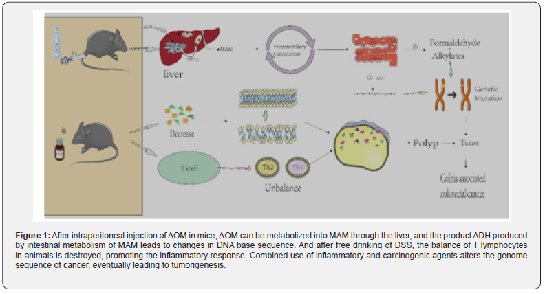
As shown in figure 1, AOM reaches the liver through blood
circulation. Under the action of cytochrome P4502E1 [40,41].
it is hydroxylated to form MAM [42]. which can spontaneously
decompose into formaldehyde and highly active alkylate after
entering the intestine [43]. In addition, some scholars have
proposed that MAM may be metabolized through another
pathway, that is, the metabolic reaction involving tissue-specific
cytoplasmic enzyme ADH in the intestine.
ADH generates reactive methylases, alkylases, or such reactive
methylases, which can promote DNA alkylation [44,45], and
subsequently induce DNA to produce O6-methylguanine adduct
[46], resulting in the conversion of G to A in the DNA base sequence.
MAM through the liver, and the product ADH produced by intestinal
metabolism of MAM leads to changes in DNA base sequence. And
after free drinking of DSS, the balance of T lymphocytes in animals
is destroyed, promoting the inflammatory response. Combined
use of inflammatory and carcinogenic agents alters the genome
sequence of cancer, eventually leading to tumorigenesis. AOM/
DSS is one of the most commonly used chemical reagents to induce
CAC in rodents [47] which can better simulate the pathological
process of the body from normal intestinal epithelial mucosa to
abnormal crypt lesions [48] adenoma and colorectal cancer [49].
Among them, DSS reagent has the characteristics of
convenient use and high efficiency [50], as a chemical reagent for
inflammatory stimulation, it continuously stimulates the mucosa
of intestinal epithelial cells during free drinking, causing repeated
inflammation in model animals. In the DSS model, sulfated
polysaccharides do not directly induce intestinal inflammation,
but act as a direct chemical toxin of colonic epithelium, leading to
epithelial cell damage. The mechanism is that oral administration of DSS
leads to the destruction of the intestinal epithelial monolayer
lining, the entry of intraluminal bacteria and associated antigens
into the mucosa, and the transmission of pro-inflammatory
intestinal contents to the lower tissues [51-53].
It is generally believed that in chronic inflammation, the
immune system is characterized by intensive infiltration of
innate immune cells (such as macrophages, neutrophils, myeloidderived
suppressor cells (MDSC), dendritic cells (DC) and NK
cells and adaptive immune cells (such as T and B lymphocytes),
further suggesting that the changes in the intestinal inflammatory
microenvironment promote tumor formation [29] In the internal
environment of the body, helper T lymphocytes (Th) play an
important role in the immune response. The dynamic balance of
Th1 and Th2 is one of the necessary conditions to maintain the
normal physiological function of the body [54]. Therefore, the
increase or decrease of Th1 or Th2 could cause immune response
[55] eventually resulting in the occurrence of inflammation
[56,57].
After oral administration of DSS in mice, the dynamic balance
between Th1 and Th2 was broken, which promoted the expression
of inflammatory factors such as TNF-α, IL-1β, IL-10 and IFN-γ,
and so on [58,59]. In AOM/DSS model animals, the continuous
production of pro-inflammatory cytokines leads to mutations in
proto-oncogenes and tumor suppressor genes APC, K-RAS and p53,
as well as genomic sequence variations, generating tumorigenesis,
progression and metastasis [60]. Myeloid differentiation factor
88 (MyD88), a molecule essential for TLR intracellular signaling,
appears to have a protective effect against AOM/DSS-induced
inflammation-associated CRC in mice [61]. Yassin M proposed that
AOM/DSS-induced inflammation led to an increase in the total
number of CD3+, TCRγδ and TCRαβ cells, including a significant
increase in CD4+TCRαβ cells, but only moderate changes in CD8αβ
TCRαβ and CD8αβ TCRαβ/TCRγδ cells. This suggests that CD4+T
cell recruitment plays a key role in regulating various parts of the
mucosal immune response during acute and chronic DSS-induced
colitis [62].
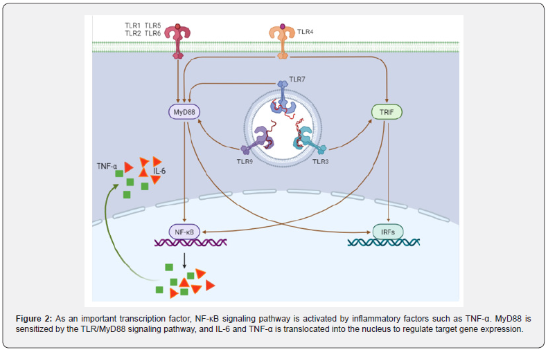
The NF-Κb signaling pathway, a central mediator between
inflammation and cancer, promoting CAC development: It
is well known that TNF-α can directly activate NF-κB signaling
pathway and induce inflammatory response. Furthermore,
MyD88 signaling molecules can also activate NF-κB-induced
kinase and indirectly activate NF-κB signaling pathway through
TLR/MyD88 signaling pathway [63,64] (Figure 2). Xie et al. [65]
synthesized a MyD88 inhibitor TJ-M2010-5, which can bind to the
TLR domain of MyD88, inhibit the production of its homodimer,
and ultimately inhibit the TLR/MyD88 signaling pathway. TJM2010-
5 can significantly reduce the incidence of AOM/DSSinduced
colitis, shorten colon length, reduce tumorigenesis, and
inhibit the production of inflammatory factors. Moreover, when
the TLR4-deficient mice and the wild-type mice were treated
with AOM/DSS at the same time, it was observed that the TLR4-
deficient mice had a lighter degree of colon cancer than the wild type
mice [66]. After the MyD88-/- mice were injected with AOM, colonoscopy
showed no tumor formation in the intestine, and
the mRNA expression of pro-inflammatory cytokines TNF-α and
IL12P40 was lower than that of the model group [67]. On the one
hand, the production of TNF-α can activate the NF-κB signaling
pathway, on the other hand, when the NF-κB signaling pathway is
activated, it can also promote the transcription and translation of
TNF-α. El-Daly et al. [68]. showed that in AOM/DSS-treated mice,
the positive staining of p65 in the nucleus of the vagus nerve crypt
of colorectal epithelial cells gradually increased with the increase
of DSS administration cycle. In addition, AOM/DSS also induced
the high expression of TLR4 and p65 proteins upstream of NF-κB
signaling pathway. Furthermore, Song et al. [69]. found that the
expression level of pro-inflammatory enzyme COX-2, which is
mainly regulated by NF-κB signaling pathway, was significantly
higher than that of the control, and the expression levels of the
other two target genes TNF-α and IL-6 also tended to increase
[70].
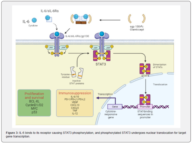
The IL-6/STAT3 signaling pathway is enhanced and
accelerates carcinogenesis in CAC: As an inflammatory
cytokine, IL-6 binds to soluble IL-6 receptors to form an IL-6/
sIL-6R complex [71,72]. which activates gp130 expression on
the cell membrane, leading to signal transducer and activator
of transcription 3 (STAT3) phosphorylation. STAT3 enters the
nucleus in the form of dimers and activates the STAT3 signaling
pathway (Figure 3) [73]. Studies have shown that p-STAT3 staining
in the colorectal region of AOM/DSS-treated mice was deepened,
and the deposition of brown granules in cytoplasm and nucleus
was enhanced with the increase of the administration cycle [74].
MiR-18a is a transcription product in the STAT3 signaling pathway,
in AOM/DSS-treated mice, it was found that the expression of MiR-
18a increased significantly with the extension of DSS treatment
cycle [75]. but the expression of PIAS3 (an inhibitor of STAT
family activation) decreased. In vitro studies have shown that the
expression level of MiR-18a is negatively correlated with PIAS3.
MiR-18a can negatively regulate PIAS3 [76]. while overexpression
of PIAS3 leads to a decrease in IL6-activated STAT3 activity. Ma
et al. reported that the proliferation and survival of precancerous
intestinal epithelial cells (IECs) were regulated by IL-6/STAT3
signaling pathway, and CAC may be related to this signaling
pathway [77].
Nrf2/ARE signaling pathway plays an inflammatory
role in CAC: In addition, the occurrence of inflammation is also
related to the activation of the antioxidant Nrf2/ARE signaling
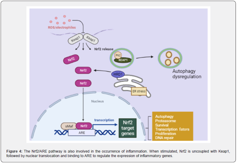
pathway. When the Nrf2/ARE signaling pathway is closed, Keap1
in the cytoplasm specifically binds to the N-terminus of Nrf2,
thereby maintaining the normal physiological function of the
body. However, when cells receive external stimuli, the Nrf2/ARE
signaling pathway is immediately activated, Nrf2 is uncoupled
with Keap1, and then enters to the nucleus and binds to the small may
protein to form a heterodimer. This process requires the participation
of transcription cofactor CREB binding protein
(CPB).
Heterodimers specifically recognize ARE and initiate
transcription and translation of target genes, resulting in
inhibition of the Nrf2/ARE signaling pathway (Figure 4). When
the antioxidant process in the body is initiated, the expression
of antioxidant proteins is significantly increased to protect cells
from damage [78,79]. Wang et al. [80]. found that the positive
expression of Nrf2 and the expression of glutathione reductase
(GR), heme oxygenase-1 (HO-1) and thioredoxin reductase
(TR) were significantly degraded in AOM/DSS treated mice. The
researchers [81]. fed mice with 0.05% phenethyl isothiocyanate
(PEITC) before and after AOM/DSS administration, found that
the incidence of intestinal adenomas in mice decreased by 37.5%
and 40%, the number of polyps reduced to 0.375 and 0.6, and the
average size decreased to 1.5 and 2.6 mm, respectively. It can be
inferred that PEITC can improve the development of intestinal
tumors, as an activator of Nrf2, PEITC can activate Nrf2/ARE
signaling pathway. In addition, some scholars performed Nrf2
gene knockout in wild AOM/DSS model mice, early model mice
and 9-week model mice [82]. It was proved that Nrf2-/- mice
showed the same phenotype as wild-type mice, but the incidence
of tumors and the proportion of adenocarcinoma were higher.
Chemical properties
DMH is a class of compound whose molecular formula is
C5H8N2O2 (molecular weight: 128.13g/mol) and provides raw
materials for the synthesis of proteins in vivo.
Modeling method
Scientists [83] injected DMH (40mg/kg) into FMH rats
three
times in the first week, and then gave 1% DSS solution to drink
freely for one week, repeating three cycles. DMH is a precursor of
the carcinogen AOM, which is not carcinogenic in itself, but can
be carcinogenic by oxidative dealkylation to form AOM [84]. The study
confirmed that DMH-treated rats had traditional tubular or villous
adenomas and serrated adenomas [85] in their colorectal
regions, and it is generally believed that most colorectal cancers
progress from adenomas.
Mechanism
As a proto-oncogene, DMH can be decomposed into
azomethane and methylazomethanol under the catalysis of liver
enzyme system. The latter substance flows to the colon tissue
through the hepatobiliary circulation and is easily decomposed
into a carcinogen methyl diazo ion [86] in the circulation. Methyl
diazo ions can methylate DNA, RNA and proteins in normal
intestinal epithelial cells. When they reach the colorectal site, they
can produce activated carbon ions, leading to oxidative stress,
over-expression of inducible nitric oxide synthase (i-NOS) and
COX-2 [87], which in turn causes colorectal cancer.
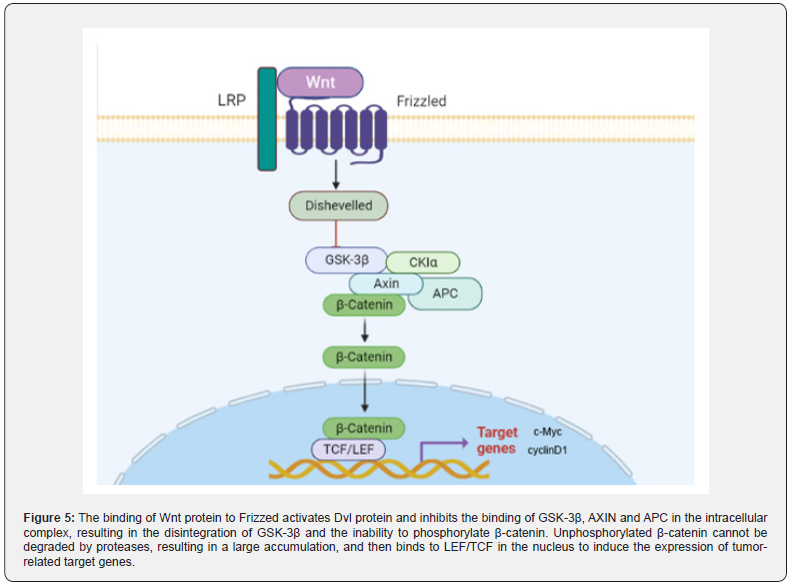
Up-regulated Wnt/ β -catenin signaling pathway promotes
CAC progression: Wnt/β-catenin signaling pathway is a conserved
signaling axis involved in a variety of physiological processes, such
as proliferation, differentiation, apoptosis, migration, invasion
and tissue homeostasis [88,89]. In the Wnt/β-catenin pathway,
the abnormal regulation of the transcription factor β-catenin
can lead to the occurrence of early cancer events [90,91]. In the
degradation complex of this pathway, glycogen synthase kinase 3β
(GSK3β) and casein kinase 1α (CK1α) mediate phosphorylation
of β-catenin, promoting its ubiquitination and subsequent
proteasome degradation [92]. The β-catenin dependent signaling
pathway is triggered by the binding of secreted cysteine-rich
glycoprotein ligands Wnts to LRP-5/6 receptors and FZD receptors.
In the presence of Wnt ligands, the binding of Wnt ligands on the
cell surface to the receptor induces disheveled (DVL), leading
to the aggregation of complexes (AXIN, GSK3β, CK1, APC) to the
receptor [93]. Subsequently, phosphorylation and inhibition of
GSK3β ensured an increase in cytosolic β-catenin concentration.
Unphosphorylated β-catenin in the cytosol migrates to the
nucleus and accumulates, interacting with T cell-specific factor
(TCF)/lymphoid enhancer binding factor (LEF) to trigger Wnt
target genes such as c-Myc [94]. and cyclin D1 (95), resulting in
up-regulation of TCF/LEF target genes [95].
In the analysis of 11 DMH/DSS-induced adenocarcinoma
mice,
Tanaka observed markedly enhanced expression of the β-catenin
gene in 10 of 11 mice [96]. Furthermore, mutations of β-catenin,
Apc and K-ras genes and cytoplasmic β-catenin expression were
found in DMH-induced mucin-depleted foci (MDF) [97,98]. MDF is a
microscopic dysplasia induced by specific colon carcinogens in the colon
of rodents. Most MDF show Wnt pathway activation,
while only one subset shows Ctnnb1 gene mutation, encoding
β-catenin [99]. It is well known that β-catenin is a key upstream
regulator of the Wnt signaling pathway. The activation of Wnt
signaling pathway can lead to the increase of β-catenin into the
nucleus (Figure 5), which leads to the increase of transcription
of tumor-related factors and induces the occurrence of tumors in
vivo.
The Slit2/Robo1 signaling pathway induces the Wnt/β
-catenin signaling pathway dysregulation to promote CAC
progression: Slit/Robo signaling pathway includes Slit secreted
protein family (Slit1, Slit2, Slit3) and specific receptors (Robo1,
Robo2, Robo3, Robo4). The Slit2/Robo1 signaling pathway was
initially found to be involved in the development of the central
nervous system. In recent years, more and more studies have
reported that it can induce tumorigenesis and participate in the
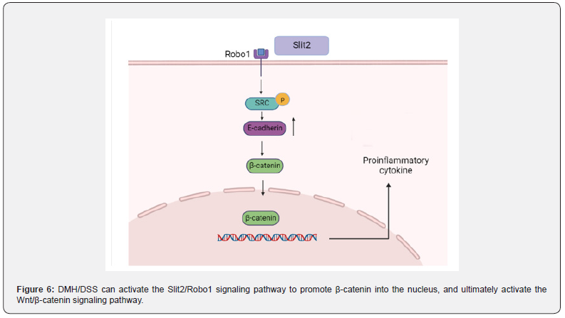
proliferation and migration of tumor cells [100]. The activation
of Slit2/Robo1 signaling pathway can be caused by ectopic
expression of Slit2 protein (Figure 6). The Slit2 and Robo1 genes
on chromosomes are located respectively at 4P15.2 and 3P12.3,
Robo1 is a specific receptor of Slit2 [101]. The extracellular IgG
region of Robo1 is an important position for binding to Slit2, and
the intracellular region is a site for interaction with related signal
molecules, which is involved in activating the downstream of
Slit2/Robo1 signaling pathway and promoting the occurrence and
development of tumors [102].
DMH/DSS can not only directly activate the Wnt
signaling
pathway, but also indirectly activate the Wnt signaling pathway
by activating the Slit2/Robo1 signaling pathway Qian et al. [103].
found that the expression of Slit2 and Robo1 in DMH/DSS mice
gradually increased with time. Morever, seven pairs of CRC tissues
and non-cancerous colon tissues were randomly selected from
patients with N0 CRC (no metastasis). The expression profiles of
Slit2 and Robo1 were evaluated by immunohistochemical staining.
It can be seen that the expression of Slit2 and Robo1 in CRC
tissues is significantly higher than that in non-cancerous colon
tissues. As long as the Slit2/Robo1 signaling pathway is activated,
it can promote the activation of Src phosphorylation, resulting in
increased expression of E-cadherin, thereby activating the Wnt/β-catenin
signaling pathway, promoting β-catenin into the nucleus,
leading to an increased expression of tumor-associated proteins.
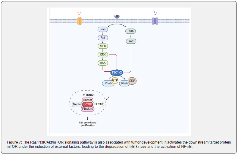
The Ras/PI3K/Akt/M TOR signaling pathway promotes
the development of CAC by activating NF-κB protein: The
carcinogenic activation of Ras protein caused by missense
mutations is a well-known marker of tumor cell proliferation.
Balb/c mice treated with DMH/DSS resulted in a significant
increase in Ras protein levels. Activated Ras protein targets MEK to
trigger ERK phosphorylation and nuclear translocation to express
genes involved in cell proliferation. The levels of activated MEK1/2
and phosphorylated-ERK1/2 was significantly increased in DMH/
DSS-treated animals. In addition, Ras oncoprotein also interacts
with PI3K and triggers Akt phosphorylation and subsequent
activation. DMH/DSS-treated animals showed significantly
increased levels of PI3K and phosphorylated Akt. The expression
of NF-κB, a downstream effector of Akt, was also significantly
increased. The authors believe that the increased expression
of NF-κB can be attributed to chronic inflammation followed by
circulatory treatment with the inflammatory agent DSS [104].
The mutation of RAS protein is induced by some external factors
[105], and mutant RAS protein interacts with PI3K and causes
autophosphorylation of PI3K. Type I PI3K is a heterodimer,
the corresponding domain of the P85 subunit on the dimer can
interact with the linker protein to recruit the corresponding
activation receptor for PI3K, thereby converting PI3K into PIP2
and then into PIP3. PIP3 binds to Akt and PDK1 containing the PH
domain, leading to the activation of Akt, which in turn regulates
the expression of its downstream target protein mTOR. On the
other hand, this binding can also activate IK-B, thereby relieving
the inhibition of NF-κB, transferring NF-κB from the cytoplasm to
the nucleus, and finally regulating the expression of inflammatory
factors in the epidermis (Figure 7).
Chemical properties
TNBS is a kind of dangerous chemical compound with a
molecular formula of C6H3N3O9S (molecular weight: 293.17).
In bioengineering, TNBS can be carried out determining aminoterminal
groups, hydrophilic modification reagents, etc.
Modeling method
Xiao et al. [37] injected AOM (10mg/kg) intraperitoneally
into C57BL/6 mice, and then 2.5mg TNBS was dissolved in 150μl
50% ethanol and injected into the mice through the rectum. The
changes of intestinal tumors in mice were observed under rectal
endoscopy. TNBS-induced colitis is very similar to CD, which can
induce transmural colitis (Th1-mediated immune response) with
severe diarrhea, weight loss, rectal prolapse [106,107], tissue
destructive and recurrent [108].
Mechanism
Normally, TNBS was used in combination with anhydrous
ethanol. By enema administration, the intestinal mucosa of
the model animals was damaged, so that TNBS penetrated the intestinal
wall, combined with the related proteins in the intestine, and caused
the intestinal epithelial mucosaltenization. Therefore,
intestinal cells are more likely to expose target proteins, stimulate
immune cells above the intestinal lamina propria [109], and make
target proteins more easily recognized by immune cells [110].
resulting in acute necrosis.
TNBS promotes the expression of the same T cytokines as CD
[111]. which is an immune inflammatory disease mediated by
Th1 [112]. However, the Th1-mediated cycle is short and mainly
mediated by Th17, which is characterized by infiltration of CD4+T
cells in the intestine [113]. and severe fibrosis of intestinal wall
cells in advanced patients [114]. Alrafas Haider [115] treated
TNBS-induced colitis with resveratrol and found that the induced
inflammatory T cells (Th17 and Th1), cytokines and transcription
factors (Foxp3 and TGF-β) were significantly decreased, and the
severity of colitis in mice was significantly increased. In general,
TNBS-induced colitis also involves Th17-related cytokines [116].
More and more evidence suggests that the growth and
proliferation of Th17 cells depend on IL-23. Therefore, some
scholars have proposed that the IL-17/IL-23 axis may also be one
of the causes of IBD [117,118]. Cubes [119]. found that I-NOS is
associated with TNBS-induced colitis and can cause intestinal
mucosal damage in model animals, which is similar to some
features of DSS-induced colitis. The pathogenesis of CD induced
by TNBS is not clear, but the mutation of NOD2 gene was found
in C57 mice treated with TNBS, and the mutation of NOD2 gene is
related to the pathogenesis of CD. Hence, it is concluded that TNBS
can induce CD-related enteritis in model animals [120].
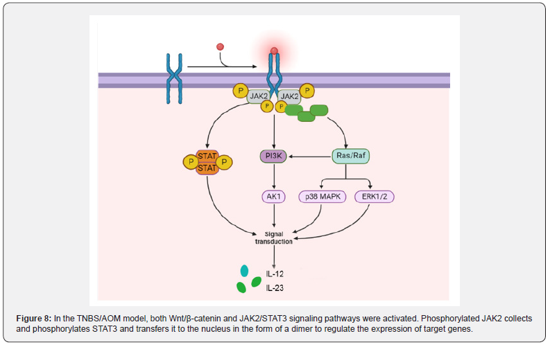
The JAK2/STAT3 signaling pathway plays an oncogenous
role in the development and progression of CAC: JAK contains
four tyrosine kinases (JAK1, JAK2, JAK3 and TYK2), while STAT
contains seven transcription factors (STAT1, STAT2, STAT3,
STAT4, STAT5A, STAT5B and STAT6). JAK2(Janus kinase 2)/STAT3
pathway is an important signal transduction pathway in the body,
which plays a role in many physiological and pathological processes
such as immunity, cell proliferation, differentiation, apoptosis and
inflammatory response [121]. Under the stimulation of certain
cytokines, JAK2 is activated, and then STAT3 is activated, which
can transmit extracellular signal into the nucleus and regulate the
expression of related inflammatory factors [122]. In the TNBS/
AOM animal model, the activation of Wnt/β-catenin and JAK2/
STAT3 pathways occurred with the development of colitis to
adenoma.
He et al. [123]. confirmed that in TNBS-treated mice,
Ang II
was significantly elevated and the JAK2/STAT3 signaling pathway was
activated to induce colitis by stimulating phosphorylation of JAK2 and
STAT3. The colitis of TNBS mice treated with JAK2/
STAT3 pathway inhibitor tofacitinib was significantly improved.
Lu et al. [124] found that TNBS can not only induce colitis, but also
accompanied by visceral allergies. Subsequently, Wan [125] found
that the expressions of JAK2, STAT3, p-JAK2 and p-STAT3 was upregulated
in TNBS-treated goats. JAK2 is one of the four members
of the JAK family located on the cell membrane, specifically binds
to some transmembrane receptors and is phosphorylated by
internal conformational changes. Phosphorylated JAK2 recruits
a large number of STAT3, leading to STAT3 phosphorylation and
nuclear translocation in the form of dimers, ultimately mediating
the expression of downstream target genes (Figure 8).
The Wnt/β-catenin signaling pathway plays a tumorigenic
role in CAC: Zhao et al. [126]. showed that the expression of
TCF, β-catenin and Wnt upstream proteins in the TNBS group
was significantly higher than that in the blank group, and the
expression of tumor-associated proteins c-Myc and cyclinD1
was also significantly enhanced. In the Wnt/β-catenin signaling
pathway, Wnt protein binds to the coiled protein (coiled gene) on
the cell membrane. After seven trans membranes [127]. the signal
is transmitted to the Dvl protein. Subsequently, the activation
of Dvl protein inhibited the binding of intracellular complexes
GSK-3β, AXIN and APC, resulting in the dissociation of GSK-3β,
and then GSK-3β could not phosphorylate β-catenin [128,129].
Unphosphorylated β-catenin cannot be degraded by proteases,
resulting in a large accumulation of β-catenin. This process
promotes the entry of β-catenin into the nucleus, where it binds to
nuclear LEF/TCF and induces the expression of tumor-associated
target genes. Therefore, TNBS-induced colitis can complete the
process from CD to CAC under the combined action of AOM.
Inflammation, endoplasmic reticulum stress and
autophagy
It is confirmed that infection and a large number of
inflammatory stimuli could cause endoplasmic reticulum stress
and activate the UPR signaling pathway, mainly by promoting Ca2+
release from the endoplasmic reticulum and ROS accumulation.
For example, pro-inflammatory cytokines IL-1β and IFN-γ can
directly down-regulate the expression of Ca2+-ATPase (SERCA2)
on the endoplasmic reticulum to depleted ER Ca2+ stores, thereby
causing ERS [130].
In addition, ROS, as an important mediator of
inflammatory
response, can cause disorders of endoplasmic reticulum folding
protein-related functions, leading to the accumulation of unfolded/
misfolded proteins in the endoplasmic reticulum and the release
of Ca2+ in the endoplasmic reticulum into the cytoplasm. High
concentration of CA2+ leads to mitochondrial dysfunction and produces
more ROS [131]. Due to the long-term existence of this
feedback mechanism in the body, intracellular ROS is continuously
generated and accumulated, and abnormally accumulated ROS
can aggravate endoplasmic reticulum stress.
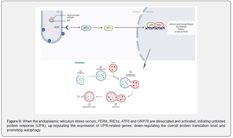
Endoplasmic is the main site for protein folding and quality
control. Any disturbance that alters endoplasmic reticulum
stress homeostasis may lead to the accumulation of misfolded
proteins, the activation of unfolded protein response, autophagy,
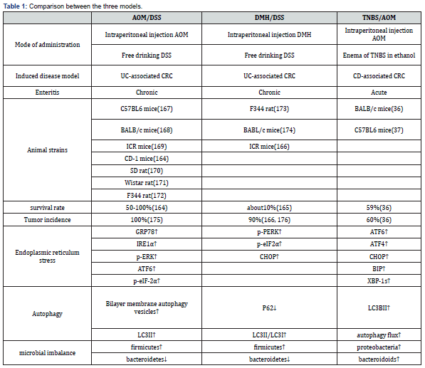
or apoptosis [132] (Figure 9). Zhang et al. [133] . treated mice
with AOM/DSS and found that the endoplasmic reticulum lumen
was enlarged, with swelling, vacuoles, and a significant reduction
in ribosomes. When the endoplasmic reticulum integrity was
destroyed, the expression of GRP78, IRE1α, p-ERK, ATF6 and
p-eIF-2α related to endoplasmic reticulum stress was significantly
enhanced. Similarly, in TNBS -induced rats [134], the expression of
endoplasmic reticulum stress-related proteins ATF6, ATF4, CHOP,
BIP and XBP-1s was enhanced. Sharma et al.confirmed that the
increased expression of p-PERK, p-eIF2α and chop proteins in rats
[135], induced by DMH can lead to endoplasmic reticulum stress.
From the mechanism of action, studies have found that
IBD-related endoplasmic reticulum stress was associated with
autophagy dysfunction [136,137]. Autophagy is a conserved
process in eukaryotic evolution through which cytoplasmic
materials are degraded in lysosomes [138]. Zhang et al. [133]
found that AOM combined with DSS could induce endoplasmic
reticulum stress in the intestine of mice and observed a large
number of double layers autophagic vesicles in the intestinal
epithelial mucosal cells of mice by electron microscopy [138]. The
expression of LC3II, an autophagy marker, was also significantly
increased. In the DMH/DSS model mice [139]. the expression
of P62 and the ratio of LC3II/LC3I were decreased. In TNBS
model mice [140], the expression of autophagy related LC3II
and autophagy flux were enhanced, and the autophagy ability of
intestinal epithelial cells was enhanced.
When endoplasmic reticulum stress occurs in the body,
autophagy function is activated [141], which in turn destroys
the intestinal mucosal barrier and affects the physiological
function of normal intestinal epithelial cells. Subsequently, the
diversity of intestinal flora decreased, flora dysfunction [142].
and inflammatory cell infiltration [143] induced IBD, then
developed into CAC. AOM/DSS, DMH/DSS and TNBS/AOM can
well induce autophagy and endoplasmic reticulum stress during
the progression of IBD to CAC.
Intestinal microbial imbalance
The intestinal epithelium is a monolayer of cells
that serves
as a physical barrier separating the mucosal immune system
from symbiotic and pathogenic microorganisms [144]. Epithelial
damage or increased epithelial permeability can destroy this
barrier, thereby activating the mucosal immune system. Increased
intestinal permeability is observed in patients with Crohn ‘s
disease before clinical onset or recurrence, suggesting that barrier
dysfunction may trigger disease progression [122,145]. The
mechanism of IBD caused by intestinal flora imbalance is mainly divided
into the following three aspects: the increased number of pathogenic
bacteria, the decreased number of probiotics, and the
imbalance of flora to break the normal immune tolerance of the
body.
At present, the common human pathogenic bacteria are
mostly found in Proteobacteria, and the bacteria belonging to
Proteobacteria play an important role in the occurrence and
development of IBD, especially Escherichia coli. When the
intestinal flora is out of balance, the number of mutual pathogenic
bacteria increases (such as adhesive invasive Escherichia coli, etc.).
Pathogenic bacteria can destroy the normal internal mechanical
barrier and immune barrier through invasion and secretion of
toxins, increase the permeability of intestinal mucosa, and create
conditions for the shift of flora. On the other hand, transferred
bacteria over activate the immune response, causing indirect
tissues to be attacked and damaged by the immune system [146].
which further increases the intestinal inflammatory response.
Relevant studies have found that the intestinal mucosal
permeability of CD patients is significantly higher than that of
normal people, resulting in a large number of bacteria and toxins
in the intestine entering the intestinal mucosa and activating
immune cells, which will trigger a strong immune response and
destroy the mucosa [147]. This may be related to a series of
reactions caused by the increase in the number of pathogenic
bacteria in CD patients (Figure 9).
There are abundant probiotics in human body, such as
Lactobacillus in Firmicutes, Bifidobacterium in Actinobacteria
and Bacteroides in Bacteroidetes. When the intestinal flora
is imbalanced, the number and richness of Firmicutes and
Bacteroidetes in the normal body are reduced, and the number
of probiotics such as Lactobacillus and Bifidobacterium is
significantly reduced. The above functions are weakened,
resulting in impaired intestinal mucosal barrier and proliferation
and translocation of pathogenic bacteria. Studies have shown
that the bacterial genus in the built-in bacteria can activate
Paneth cells to secrete antimicrobial peptides to produce normal
antibacterial effects. When the disease causes a significant
increase in pathogenic bacteria and a decrease in bacterial genus,
the production of antimicrobial peptides is reduced, which further
causes excessive immune activation and damage to the intestinal
mucosa [148].
Physiologically, there is a symbiotic relationship between
intestinal microorganisms and hosts, which depends on a variety
of immune mechanisms, such as intestinal mucus secreting
immunoglobulin IgA and releasing antimicrobial peptides [149].
In IBD patients, the intestinal microecology is not regulated, and
the imbalance of intestinal flora increases the expression and
release of inflammatory factors. The normal immune balance of
the intestine is broken [150], and the abnormal immune response
is activated, resulting in intestinal mucosa and tissue damage.
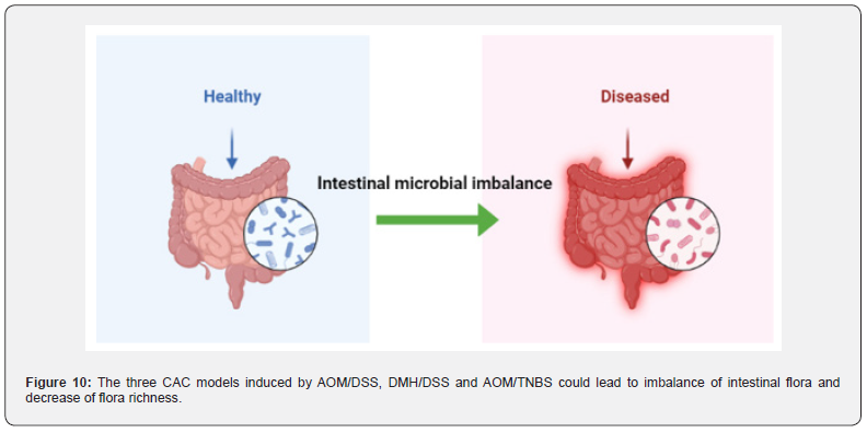
In AOM/DSS [151] and DMH/DSS [152] treated model animals,
the abundance of Firmicutes increased and the abundance of
Bacteroidetes decreased, resulting in a decrease in microbial
diversity in vivo. In TNBS-induced model animals [153], the
intestinal flora composition, species richness and diversity of the
model group were significantly different from those of the control
group, and the Proteobacteria and Bacteroidetes were significantly
increased. After transplantation of fecal microbiota (FMT) in mice,
the abundance of these two floras returned to normal levels and
microbial diversity increased significantly by observing Shannon
curve, PD forest index and CHAO1 index [154] (Figure 10).
Advantage and disadvantage
AOM/DSS is the most widely used chemical reagent among the
three models [155]. It is easy to use, easy to operate, and has a high
tumor formation rate that can target tumor formation in animal
colorectal regions. At the same time, it has the histological order
change of ‘inflammation-dysplasia-carcinoma’. However, there are
still some shortcomings in this model. For example, there is a lack
of research on cancer stem cells in this model, and the mutations
of tumor suppressor genes APC and P53 are rarely seen [156]. CRC
has a low tendency of distant metastasis, and the life span of mice
is shortened after illness, so it is not suitable for tumor invasion
related research [28].
DMH/DSS-induced colon tumors in rodents are very close
to human colon cancer in morphology, growth pattern and
clinical manifestations [157]. And DMH-induced colorectal
adenocarcinoma in mice often invades the submucosa and
muscular layer, while AOM-induced colorectal adenocarcinoma
does not exhibit such biological and histological properties
[158]. However, the main disadvantage of the model using
DMH is that multiple injections of DMH and long experimental
periods are required to induce colon tumors in experimental
animals. Secondly, after DMH is injected into the body, there are
many uncontrollable factors in the process of producing AOM by
oxidation, and its feasibility is not as high as AOM.
TNBS/AOM, in the construction process, due to TNBS is
a
semi-antigenic substance, it needs to be combined with ethanol to
form a complete antigen, so some researchers use rectal perfusion
construction. This model is based on the increased membrane permeability
that occurs in IBD, which contributes to the entry of the luminal
antigen that immune system cannot fully eliminate,
namely haptenization [159]. The main advantages of the model
include simple and low-cost protocol and repeatable colonic
injury, short duration of the experiment, persistent injury and
ulcers with inflammatory cell infiltration. Because of the lack of
practice of this technique, the presence of feces in the colon, the
anatomical localization of the descending colon, and the volume
of the injection rate administered may cause rectal reflux of TNBS,
leading to defects in the induction method or increased variability
in animal disease.
Pathology
Clinically, IBD patients are also divided into UC and CD.
According to Lin et al. [160], UC patients typically present with
abdominal pain, increased frequency of diarrhea, and bloody
mucopurulent diarrhea. Radiological examination showed
obvious edema and hemorrhage of colonic mucosa, diffuse
thickening of intestinal wall, occasional cryptitis and deformation
of crypt dissection. In CD patients, intestinal wall thickening often
occurred in the ileum [161], mucosal thickening and adjacent
mesenteric fluid with severe submucosal fibrosis [162]. Saade C et
al. [163] reported that approximately 91% of CD patients develop
an inflammatory response in the ileum.
In the AOM/DSS-induced mouse UC model, Zhang et al. [133]
found that the mice in the model group had pathological symptoms
such as diarrhea, vertical hair and anal prolapse, and bloody stools
appeared on the 6th day of modeling. Adenoma was diagnosed by
histological examination. In Li ‘s experiment, mice in the AOM/
DSS treatment group showed glandular structure disorder and
inflammatory cell infiltration. In addition, researchers found
that about 80% of mice [164] in the AOM/DSS model group
developed adenocarcinoma. In DMH/DSS-induced mice, polyploid
adenomatous hyperplasia with malignant transformation of
colon, polypoid adenoma [165], thickening of intestinal mucosa,
inflammatory cell infiltration [83] bloody stool and rectal prolapse
[166].
In TNBS/AOM-treated mice [167], lesions occurred mostly in
the distal colon, with colonic crypt abnormalities, accompanied by
colorless or polypoidal adenomas, and eventually adenocarcinoma
[36]. In addition, some scholars found that AOM/TNBS-induced
mice often appear diarrhea [168], bloody stools, colonic mucosal
ulcers, submucosal edema and other obvious symptoms of enteritis
[37]. Broadly speaking, UC and CD patients will first appear in the
body’ s clinical inflammatory response, and then accompanied by
abnormal growth of the crypt. However [169], in the three models
of AOM/DSS, DMH/DSS and TNBS/AOM, the atypical hyperplasia
of intestinal crypt usually appears first, followed by inflammatory
cell infiltration and high expression of proinflammatory factors
[170].
Various available animal models provide important tools for
studying the complex development and pathogenesis of diseases
[171]. These models could be used to provide new insights into
etiology [172], pathophysiological mechanisms and treatment
of human cancers [173]. For example, animal models help us
understand the sequential acquisition of genetic and epigenetic
changes observed in humans, and the consequent changes in
cell behavior and tumor biology [3,159]. Many models are also
provided to show the transfer process and the sensitivity to
treatment characteristics [174].
Animal studies cannot replace human clinical trials [175],
but they must be used in preclinical studies in time so that
human diagnosis and treatment-oriented trials can become more
concentrated and have a greater chance of success [176]. In fact,
these models are not only valuable tools to reveal new mechanisms
of CAC pathophysiology [177], but also promising tools to advance
our understanding of tumor responses to novel chemoprevention
and treatment strategies [178].
Among the three models, AOM/DSS animal model is the
most widely used [179,180] the most in-depth studied and the
most reductive chemical reagent, which can well simulate the
pathogenesis of IBD to CAC [181]. Its operation is simple and
convenient [28, 30,182,183] and the success rate and tumor
formation rate are higher than the other two modeling methods,
and the mechanism of AOM/DSS in vivo is relatively clearer than
the other two chemical reagents. Therefore, most of the animal
models used for the IBD to CAC process are mainly AOM/DSS [28].
However, the process from IBD to CAC is a complex pathological
process induced by multiple factors. Although the model of
AOM/DSS-induced IBD to CAC is relatively mature, the effects of
diet and environmental factors on the pathogenesis are still not
involved [184]. Secondly, the occurrence of cancer is inevitably
accompanied by the process of tumor metastasis, but this feature
has not been found in AOM/DSS-induced model animals. Thirdly,
like any model, the CAC animal model induced by AOM/DSS has
its limitations. For example, Kras or p53 mutations are typical in
humans, but have not been detected in this mouse model [185]. In
contrast, Kras mutations have been observed in CAC rats model
using AOM reagent alone [186,187]. In addition, few literatures
have compared the pathological features of AOM/DSS, DMH/DSS
and TNBS/AOM with clinical related diseases.
If the future technology can combine dietary and
environmental
factors with the three models of CAC, the clinical tumor
metastasis will be modeled in animals, and the clinicopathological
features will be compared with the pathological features in the
pathogenesis of model animals. It is believed that the pathogenesis
of IBD to CAC induced by these three models will be closer to the
process of human IBD to CAC, and its pathogenesis will be more
comprehensive, providing more reasonable treatment for such patients in
the direction of clinical treatment.
To Know more about Advanced Research in Gastroenterology & Hepatology
To Know more about our Juniper Publishers