Tuesday, August 10, 2021

Bacterial and Fungal Infections in Critically Ill Patients with Covid-19 Pneumonia: An Observational Study - Juniper Publishers

 Anesthesia & Intensive Care Medicine - Juniper Publishers

Abstract

Objectives: The aim of this study is describe and analyse bacterial and/or fungal infections in critically ill patients with coronavirus diseases 2019 (COVID-19) pneumonia during the first pandemic period and compared them with historic cohort of influenza pneumonia patients. Design: Multicentre, observational, and retrospective. Setting: Three ICUs of Spain Patients: Critically ill patients with COVID-19 and influenza pneumonia which required ICU admission were recruited from October 2018 to April 30th, 2020. We described population, distribution of infections/microorganisms and risk factors related. Main variables of interest: Exposure: COVID-19 infection. Outcome: Superinfection, Overall mortality 90 days Measurements and Main Results: 58 critical care patients with COVID-19 and a further 48 patients with influenza pneumonia were included. COVID-19 patients had significantly less respiratory co-infection (3 vs 29; p˂0.001). Superinfections were not significantly higher in patients with COVID-19 (46 vs 29%; p=0.06). Gram negative bacteria were significantly higher than in influenza pneumonia patients (48 vs 27; p=0.02) and Pseudomonas aeruginosa (27%), and lower respiratory infections (42 vs 25; p=0.07) were the most frequent aetiology and source of superinfection in COVID-19 patients. In adjusted analysis, days of invasive mechanical ventilation (OR 1.08; CI 1.01-1.16; p=0.04) and SOFA score at admission (OR 1.48; CI 1.13-1.95; p=0.04) were statistically associated with superinfection. Superinfection (HR 4.76; CI 1.63-13.91; p=0.004) was related in crude analysis with overall mortality at 90 days. Conclusion: The incidence of coinfections is low in critically ill patients with COVID-19 pneumonia and significantly less frequent than in patients with influenza pneumonia. Empirical treatment at admission should be closely revaluated. Incidence of superinfection is high, if is suspected, empirical antipseudomonal treatment should be considered specially in lower respiratory infections, considering local flora. Severity of illness and supportive treatments seems to be the clearer factors related to superinfections.

Keywords: Coronavirus infection, Pneumonia, Infection, Coinfection, Superinfection, Critical care

Introduction

Information about bacterial and fungal infections in critical patients with Coronavirus disease 2019 (COVID-19) is increasing after one year of the pandemic outbreak. Some explanations of this fact are the most immediate priorities for hospitals providing acute medical care (intensivist have habilitated up to 300% more critical patients’ beds in hospitals), keeping critically ill patients alive, protecting staff, and optimizing care of critically ill non-COVID patients in this context [1-3].

Coinfection in patients with COVID-19 seems to be low according to published data, but complications with superinfections in these critically ill patients shows important variability in different studies [4]. While some reports show low incidence of hospital acquired pneumonia of 11%, others show an increase of incidence of more than 50% of lower respiratory infections or high incidence of blood stream infections in these patients [5,6].

The aim of this study is to describe bacterial and/or fungal coinfections and superinfections in critically ill patients with COVID-19 pneumonia, compare them with critical care patients with influenza pneumonia and evaluate the risk factors associated. This knowledge may be useful to improve management of these patients, optimize antibiotics policy and to adapt initial protocols.

Patients and Methods

This is an observational, retrospective, cohort study carried out at 3 hospitals in the south of Spain. We evaluated all adult patients with viral pneumonia due to SARS-VoV-2 and influenza who required ICU admission from October 2018 to April 30th, 2020. Eligibility criteria included were age ≥18 years old, confirmed laboratory test of SARS-COV-2 or influenza (A or B) infection, and compatible chest X ray or computed tomography infiltrates. Patients with limited therapeutic effort at admission and ICU hospitalization duration lasting ˂48 hours were excluded.
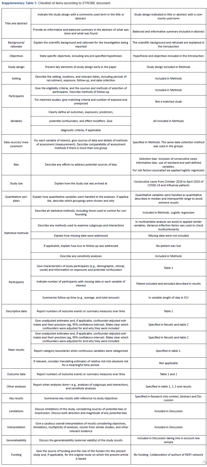
The study was approved by the local Ethics Committee of the Reina Sofía University Hospital of Córdoba (Code 4706), which were exempted from the need to seek written informed consent due to the observational and retrospective nature of the study. STROBE recommendations for observational studies were followed and checked (Supplementary Table 1) [7]

Clinical data was recorded in a standardized protocol and anonymized in a database. Variables evaluated were: (i) demographics, (ii) previous conditions, (iii) clinical characteristics, (iv) severity of illness (APACHE II and SOFA score) (v) analytic and microbiological studies, (vii) pharmacologic and support therapy, (viii) and outcome variables (microbiologically confirmed coinfection and superinfection during ICU stay, type of infection, microbiology characteristics, overall mortality at 30 and 90 days and died with active infection). Corticosteroids when administered were given at maximum doses of 1mg/kg/day of methylprednisolone. All patients admitted during this period were followed until ICU discharge and overall mortality 90 day was assessed by phone call in discharge patients.

To standardize the classification of an infection as present on admission (co-infection) or a healthcare-associated infection (superinfection), we used the CDC/NHSN criteria. Superinfection is defined as localized or systemic condition resulting from an adverse reaction to the presence of an infectious agent(s) or its toxin(s) that was not present on or after the 3rd hospital day (day of hospital admission is day 1) [8]. Diagnosis of lower respiratory infection was based on two of the next criteria: temperature up to 38.5 ˚C or leucocyte account (≥12.000 or ˂4.000 cells per μL), and purulent secretions. Episodes need microbiological confirmation with isolation in the endotracheal aspirate. Ventilator associated pneumonia was defined with the above criteria and the presence of new or progressive infiltrates on radiological explorations after 48 hours of mechanical ventilation [6]. ICU bloodstream infection for typical skin contaminants (e.g. coagulase-negative Staphylococcus) were included only if ≥2 blood cultures showed the same phenotype on separate occasions, or 1≥ blood cultures for clinical sepsis and no other infectious process; catheter related bloodstream infections were documented by quantitative tip culture [9]. All episodes and criteria were critically reviewed by two investigators (A.M and J.R.G).

SARS-COV-2 testing was performed at Microbiology Reference Laboratory (Reina Sofía University Hospital/IMIBIC). VIASURE SARS-CoV-2 Real Time PCR Detection Kit (CerTest Biotec), a multiplex real-time PCR detecting ORF1ab and N genes, was used for diagnosis. Influenza A/B virus were also investigated using GeneXpert® (Cepheid) or the BioFire® FilmArray® Respiratory 2 plus Panel (Biomerieux).

Continuous variables were compared using the Mann- Whitney U or T Test of Student Test and categorical variables were compared using the Chi Square Test or Fisher’s Exact Test as appropriate. Ventilator associated pneumonia and blood stream infections incidence rate were calculated as the number of events per 1000 patient-days at risk; the 95% confidence interval (CI) for the incidence rate estimate was obtained using mid-P exact test. In patients with COVID-19, multivariate analyses were performed using logistic regression for study variables related with superinfection and Cox regression for overall mortality 90 days. In logistic regression for variables related to superinfection, each multivariate analysis included one variable for every ten events; they were selected those with p˂0.1 (model 1) or those with clinical interest (model 2 and 3). Due to prolonged length of ICU stays of COVID-19 patients we decide overall 90 days mortality over overall 30 mortality. In Cox regression analysis for overall mortality 90 days, superinfection variable was included as time-dependent; patients have an additional transient state before superinfection event, not taking timing, this time, into account would cause time-depending bias [10].

Results

Description of patients with COVID19 and Influenza pneumonia

During the study period 116 patients were evaluated. 62 patients with highly suspected COVID-19 pneumonia were identified; 58 met inclusion criteria, 3 patients with COVID-19 disease like symptoms were not confirmed in laboratory RT-PCR of SARS-COV-2 and 1 patient with limited therapeutic effort at admission was excluded. There were 48 influenza pneumonia patients evaluated and all of them were legible without exclusion criteria.

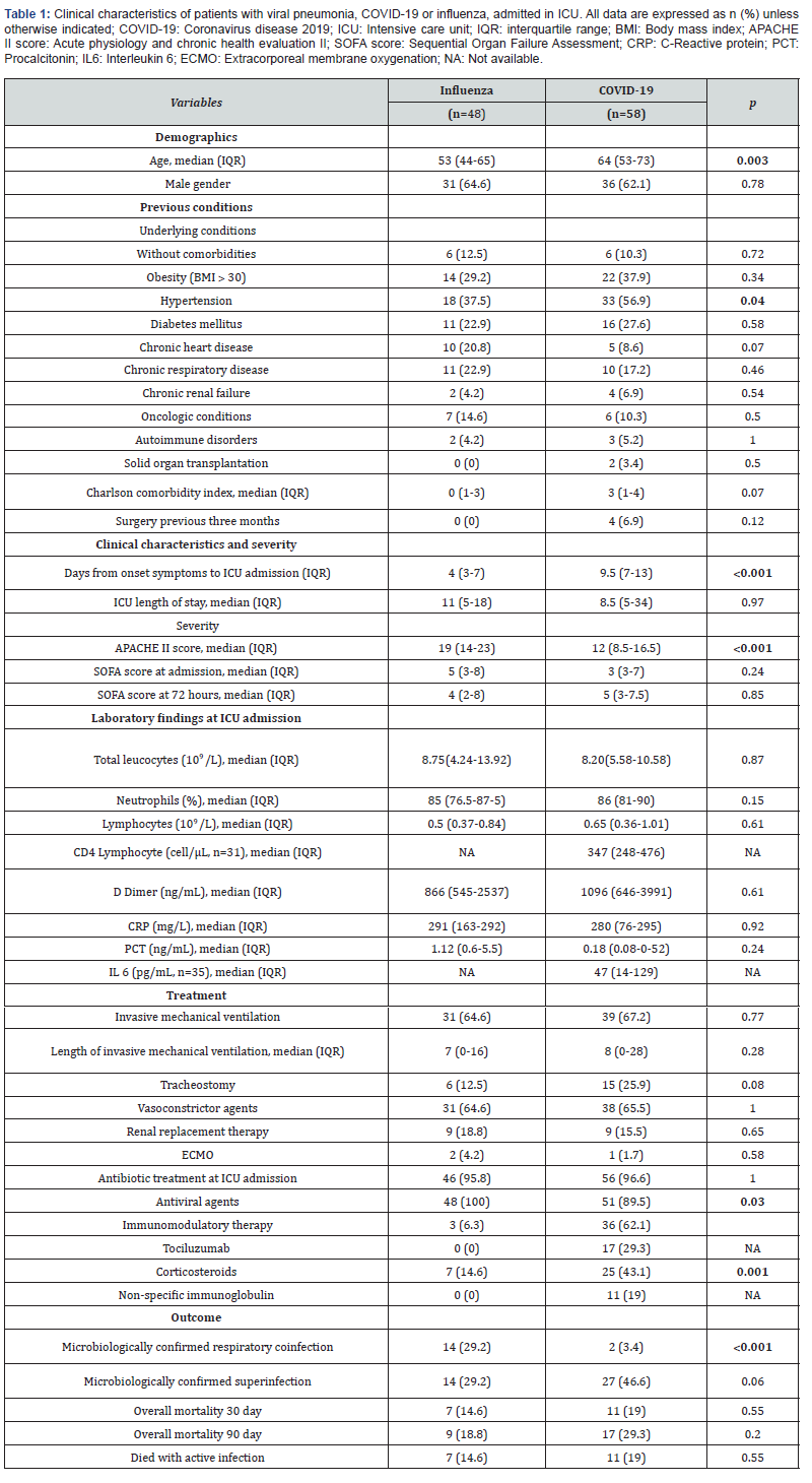
Clinical characteristics of COVID-19 and influenza pneumonia cohorts are described in Table 1. Critically ill patients with COVID-19 pneumonia were median age 64 years old. Only 10% (6) of patients did not have comorbidities while the most frequent were hypertension and obesity with 56% (33) and 37% (22) respectively. At admission, laboratory results showed the median of D Dimer of 1096 ng/ml, median of 347 cell/ μL of CD4 and 47 pg/mL of IL6.

Patients with COVID-19 in comparison with those with influenza pneumonia were significantly older (64 vs 55; p=0.004), with higher incidence of hypertension (56 vs 37; p=0.04), more days from onset of symptoms to ICU admission (9 vs 4; p˂0.001), treated with corticosteroids (43 vs 16%; p=0.002), but have less APACHE II score (12 vs 19; p˂0.001).

Co-infection and superinfections

Patients with COVID-19 pneumonia, in comparison, had significantly less frequent respiratory co-infection (3 vs 29; p˂0.001) (Table 2). 96% of patients with COVID-19 were treated with antibiotics at admission. There were 3 cases of Coagulase Negative Staphylococcus in COVID-19 patients at ICU admission.
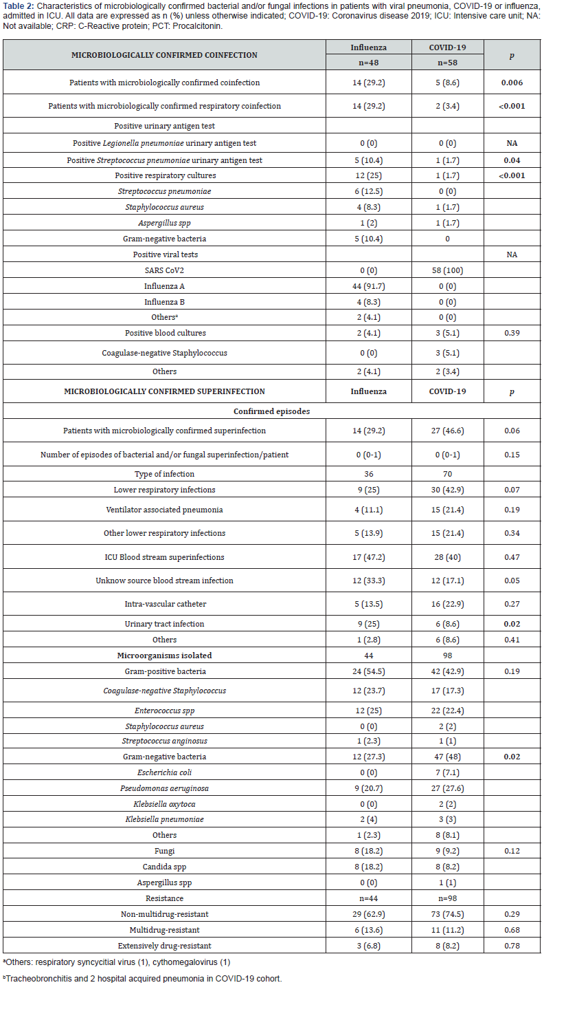
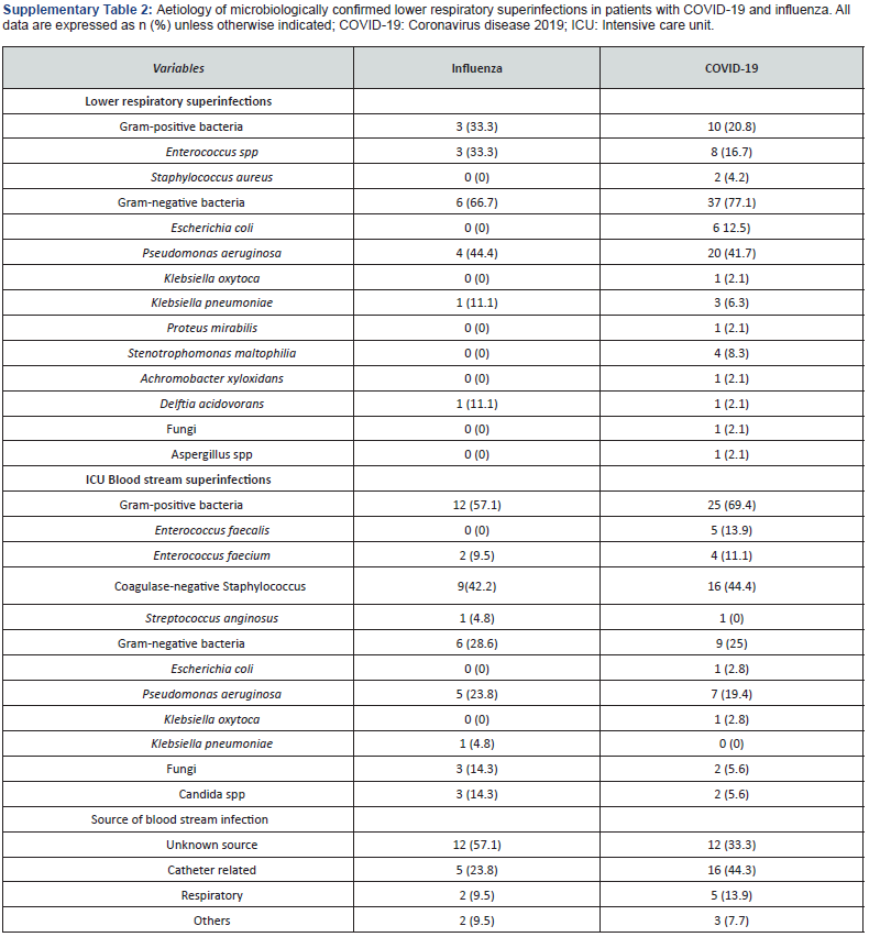
Superinfections were not significantly higher in patients with COVID-19 (46 vs 29%; p=0.06). The most frequent microorganisms isolated in this cohort was Pseudomonas aeruginosa (27%), and Gram-negative bacteria were significantly higher than in influenza pneumonia patients (48 vs 27; p=0.02) (Table 2). Lower respiratory infections were the most frequent superinfections in COVID-19 patients, and they were not significantly higher than influenza pneumonia patients (42 vs 25; p=0.07); gram negative (77%) and Pseudomonas aeruginosa (41%) were the most frequent microorganisms related in this infection. (Supplementary Table 2) Incidence rate of ventilator associated pneumonia was not significantly higher in this group with 13.2 (CI95% 7.24-21.8) per 1000-days-ventilated COVID-19 patients in comparison with 7.1 (CI 1.9-18.3) per 1000-days-ventilated patients in influenza pneumonia cohort (p=0.28). ICU blood stream superinfections were similar in both cohorts (40 vs 47%; p=0.47) with and incidence rate (including secondary bacteraemia) of 25.8 (CI95% 18.1-35.8) in COVID-19 patients and 24,3 (CI95% 15.1-37.2) per 1000-days-at risk in influenza pneumonia cohort (p=0.84). Grampositive bacteria and Coagulase-negative Staphylococcus were in both cohorts the most frequent bacteria (Supplementary Table 2). The rate of blood stream candidemia was 5 and 14%.
Variables related with bacterial and/or fungal superinfection in critical care patients with COVID-19

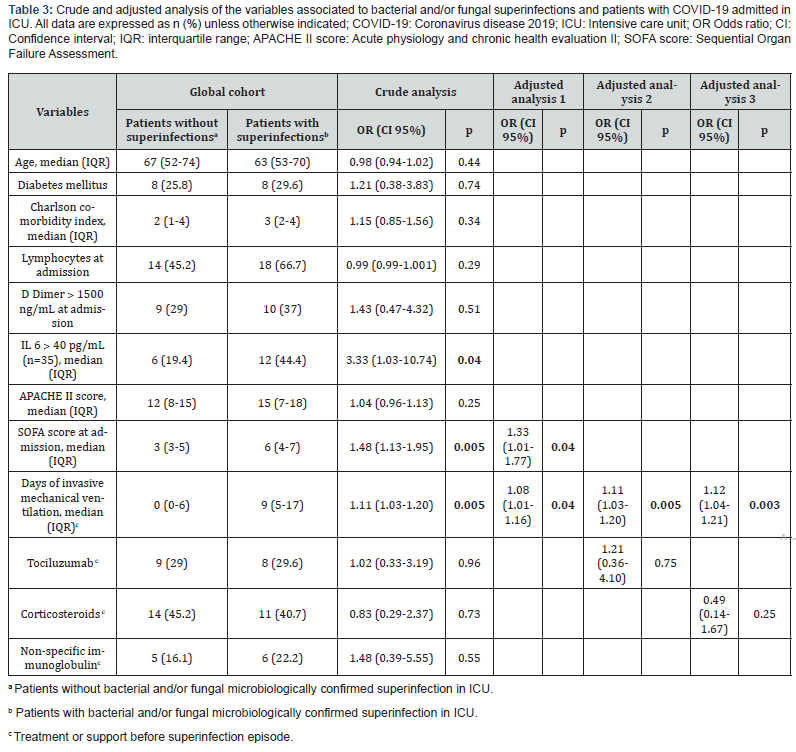
The crude and adjusted analysis of the association between selected variables and superinfection in COVID-19 patients is shown in Table 3. In the crude analysis, variables statically significant related to superinfection were the severity of the illness at admission, SOFA score (Odds Ratio (OR) 1.48; CI 1.13- 1.95; p=0.005), interleukin 6 (OR 3.33; CI 1.03-10.74; p=0.04) and days of invasive mechanical ventilation (OR 1.11; CI 1.03- 1.20; p=0.005). In adjusted analysis, days of invasive mechanical ventilation (OR 1.08; CI 1.01-1.16; p=0.04) and SOFA score at admission (OR 1.48; CI 1.13-1.95; p=0.04) were statistically associated with superinfection. Treatments with tociluzumab (OR 1.21; CI 0.36-4.10; p=0.75) or corticosteroids (OR 0.49; CI 0.14- 1.67; p=0.25) failed to show any relation in the crude and adjusted analysis.
Prognosis in COVID19 patients: Impact of superinfection

Patients with COVID-19 showed higher overall mortality 90 day but there were not statistical differences in comparison with patients with influenza infections (29 vs 18%; p=0.20). Eleven patients died in treatment with active infection in COVID-19 cohort. Survival analysis of 90-day overall mortality did not show differences between COVID-19 and influenza pneumonia (p=0.20). (Supplementary Figure 1)
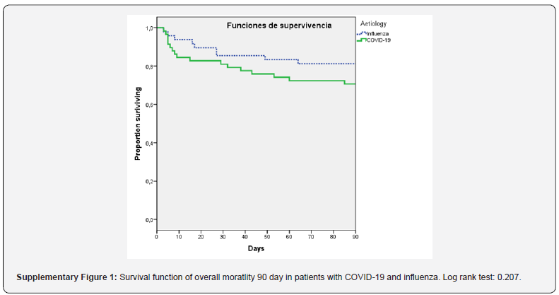
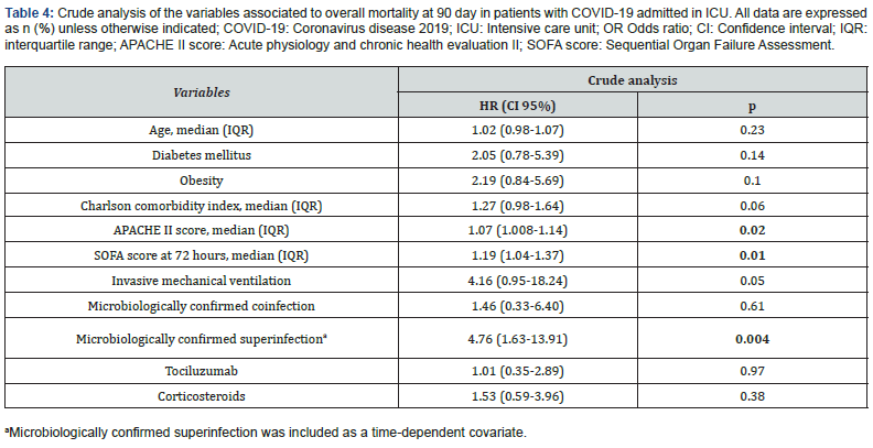
Table 4 shows that variables that were related in crude analysis with overall mortality at 90 days were severity of illness, APACHE II score (HR 1.07; CI 1.008-1.14; p=0.02), SOFA score at 72 hours (HR 1.19; CI 1.04-1,37; p=0.01) and superinfection (HR 4.76; CI 1.63-13.91; p=0.004) when was explored as timedependent variable.
Discussion

We found that in our cohort of critically ill patients with COVID19 pneumonia, patients experienced low coinfection rate (3%) and high incidence of superinfections (46%). Gram negative infections were significantly higher in comparison with those with influenza pneumonia and lower respiratory infections and Pseudomonas aeruginosa were the most frequent source and microorganism related.

Microbiologically confirmed respiratory co-infection of COVID-19 patients of our cohort was significantly lower (3%) than patients with influenza pneumonia. Similar findings are noted in other studies, so we could suggest that coinfection is unlikely to be common in patients with COVID-19 upon admission to the ICU [11,12]. On the other hand, we are aware that coinfection could be influenced by seasonal factors and it is unknown if these results could change during other periods. Considering the low coinfection rate, empiric antibiotic treatment should be closely revaluated once bacterial and/or fungal bacterial infections has been ruled out to reduce resistant emergence. We found 3 cases at admission of bloodstream infections by Coagulase-negative Staphylococcus; in the context of fulfil bloodstream infection criteria and clinical systemic inflammatory response they were treated. We could not rule out that those results as contaminations.

The published incidence of superinfections in critical care patients with COVID-19 is highly variable. While some reports show low incidence of lower respiratory infections less than 30%, other studies point to SARS-CoV-2 as an independent risk factor and to increase its´ incidence (˃50%) [5,6,13,14]. In our study, the incidence of lower respiratory infection is elevated (42% of lower respiratory infection and 21% of ventilator associated pneumonia) and higher than those patient with influenza pneumonia. Even though we found lower incidence rate of ICU bloodstream infection than other reports like Giacobbe et al. [15] study (47 vs 28 episodes per 1000 patients-days at risk in our COVID-cohort), and was not significantly higher than influenza patients, still remains high with up to 20% incidence of catheter related blood stream infections [15].

Some reasons that could explain this high variability between studies may be: different pressure of ICU admissions and its´ impact in quality care, publication bias (tend to report good results), different severity of illness and patients characteristics, the effect of mortalities differences in superinfection incidence (competitive risk bias) and exclusion in some studies of patients that are still admitted to hospital or ICU at the end of the studies follow up, which could corresponded with long term patients with high rate of complications [12-14,16].

Critical COVID-19 patients are exposed to important risk factors for superinfection that can explain the high rate established like advanced age, underlying systemic diseases, acute respiratory distress syndrome, mechanical ventilation, catheters and prolonged hospital and ICU stays [17,18]. In our case we found that the severity of presentation (SOFA score at admission) interleukin 6 and supportive treatment (days of invasive mechanical ventilation) showed to be more important factors associated with superinfections in that of critical care patients with COVID-19. Even though we were unable to assess if immunomodulatory treatment was related, the study is not powdered enough to test this hypothesis; published data shows controversial findings related corticosteroids and tociluzumab: the balance of the result of theatrically reduction of supports with immunomodulatory treatment (e.g. mechanical ventilation and prognosis) and the increase of the exposure to superinfections is still unknown [9]. Some studies point to SARS-COV-2 as an independent risk factor for exposure to lower respiratory and blood stream infections. Suggested explanations for this hypothesis are endothelial dysfunction, widespread thrombosis, histologic findings of acute fibrinous and organizing pneumonia and gut biota disruption [6,9].

In some papers increasing Pseudomonas aeruginosa superinfections in COVID-19 patients were noted [6,19]. In our case, Pseudomonas aeruginosa was too the main pathogen isolated (27%). Future studies will assess if COVID-19 clinical evolution and its´ histopathology lung damage/ADRS may predispose to these kinds of infections; despite this, we found that gram negative infections were significantly higher in COVID-19 patients in comparison with influenza pneumonia, local flora may play an important role in this issue.

In our case we can show that superinfection was independently associated with overall mortality at 90 days in crude analysis, but future studies will clear the complex relationship between severity of illness, superinfection, treatments, and mortality.

Our study has obvious limitations. Its´ observational design is exposed to certain biases by its nature, even though we tried to minimize them by applying strict definitions and following STROBE recommendations for observational studies. We are aware of the important limitation of sample size; that is important in the precision of our estimate, so it must be taken with caution. Epidemiology may be influenced by local flora. This cohort of critical care patients with COVID-19 were admitted to ICU before RECOVERY trial results, so more than one half of patients did not receive corticosteroids, it can impact in the future patients and superinfections events [20]. But considering the context of the high variable data reports, these results in this critical care cohort specifically focused on bacterial and/or fungal infection in COVID-19 patients may be useful discussing issues such as the co-infection and superinfection, it´s impact and epidemiology and treatment approach.

Conclusion

The incidence of coinfections is low in critically ill patients with COVID-19 pneumonia and significantly less frequent than in patients with influenza pneumonia. Empirical treatment at admission should be closely revaluated. Incidence of superinfection is high, if is suspected, empirical antipseudomonal treatment should be considered specially in lower respiratory infections, considering local flora. Severity of illness and supportive treatments seems to be the clearer factors related to superinfections.

To Know more about Anesthesia & Intensive Care Medicine


To Know more about our Juniper Publishers

Click here: https://juniperpublishers.com/index.php  

Monday, August 9, 2021

Applications of Thromboelastography in Acute Ischemic Stroke Patients - Juniper Publishers

 Neurology & Neurosurgery - Juniper Publishers


Abstract

Thromboelastography is a test that can be carried out beside the patient, providing valuable information on the state of coagulation. The main utility of TEG is that it allows integration of conventional coagulation tests and platelet function, achieving a larger understanding of the physiology of haemostasis. According to TEG patterns it is possible to accurately identify the underlying defect in the coagulation cascade and allow to ease patient treatment decisions. There are some reports that this technique can be used in neurological patients with acute ischemic stroke. There are some contradictory or indirect and inconclusive reports related to the applications of several tests of thromboelastography in acute ischemic stroke patients. The aim of the article is to review the available reports on the use of thromboelastography in acute ischemic stroke patients.

Keywords: Thromboelastography; stroke; Acute ischemic stroke; Coagulation; Platelets

Introduction

Thromboelastography (TEG) is a test that can be carried out beside the patient, providing valuable information on the state of coagulation [1]. TEG measures the dynamics of coagulation and it is normally used during blood transfusion therapy to begin earlier and directed towards more specific disorders such as coagulation factor deficits and/or platelet deficiency or dysfunction [2]. TEG include in vitro analysis of the relation between different components of coagulation, and thus observing interactions among platelets (PLT), fibrinogen (FIB) and coagulation proteins from a general standpoint. The main utility of TEG is that it allows integration of conventional coagulation tests and platelet function, achieving a larger understanding of the physiology of haemostasis. According to TEG patterns it is possible to accurately identify the underlying defect in the coagulation cascade and allow to ease patient treatment decisions. [3] There are limited data about TEG in Acute Ischemic Stroke (AIS). The aim of the article is to review the available reports on the use of TEG in AIS patients.

An investigation of recent published literature was conducted. Authors based on PRISMA [4]. Briefly, a database search (date of search 10.05.2021) including PubMed, CENTRAL and Google Scholar databases. The following keywords were applied thromboelastography, TEG, acute ischemic stroke. References of retrieved studies were searched manually for additional studies and reviews. Reviews were also considered by source of citations of relevant studies and interpretation of their results. Duplicate and multiple citations and reviews not containing any relevant information were excluded. Eventually, 10 original reports [A-K] directly related to the applications of TEG in AIS patients were considered eligible for inclusion in the review. Elliot et al. [5] designed a study that was aimed to providing initial TEG data in stroke patients before and after tissue plasminogen activator therapy and to providing the necessary preliminary data for further study of TEG's ability to identify clot subtype and predict response to tissue plasminogen activator therapy. AIS patients had shorter R (means the time until the first clot is detected), greater α angle (means the rate of fibrin deposition and cross-linking) and shorter K (the time from the end of the test until the clot reaches 20 mm, which is the rate of clot formation), which indicating faster clotting. Additionally, a subset formed clots with stronger platelet-fibrin matrices. Treatment with tissue plasminogen activator resulted in reduction in all indices of clot strength. TEG demonstrates that many AIS patients are hypercoaguable.

Results from another study [6] aimed to investigate the application of TEG on admission for predicting early neurological deterioration (END) in patients with AIS and its potential correlation with the evolution of ischemic lesions. Of the 246 eligible patients, END was identified in 29.3% patients. Results showed that decreased R time on admission TEG is associated with END within 3 days in patients with AIS. Yu et al. [7] suggested there are limited data on TEG as a predictive tool for hemorrhagic transformation in patients with AIS. Authors designed a study to investigate whether TEG values on admission could predict hemorrhagic transformation in patients with AIS. TEG was performed in 205 patients with AIS. The primary outcome was hemorrhagic transformation, defined as the presence of blood in brain on follow-up imaging study and secondary outcome is neurological deterioration, defined as a 2-point increase on the National Institutes of Health Stroke Scale (NIHSS) within 72 h of stroke onset. In patients with AIS, TEG value of R < 5 min can identify patients who have an increased risk of hemorrhagic transformation during hospitalization. One of the studies [8] was designed to provide initial data to compare TEG with the Conventional Coagulation Test (CCT) to analyze the coagulation function of antiplatelet drugs in such patients. The retrospective cohort study included 240 patients who received endovascular therapy. The baseline and clinical characteristics of these patients were collected with respect to TEG and CCT on day 5 after aspirin and clopidogrel post-endovascular interventions. The correlation and agreement of these 2 detecting methods were analyzed. Parameter FIB derived from CCT might be advantageous for evaluating early neurological deterioration, while parameter R detected by TEG might be superior for evaluating symptomatic intracranial hemorrhage.

Quan et al. noticed [9] that lung cancer related hypercoagulability could increase the risk of ischemic stroke. Routine coagulation tests may have limited capacity in evaluating hypercoagulability. The aim of this study was to investigate the ability of TEG in the identification of hypercoagulability in patients with Lung Cancer and Cryptogenic Ischemic Stroke (LCIS). Therefore, TEG could identify hypercoagulability in LCIS patients and healthy controls. Identification of hypercoagulability in lung cancer patients by TEG may be helpful to prevent the occurrence of LCIS. Yan et al. [10] was designed the study evaluating ticagrelor versus clopidogrel on platelet reactivity measured by TEG in patients with minor stroke or transient ischemic attack (TIA). Platelet reactivity was assessed by TEG using test to platelet mapping. Patients were divided into carriers and non-carriers according to the carrier status of CYP2C19 loss-of-function (LOF) alleles. The primary outcome was the proportion of patients with High On-Treatment Platelet Reactivity (HOPR). Ticagrelor was superior to clopidogrel in inhibiting platelet reactivity measured by TEG platelet mapping among patients with acute minor stroke or TIA, particularly in carriers of the CYP2C19 LOF alleles. Large randomised controlled trials are needed to confirm our findings.

In this case, researchers [11] used different laboratory testing in a patient with ischemic stroke and reduced prothrombin Time (PT) to identify an in-vitro effect of Lupus Anticoagulant (LA) excluding an in-vivo bleeding disorder. LA owns procoagulant properties in vivo and prolongs phospholipid-dependent clotting times in vitro. The prolonged in vitro clotting time can be misinterpreted as a bleeding disorder. The activity of various coagulation factors was evaluated both with recombinant thromboplastin Innovin (Siemens Healthcare) and reagent tissue extracted thromboplastin Thromborel® (Siemens Healthcare). Researchers conclude, that different thromboplastin reagents, plasma mixing tests and thromboplastin independent coagulation tests may be helpful to differentiate LA and in vitro changes from in vivo factor deficiency in patients with LA.

Koch et al. [12] designed a study using canine model with ischemic stroke. In this case, authors reported hemostatic parameters, including D-dimer and TEG along with clinical and imaging findings for five dogs diagnosed with ischemic stroke. A hypercoagulable state was identified in two dogs based on the results of the TEG and was suspected in the remaining three cases based on a shortened TEG clot reaction time. The demonstration of a possible hypercoagulable state, as identified by the TEG, is an interesting finding which should be explored further to help reveal predisposing hypercoagulable conditions in dogs with ischemic stroke. Wiśniewski et al. [13,14] demonstrated prospective study about hypercoagulability as measured by TEG. Authors investigated whether a hypercoagulability detected by TEG may be associated with larger size of acute ischemic infarct. In the entire group, they reported that subjects with a large ischemic focus had a higher diameter of a clot than subjects with a small ischemic focus. Stroke subjects with hypercoagulability had significantly higher probability of a larger size of acute ischemic focus compared to normal-coagulable subjects. Authors conclude that TEG, based on the parameters related to clot strength, may have clinical utility to identify the risk of the extensive ischemic infarct.

Thromboelastographic changes in patients experiencing an AIS and receiving alteplase [14,15] was investigated. It was a prospective cohort study enrolled patients who presented to the emergency department with symptoms of AIS and received intravenous alteplase. Blood samples were obtained before alteplase administration and at 30, 60, 90, 120, and 150 minutes after alteplase administration. The maximum inhibition of fibrin buildup was seen at 30 minutes after the start of alteplase infusion, and the lowest clot strength was observed at 60 minutes after initiation of alteplase. Most patients return to near baseline parameters within 150 minutes of alteplase initiation; however, two patients did not return to their baseline values within the 150-minute time frame. This study proved that TEG is a useful tool for determining changes in the coagulation system of patients whom have received recombinant tissue plasminogen activator.

Discussion

The above-mentioned studies proved the potential benefits of a comprehensive coagulation assessment for stroke evaluation. The summary are presented in table 1. Several studies have highlighted the potential usefulness of TEG coagulation monitoring and hypercoagulability detection in predicting clinical outcomes and prognosis in ischemic stroke subjects. TEG measures the dynamics of clot formation and dissolution and provides more complex and integrated measurement of venous blood coagulation properties than conventional tests. Therefore, most of presented studies of TEG may contribute to a more reliable and accurate assessment of hypercoagulability which remains the undoubted pathological cause of ischemic stroke.

The most frequent parameter in the studies which indicated a prognostic or diagnostic value in patients with AIS was R. From a clinical point of view, it’s crucial whether a hypercoagulability that could be detected by TEG. It is a hypothesis, that an individual’s tendency to form firm and large clots in a short period of time may promote the expansion of the ischemic focus in the brain. Hypercoagulability still remains the baseline background of a higher risk of cerebrovascular events, including ischemic stroke. In one study, Quan et al. [9], hypercoagulability was also defined by the Maximum Amplitude (MA), however, there remains a broad variability in the definition of hypercoagulability as determined by MA and thus limits its predictive ability. Some studies proved, this tool may be also useful for monitoring the hemostatic processes in stroke subjects undergoing the alteplase therapy and in predicting the risk of hemorrhagic transformation after intravenous thrombolysis.

Conclusion

In conclusion, in this review we emphasized that there are some reports which indicate the use of several parameters of TEG in AIS patients. The most frequent parameter in the studies which indicated a prognostic or diagnostic value in patients with AIS was R, which may define hypercoagulability in this group of patients. Authors also point to other clinical areas among AIS patients that should be further explored. Therefore, our novel findings contribute to the expand the existing knowledge on predictive values of TEG and support its clinical usefulness in the acute phase of ischemic stroke. Nonetheless, it is crucial to further investigation in this field, including large cohort prospective studies.

To Know more about Neurology & Neurosurgery


To Know more about our Juniper Publishers

Click here: https://juniperpublishers.com/index.php  

Friday, August 6, 2021

The Needs and Applications of Delivery Systems to Fortify Food with Active Ingredients - Juniper Publishers

Nutrition and Food Science - Juniper Publishers

Introduction

Consumers are increasingly demanding a healthy diet rich in active ingredients such as flavors, vitamins, minerals, antioxidants, lipids and so on [1-3]. However, on the one hand, the compositions of active ingredients in natural food are usually limited; on the other hand, some agents may be high in raw food, but their bioavailability is very low [4-6]. For example, β-carotene has received increasing attention during the past 30 years due to its beneficial health effects such as antioxidant and anticancer activities as well as its ability to reduce the risk of heart disease and certain chronic disease [7]. However, humans cannot synthesize β-carotene in their bodies, and they must obtain it from the foods they consume. Many vegetables and fruits (peppers, tomatoes, carrots, mangoes, and kale) are rich in β-carotene, but literatures show that only a minor part (<10%) of the carotenoids in these raw foods is absorbed [8,9]. In addition, many active ingredients including β-carotene also have many other disadvantages such as low water-solubility, physical or chemical instability, undesirable flavor, and so on [10-12]. All of them make active ingredients difficult to directly incorporate into final food products. Therefore, development of strategies to incorporate active ingredients is necessary if such foods are to keep successful and initiative in the marketplace.

Luckily, these challenges can be overcome by entrapping active ingredients into suitable delivery systems. During the past decades, delivery systems (such as liposomes, emulsions, lipid particles, microcapsules, beads, etc.) have been effectively designed and utilized in food industry to encapsulate, protect, and deliver functional components before introduced into final food products [13-16].

Among of them, emulsions especially for oil-in-water emulsion have attracted the interest of many research groups in food and pharmaceutical fields due to their favorable properties such as good biocompatibility, easy design and preparation [3,17]. To obtain uniform emulsion, the oil phase dissolved with bioactive agents is mixed with emulsifier water solution, blended and then passed through a homogenizer. In this system, emulsifiers play an important role in the formation of emulsion and its stability thereafter. To be an effective emulsifier, ingredients should exhibit perfect emulsifying activity [18]. That means it should quickly adsorb to the surfaces of small oil droplet form an interfacial coating and appreciably decrease the interfacial tension during homogenization [3]. Synthetic emulsifiers such as Tween 20, tween 80 are the traditional emulsifiers for many products. With the increasing demand for food and beverage with natural ingredients in recent years, many food manufacturers are trying to replace synthetic emulsifiers with natural and sustainable alternatives. To date, researchers have found a lot of natural emulsifiers (such as protein, polysaccharides, phospholipids, and bio surfactants), which are able to form and stabilize oil droplets [18-21]. Chung et al., [18] found that emulsion prepared by using quillaja saponin as a natural emulsifier could produce whitish milk similar to a commercial liquid creamer. Lin et al., [22] have prepared β-carotene loaded emulsion by using modified starch. He et al., [4] have fabricated curcumin emulsions containing Konjac glucomannan stabilized with whey protein isolate and achieved a controlled and sustainable release of bioactive compounds from emulsions. Lv et al., [20] have compared the effectiveness of a number of natural emulsifiers (whey protein, gum arabic and quillaja saponin) on the production and stability of vitamin E fortified emulsions and found that whey protein isolate and quillaja saponin were more effective at forming emulsions containing fine droplets than gum arabic, while quillaja saponin based and gum arabic based emulsions exhibited better resistance to pH change. Due to the above-mentioned advantages, emulsions are already widely utilized in the food industry, e.g., in dressings, sauces, soups, beverages, dips, creams, and desserts [6,19,22].

Liposome, as one of the most common used formulations, also has great potential to embed bioactive agents. A liposome is a self-assembling and cell-resembling delivery system with concentric bilayer structure. The size of liposome is typically about 10 to 1000nm with the structure varying from a balloonlike unilamellar to an onion-like multiple structures [3,12]. There are different hydrophilic and hydrophobic regions in liposomes separated by surfactant substances (such as phospholipids) [3]. Therefore, both hydrophilic and lipophilic molecules and even amphiphilic molecules can all be entrapped in the special bilayer. Since liposomes were first described in the 1960s by Alec Bangham, they have been beneficial in medical, cosmetic, and agricultural fields, and have become integral to food research. More recently, liposomes have been widely used to encapsulate proteins/peptides/enzymes, polyphenols/flavones, essential oils/fatty acids, vitamins, energetic substrates, and minerals [3,23-25]. Active agents in the form of liposomes could then be effectively added into different food products: dairy products (milk, cheese, and yogurt), drinks (juices and milk drinks), meat (pork, beef, and rabbit), and other products (chocolate, tofu, etc.) [12,27-32].

Delivery systems containing bioactive agents could be directly added into food material with simple blending or homogenization and bring a number of benefits to food industry: supplying good protection for bioactive agents against environment stress, masking off-flavor (bitterness, astringency), improving storage and handling characteristics, extending shelf life, besides increasing bioavailability. However, the food system microenvironment (pH, intrinsic component, and ionic strength etc.) and emulsifier properties used are critical factors needed to take into account if appropriate final food is wanted [3,6]. For example, whey proteinbased emulsion could effectively inhibit oxidation of fish oil in milk, but is less stable in yoghurt and dressing [18].

Conclusion

A number of challenges associated with active ingredient need to be addressed when incorporated into food. Suitable delivery systems such as emulsions, liposomes, etc. could be designed to encapsulate active agents followed by simple blending or homogenization. However, whether appropriate final food could be obtained is dependent on food and emulsifier properties.

To Know more about Nutrition and Food Science


To Know more about our Juniper Publishers

Click here: https://juniperpublishers.com/index.php  


Thursday, August 5, 2021

Using Online Simulation to Replace Clinical Practice during COVID-19 - Juniper Publishers

 Nursing & Health Care - Juniper Publishers

Abstract

In this article, we describe the use of computer-based simulations and other technology to replace clinical hours for undergraduate nursing students during the COVID-19 pandemic. We describe the technology used and how we prepared for implementation. Most importantly, we describe how we facilitated a full clinical day online using the technology and the feedback from students about the experience. To our knowledge, this is the first example of how to facilitate a fully online clinical day using these technologies. As such, this information will guide educators as they design online instruction to overcome the restrictions related to the pandemic while continuing to ensure quality education and achievement of course outcomes.

Short Communication

Simulation-based education has been used extensively in undergraduate nursing education to replace or deepen the understanding of complex nursing concepts [1]. Historically, a variety of different levels of fidelity (realism) have been used to achieve learning needs, with all levels of fidelity (low, moderate, and high) having a strong positive impact on learning experiences [2]. With the restrictions applied to clinical practice because of the COVID-19 pandemic, students from nursing programs were prohibited from attending in-person simulation labs and clinical settings. New instructional delivery was implemented to provide an opportunity for students to replace clinical skills using innovative simulation experiences that accommodate COVID-19 restrictions. Two types of software from Elsevier (https://evolve.elsevier.com) were incorporated to provide optimal learning experiences. The first, Simulation Learning System for RN® (SLS), which is designed to support the delivery of high-fidelity simulations, was used to provide the framework for unfolding simulations that the instructor delivered using images of a simulation set up, a concept map creator, and an electronic health record. The second software, SimPractice, is a screen-based simulation software where students interact with a virtual client and make decisions about and provide nursing care. Zoom meeting software was used as a means to create telepresence and facilitate fully simulated clinical days. Simulation days were designed intentionally to provide the opportunity to apply the knowledge, skills, and behaviours required to achieve course outcomes.

Instructors and students were required to have a strong internet connection and laptops that met the required specifications. Students and faculty received training sessions about using the new software, before implementation. Furthermore, individualized support was provided as needed by two senior instructors with experience running this software. There was no need for Zoom software training as the students and instructors have previously used it. One example of the online clinical simulation day is for second-year students, where the concepts of “self-concept” and “interprofessional communication” were incorporated. Students were instructed to wear their uniforms and approach the day as they would a shift in the actual clinical setting. The day began with students watching a YouTube video about self-concept followed by a group discussion to reflect critically on the video. The students then drew a mandala that reflected a stressful event in their life and how they coped with that stress. After this activity, the instructor facilitated a group discussion of how positive and negative self-concepts can affect stress management. In order to incorporate related psychomotor skills, students completed a computerized simulation of taking vital signs using SimPractice. The instructor debriefed the students to help them make a connection between vital signs and stress. Interprofessional communication was addressed through a role-play activity involving two students in each Zoom breakout room. Following this, the students practiced providing a handoff report in an SLS simulation. At the end of the day, students submitted a reflection on their performance, which included specific examples of how they met evaluation criteria and how they would improve their professionalism, preparation, knowledge, and participation. The instructor then provided individual formative feedback to students.

Students enjoyed the learning experience and were appreciative of the efforts to provide practical strategies to meet learning outcomes. Carefully developed online clinical days that incorporate simulation technology may be used to replace some clinical experiences. However, several psychomotor skills that need physical contact with patients should be performed in a clinical setting. Online simulation can support clinical education but does not entirely replace it.

To Know more about Nursing & Health Care


To Know more about our Juniper Publishers

Click here: https://juniperpublishers.com/index.php  


Wednesday, August 4, 2021

Bioanalytical Approach-Ageing, Exercise and Oxidative Stress - Juniper Publishers

 Complementary Medicine & Alternative Healthcare - Juniper Publishers

Abstract

Abstract: Exercise interferes with the ageing, causing changes in markers of the antioxidant system, such as nitric oxide (NO) and uric acid.

Aim: The objective of this study was to evaluate if regular exercise affects the ageing process by causing changes in the antioxidant markers, nitric oxide and uric acid, in individuals aged ≥ 65 years, subjected to an exercise plan.

Methods: The study involved 12 participants distributed in an experimental and control group. The experimental group performed exercises guided by a physiotherapist during 12 weeks. Three blood samples were collected from participants: T0, at the start of the study; T1, after 12 weeks of exercise and T2, 12 weeks after finishing the exercise. NO and uric acid were quantified with commercial kits. The statistics were performed using SPSS and the ANOVA and Student t-tests were applied.

Result: In the experimental group, the mean values of NO showed an increase between T0 and T1, with a slight decrease between T1 and T2. Similar effects were observed for the uric acid concentration in the experimental group, with statistical significance from T0 to T1.

Conclusion: After exercise, there was an increase in blood uric acid and NO levels, highlighting the importance of moderate and controlled exercise practice. Therefore, exercise seems relevant and beneficial in activating antioxidant mechanisms.

Keywords: Ageing; Exercise; Nitric oxide; Uric acid; Ageing process; Signalling pathway; Postponement; Biological process

Introduction

Ageing is an active, irreversible biological process, including morphological, functional, biochemical and physiological changes. These changes have an impact on the health and quality of life of the elderly, leading to a progressive functional loss of the organism. In the process of ageing, there is a decrease in the metabolic rate caused by the reduction of energy exchanges, which is related to the presence and action of free radicals [1]. The accumulation of free radicals in the body leads to a phenomenon called oxidative stress. This process entails a disruption of the redox signalling pathway, which is fundamental to the aerobic system and metabolism. Free radicals are produced as part of the normal physiology and in disease. Oxidative stress is involved in a variety of chronic and degenerative diseases, such as arthritis, autoimmune disorders, cardiovascular and neurodegenerative diseases, as well as in cancer, inflammatory conditions, ageing and even following intense physical exercise [2-4]. The fundamental aspect in the prevention of oxidative stress is the correlation between the ability to inhibit or delay the onset of, for example, cancerous cells and the antioxidant activity of substances able to block the free radicals. The same principle accounts for the postponement of cellular ageing [5]. Ageing is associated with a functional decline and performing physical exercise can play a role in the reduction and prevention of ageing [6]. Although living organisms have a biochemical defence mechanism which protects them against against potentially harmful radicals, certain events of oxidative stress cannot be inhibited by these mechanisms. One example is exercise: it initiates oxidative stress, however, this activation is important to induce an activation of the antioxidative mechanisms. Training can, therefore, have a positive or a negative effect on oxidative stress, depending on the frequency, intensity, duration and exercise type practised as well as on the initial fitness levels of the individual [7]. In general, the physical activities should promote the production of antioxidative molecules to such a level, that the oxidative stress caused by the exercises is neutralised. Ideally, the produced anti-oxidant levels are even higher, thereby also reducing oxidative species induced by other events (for example inflammation, cancer, ageing) [1,8].

A common approach to assessing oxidative stress in biological systems involves the measurement of the increase or decrease of a redox-sensitive molecule that responds to this kind of stress. Usually, reliable markers are chemically unique and easily detectable. In addition, the marker should be rapidly increased during periods of oxidative stress and immediately decreased when the event has passed. Nitric oxide (NO), uric acid and enzymes are examples of such markers. In addition, these markers can be rapidly altered during oxidative stress periods, taking into account the surrounding environment [9,10]. Biological antioxidants of low molecular weight have the ability to prevent oxidative damage by direct and indirect interactions with reactive oxygen species (ROS) [8,11]. Uric acid is a secreted form of a derivate from the purine catabolism. These derivates are components of cellular molecules involved in the energy household, such as adenosine triphosphate (ATP), deoxyribonucleic acid (DNA) and ribonucleic acid (RNA). Uric acid is produced in the liver and secreted in the urine. The antioxidative capacity of uric acid is the protection of DNA and lipids from ROS and reactive nitrogen species (RNS). Enhanced levels of uric acid are often found in individuals with atherosclerosis, where the inhibition of oxidative stress is of utmost importance [5,6]. NO is a highly reactive mediator shown to play different roles in a variety of different biological processes and in ageing. NO is a free radical of high biological importance. An increase in the levels of NO can lead to stress responses, whereby NO reacts with other radicals, such as the superoxide anion. NO synthase (NOS), when activated, produces NO and L-citrulline from L-arginine using nicotinamide adenine dinucleotide phosphate (NADPH) and oxygen. There are tissue-specific constitutive isoforms of NOS: neuronal (nNOS) and endothelial (eNOS). The other form is the inducible type of NOS (iNOS), which is found to be expressed in various cell types and is induced under pathological conditions [12,14]. According to some studies, exercise promotes an increase in NO concentrations, thereby improving endothelial function with increased oxygen consumption [12,15]. The main objective of the presented study was, to evaluate if exercise performed by aged individuals (≥ 65 years of age) still affects the levels of the antioxidants NO and uric acid.

Material and Methods

The study cohort was composed of 12 individuals (control group n=6; experimental group n=6) with a mean age of 84 years who lived in a home care situation. The sole inclusion criteria for participation in the study was the age of the individuals (≥ 65 years of age). The exclusion criteria were the presence of a physical disability that prevented the accomplishment of the exercise plan or the presence of a marked cognitive deficit.

The exercise plan was adapted from the HIFE Program [16]. This plan involved strength training exercises of the lower limbs, of static and dynamic balance and gait. The intensity was defined individually, and each participant was encouraged by the physiotherapist to perform the exercises in a progressive way in order to reach a high intensity. The exercises were performed for 12 weeks, one session per week. The individuals were evaluated at three different time points: T0 (before starting the exercise plan), T1 (immediately after finishing the exercise program) and T2 (12 weeks after finishing the exercise). In addition to the assessment, the Late-Life Function and Disability Instrument (LLFDI) scale was adapted and validated for Portuguese culture by Cavalheiro and coauthors (2003), for the measurement of physical function and disability and the Six Item Cognitive Test Commitment (6 CIT) to assess cognitive ability [17]. The project was presented to each of the participants. All signed an informed consent form before participating in the study. Work was carried out in accordance with the principles of the Helsinki Declaration. A venous blood sample was taken by venipuncture at 3 different time points (T0, T1, T2) and collected in test tubes without supplements. All the samples were centrifuged 1800g at 4°C for 10min, followed by separation of the serum and stored at -23°C. For quantification of NO and uric acid the following kits were used: Total Nitric Oxide and Nitrate/ Nitrite Assay (R&D Systems Europe, Abingdon, United Kingdom) and Liquick Cor-UA (Cormay, Łomianki, Poland), respectively. For NO the spectrophotometric reading was performed at 540 nm using a ThermoScientific Multiskan GO (Japan). The uric acid levels were detected at 546nm using a Tokyo Boeki Prestige 24i (Diamond Diagnostic, Japan). The techniques were performed according to the manufacturers’ instructions. All statistical analyses were performed using the statistical package IBM SPSS Statistics 22.0. After testing, the data was statistically analyzed by the use of ANOVA and Student’s t-tests. The results are presented as the mean ± standard deviation (SD) and were considered statistically significant at p <0.05.

Results

After analysing demographic data, the experimental group had an average of 85.0±2.4 years and the control group had an average of 83.0±3.9 years at the beginning of the study. Both groups consisted mainly of female subjects (5 females and 1 male). Concerning the existence of chronic pathologies, all participants reported having dyslipidemias, respiratory or cardiac insufficiencies, pathologies of the central nervous systems and Diabetes Mellitus. With respect to the use of medication, all individuals in the experimental group and 83.3% of the control group reported taking at least 4 tablets per day.

Quantification of uric acid

The uric acid measurements showed that differences occurred between the values measured at the different time points in both the control and the experimental group. After exercise, in the experimental group, a mean value of 6.20±1.78 mg/dL was detected at T1, compared to 5.47±1.64 mg/dL at T0 and a concentration of 6.03±2.0 mg/dL at T2. The control group had a mean value of 5.80±1.0 mg/dL at T1 compared to 5.44±0.95mg/ dL at T0 and finally 5.34±1.15 mg/dL at T2 to uric acid (Figure 1). Using the ANOVA statistical test, we did not obtain significant differences between the various time points (T0, T1 and T2), in the experimental and control group. However, when applying the Student’s t-test between two time points, the experimental group revealed a statistically significant increase (p = 0.03) from T0 to T1 (Figure 1).


Quantification of NO

The NO data showed that there was no statistically significant difference between the different time points analyzed in the experimental group. This was also due to the large variation of NO in the serum samples. At T1, a mean value of 22.86±12.48 μmol/L was detected after the intervention. At T0 the NO levels were 12.72±1.55 μmol/L and at T2 these were 15.28±3.55 μmol/L. The NO concerning the control group; at T1 was 21.67±12.52 μmol/L compared to T0 with 18.84±15.0 μmol/L and 29.98±8.52 μmol/L at T2. There was no statistical difference between the 2 groups, nor between the time points analyzed.

Discussion

The theory about the action of free radicals as the main cause of the ageing process was proposed by Harman in 1955. This theory is currently being confirmed by a large number of scientific publications [18]. Therefore, the biological ageing of humans is associated with an increase in free radicals that can cause oxidative changes in proteins, lipids and DNA. Due to the increased oxidative stress, exercise is constantly used as an oxidizing stimulus, which is under normal circumstances directly counterbalanced by antioxidative measures. The health benefits related to regular exercise are widely described in the literature. Some of the major mechanisms are related to the prevention of DNA damage involved in various degenerative diseases and in the ageing process. Thus, individuals who practice moderate exercise tend to exhibit an increase in antioxidant markers compared to non-practitioners. Therefore, the human body adapts to training intensities through a defense network that regulates and protects against oxidative reactions to ensure survival [3,7,19]. The present study utilized 3 different time points (T0, T1 and T2), in order to investigate whether the performance of an intervention plan with several exercises training strength, endurance, balance, flexibility and gait for 12 weeks was able to induce the production of antioxidant species [16]. The uric acid analysis showed an increase in the average value from T0 to T1 and a tendency to decrease from T1 to T2. This observation was similar for both groups (experimental and control), which was unexpected, as the control group was not instructed to change their behavior. This may simply be associated with feeding by purine metabolism [5,6]. However, the effect on the experimental group was significantly more pronounced, indicating that exercise induces the formation of antioxidant uric acid and shows that it is a good marker for detecting antioxidant effects in humans. In the present study, the results of the NO levels revealed slight changes in the mean values of T0 to T1 and of T1 to T2, but without statistical significance and the strongest increase was observed in the control group and not in the exercise group. Moreover, the analysis at T0 already indicated a difference between the two study groups, which was not observed for the uric acid measurements. There are several factors that may have affected these results, such as the effects of the seasons on activities in general, differences or alterations in the intake of medication or also difficulties in the analysis of NO as a marker, since it is unstable and can react with oxygen in the medium transforming into another compound. Therefore, NO might not be the most suitable marker to detect antioxidative effects in humans. Recent studies have shown that exercise increases the production and availability of NO in the body. Thus, exercise can, among others, induce positive improvements of the cardiovascular, hepatic, skeletal and muscle systems. Souza et al (2009) observed in rats that swim training for 8 weeks promoted an increase in plasma nitrite and, consequently, an increase in endothelial function and performance [20]. Many studies state that the participation of the elderly in physical activity programs is an independent way to reduce and prevent functional decline associated with ageing [6,7] Nelson et al (2007) indicated the need to choose the type and amount of exercise to improve the quality of life of elderly individuals, taking into consideration the modality, duration, frequency, intensity and progression [21]. Individuals who undergo intense and prolonged exercises, exhaustive training and very high frequency may exceed the capacity of the endogenous antioxidant system, thereby promoting severe muscular injuries, with consequent induction of inflammatory processes and oxidative stress [4,22]. Thus, it is important to emphasize that while the exercise plan stimulates the antioxidant activity, there is a need for a longer intervention of the physiotherapy team, so that the values of the markers can be more precise, and the project reproduction and markers, especially uric acid, may be utilized with a larger sample for a better evaluation.

Conclusion

This study is one of the first to detect the effects of exercise on antioxidants in the serum of elderly people, showing the importance of life-long physical activities adapted to personal needs. The data confirms that oxidative stress is a factor that has a great impact on the health and quality of life on humans. Strategies, such as the practice of moderate and controlled exercise, should be instituted as an important factor in the activation of antioxidant mechanisms. This results in a later adaptation of the organism, increasing the efficiency against ageing.

To Know more about Complementary Medicine & Alternative Healthcare

Click here: https://juniperpublishers.com/jcmah/index.php


To Know more about our Juniper Publishers

Click here: https://juniperpublishers.com/index.php  


Tuesday, August 3, 2021

A Neurological Presentation of Intravascular Large B-cell Lymphoma: a Rare Cause of Multi-Territory Strokes - Juniper Publishers

 Juniper Online Journal of Case Studies - Juniper Publishers

Abstract

Intravascular Large B-Cell Lymphoma (ILBCL) affects one in a million people. Here it presented as dysphasia and leg weakness in a forty-five-year-old woman with a history of mental health issues. The initial investigations showed multi-territory infarcts for which we were unable to find a treatable cause. Her condition progressed and she unfortunately died. Although rare, ILBCL ought to be included in differential diagnoses of multi-territory strokes, however, the diagnosis is commonly made post-mortem. Here we discuss common presentations of ILBCL. We note the proximity of her worsening psychiatric symptoms to the time of her death.

Keywords: Lymphocyte cells; Psychiatric symptoms; Intracerebral haemorrhage; Neurological manifestations; Intravascular large B-cell lymphoma

Introduction

Intravascular Large B-Cell Lymphoma is a rare subset of Non-Hodgkin’s Lymphoma that involves the pathological proliferation of lymphocyte cells within capillaries and other small blood vessels [1], causing their occlusion.

As the clonal proliferation is intra-vascular there is relative sparing of the surrounding tissue, making it more difficult to diagnose. The presenting symptoms vary according to the organ affected, although the skin and nervous system are common. When occlusion occurs intra-cerebrally the result is often a stroke.

The importance of identifying the aetiology of stroke in the afflicted patient is well recognised, as it can help direct management and treatment. This is particularly relevant to patients with multi-territory strokes, recurrent strokes, and those who are affected at a young age.

Our patient had worsening psychiatric symptoms leading up to her final admission and diagnosis. We note the proximity of these events, as psychiatric symptoms of ILBCL are poorly documented.

Case Report

A forty-five-year-old Caucasian woman was seen in the emergency department at a tertiary hospital in the UK. She presented with a two-day history of mumbling speech and acting withdrawn, left-sided leg and facial weakness, and right arm spasms. She had a background of depression and had presented acutely to community and hospital psychiatric services 5 months prior to this admission (we do not know the nature of that presentation). Her current medication included olanzapine 10mg daily and venlafaxine MR 300mg daily. Initial examination demonstrated an up-going right plantar response but no other focal neurology.

A CT head scan revealed low attenuation signal changes in the right cerebellar and right frontal lobes. Although her presentation appeared more psychiatric than organic, she was kept in to investigate her symptom complex and CT findings.

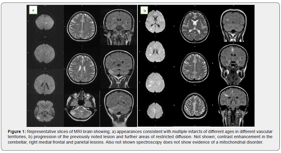
A subsequent MRI head demonstrated multiple areas of restricted diffusion in different vascular territories (Figure 1a). There were also foci of high T2 signal changes in the supra-tentorial white matter, with some demonstrating restricted diffusion. Considering the different territories, and different ages of these lesions, a cardio-embolic source was the most likely cause of these infarcts. Her trans-thoracic echocardiogram (TTE) was normal, as was her carotid Doppler. A lumbar puncture showed normal CSF. At this point she developed episodes of seizures corroborated by EEG.

We undertook investigations for lymphoma, and the blood tests showed little except a raised lactate dehydrogenase (LDH 400iU, plasma viscosity normal, blood film normal, haemoglobin 115g/L). We also discussed the possibility of vasculitis. However, her blood results and MR Angiogram did not show any positive findings.

A full-body CT scan was performed to look for other neoplastic lesions, but this was normal. We started high-dose corticosteroids as an empirical treatment for vasculitis.

Two weeks after this, she started to deteriorate. Her seizures became more frequent and her dysphasia more pronounced. A repeat MRI brain (Figure 1b) showed an increase in size of the lesions in the medial frontal and parietal lobes. New lesions in the corpus callosum and left thalamus showed restricted diffusion. The cerebellar, right medial frontal and parietal lesions enhanced with contrast. Long- and short-term MRI head spectroscopy did not show any evidence of a lactate peak to suggest mitochondrial disorder. This picture was consistent with a diagnosis of vasculitis.

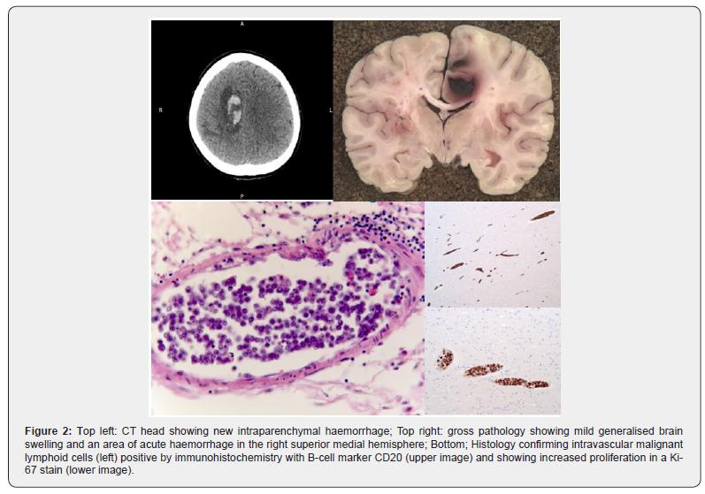
Her condition continued to decline. Her GCS dropped to 7 and did not fluctuate, a repeat CT head revealed a right-sided intracerebral haemorrhage (Figure 2). Given the multitude of insults and progressive nature of her symptoms the difficult decision was taken to adopt a palliative approach for her symptoms. She died a day later.

Discussion

Intravascular Large B-Cell Lymphoma is a rare subset of non-Hodgkin’s lymphoma (NHL), affecting one in a million people [2]. First characterised in 1959 [3], it is known colloquially as “the great imitator” [2] due to its diverse range of presentations. It causes disease by a clonal expansion of lymphocytes within small blood vessels [1] resulting in end-organ damage. In retrospective analyses of case reports, around 90% of intravascular lymphomas are of B-cell origin. Unlike other NHL presentations, there is relative sparing of the lymph nodes [4].

There are two forms of ILBCL, Western and Asian. The former presents, as in this case, with neurological or dermatological symptoms [4]. The Asian variant typically presents with multi-organ failure, pancytopenia, fever and bone marrow involvement.

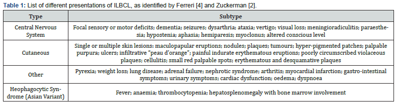
Common manifestations of the Western variant are dermatological (nodes, plaques, nodules, purpura etc.) and neurological (Table 1). Neurological manifestations are present in two thirds of patients and can include mental status changes, seizures, paralysis, aphasia, dementia, motor deficits, neuropathies, paraesthesias and cerebral haemorrhage as well as many others [5].

One meta-analysis [2] published a table of presenting features from seven major studies. Of the 154 patients, 54% presented with CNS involvement, 45% with cutaneous involvement, and 41% with fever. Two of the most common biochemical findings associated with ILBCL are a raised lactate dehydrogenase (LDH) in up to 90% of cases, and anaemia in two thirds of cases [4]. There may also be a raised erythrocyte sedimentation rate, weight loss or fever.

The diagnosis of ILBCL is made by biopsy [6] of affected tissue, which typically shows malignant lymphoid cells in the lumen of the small blood vessels. One of the difficulties in diagnosing this disease comes from the intravascular location of the pathological cells, and the sparing of surrounding tissue making it difficult to identify pathological cells on biopsy (Figure 2). Bone marrow involvement with positive biopsies have been shown in about one third of patients [4]. There is also evidence that for all presentations, a biopsy of macroscopically normal skin is over 80% sensitive as a test for ILBCL [6], providing an alternative to brain biopsy.

Contrasting theories exist as to why these B-cells have a predilection for capillaries. One theory posits the pathological expression of certain cluster differentiation factors (CD11a and CD49d), which enhances binding to capillary epithelium expressing ligands for these factors (CD54 and CD106) [7]. The other suggests that an absence of surface molecules (CD29 and CD54) disrupts lymphocyte homing and leads to an aggregation in small vessels [8]. It may be that as well as understanding the pathogenesis of this illness, elucidation of any aberrant surface molecule expression may also aid the development of target-specific treatments.

If it is identified early, ILBCL can be treated with anthracycline-based chemotherapy regimens. Typically, cyclophosphamide, hydroxydaunorubicin, oncovin, and prednisolone (CHOP) therapy is used –with rituximab in patients with specific B-cell markers. This shows better response and higher rates of remission than alternative regimes [9]. However, only 33% of treated patients survive 3 years [9].

Cutaneous presentations of ILBCL have a better prognosis than other variants [4]. Factors indicating a poor prognosis include elevated LDH and advanced disease with presence in several sites [10].

When considering differential diagnoses it is important to consider ILBCL as a well-recognised cause of stroke (ischaemic or haemorrhagic) despite its low incidence.

This case is important as it demonstrates a specific neurological presentation of ILBCL and illustrates the difficulty of making a diagnosis, as well as the aggressiveness of the condition. We should also not dismiss worsening psychiatric conditions as being a manifestation of the illness – however unlikely – given the close temporal association.

Learning Points

a) Intravascular Large B-Cell Lymphoma often presents with neurological symptoms (in North America and Europe) and is a rare cause of multi-territory infarcts.

b) The intravascular confinement of the clonal expansion of pathological B-cells causes end organ damage by ischaemia.

c) Diagnosis is made by tissue or bone marrow biopsy; but being intravascular it is usually made post-mortem.

d) Biopsy of macroscopically normal skin also has the potential to be diagnostic.

e) Raised LDH and anaemia are common findings.

To Know more about Juniper Online Journal of Case Studies

Click here: https://juniperpublishers.com/jojcs/index.php


To Know more about our Juniper Publishers

Click here: https://juniperpublishers.com/index.php  


Monday, August 2, 2021

Differentiation of Cut Damage caused by Sharp Objects on Cotton Clothes using SEM Analysis - Juniper Publishers

 Forensic Sciences & Criminal Investigation - Juniper Publishers

Abstract

Cut damage due to Sharp objects on different types of clothing is occasionally referred for analysis to forensic laboratories to correlate crime with criminal and/or crime scene in different kinds of cases related to stabbing, sexual assaults, robbery etc. Such examination is essential to help the investigation & to corroborate even the events of crime. Present study is focused on the correlation of the damaged clothing with the specific object/ weapon which was used and caused the damage. The end areas of the cutting damage on clothing are the most important factors for the correlation of clothing with used weapon(s) therefore morphological characteristics of the fiber-end of damaged clothing were examined using scanning electron microscope by observing different features e.g., frayed ends, fiber distortion etc. It was noticed that differentiation can be made between cut caused by razor, Paring knife, Lazer and/or Steak Knife by SEM studies.

Keywords: Clothing damage; Fiber; Knife; Scanning electron microscope; Weapon

Introduction

Examination of damaged clothing is an integral part of crime investigation and is carried out in various types of criminal cases including stabbing, sexual assaults, robbery etc. The clothing related to such types of cases can be used to corroborate the events of crime as clothing damage analysis has potential to correlate crime with criminal and crime scene. Moreover, it is an established fact that the examination of clothing not only provides evidence with person or locations but explains as to what happened during the crime event. The examination of fabric damage is a common task for the forensic scientist, predominantly in cases of homicide and alleged assault. The damage on clothing can occur because of the four major effects- Physical, mechanical, chemical, and thermal effect. The physical and mechanical effect includes the damage by sharp weapon, blunt object, abrasion or frictional force, mechanical strain (pulling the textile by great force) etc. [1]. Different methods for the analysis of damage to clothing-cuts and tears have been reported [2-5]. The present study is focused on Scanning Electron Microscopic Studies on Cut damage due to Sharp objects on Cotton Clothes.

Materials and Methods

Preparation of simulated samples

The clothing samples selected for this study consisted of cotton fabric. Cotton clothes specimens were mounted on a round wooden cloth holder to ensure proper stretch. Four different kinds of objects/weapon like Paring knife, Steak knife, laser knife and razor were used to make simulated samples. Each cloth sample was fixed over a holder and cut were made using the object. Paring knife had a blunt upper edge and a plain sharp lower edge (length = 203mm, blade = 100mm, thickness =2mm and mass= 34.1g). The Steak knife had a clip shaped upper edge and a scalloped lower edge with serrated blades (length=210mm, blade length = 105mm, thickness =1.8mm, and mass =34.0g). The laser knife was 170mm long, with a blade length of 80 mm and a mass of 15.0 g. It was thin at spine and very thin behind the edge. The razor used had a flat cutting edge with pointed tip. (Length= 110mm, blade length= 50mm, thickness =0.5mm and mass =3.0g) Table 1.

Examination of samples

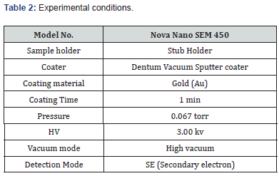
The examination of damaged clothes was designed to three level observation, morphological level, yarn level and fiber level. Fiber is a basic raw material (natural or man-made) used to make yarn and yarn is a continuous length of interlocked fibers. Initially cut portions/ends were examined under compound microscope and the results obtained are given in image 1-5. No distinct features could be noticed therefore these samples were subjected to scanning electron microscopic (Nova NanoSem 450) examination. This microscope is a modern field-emission scanning electron microscope with 1nm ultimate resolution, large sample chamber with advanced motorized stage and navigation supported by an in-chamber camera. High- and low-vacuum operations can be possible with this instrument. Samples were placed on stub-holder and were kept inside Denton Vacuum (Sputter Coater). Vacuum was generated at 0.067 Torr and Coating was done using Au metal [6] Table 2.

Result and Discussion

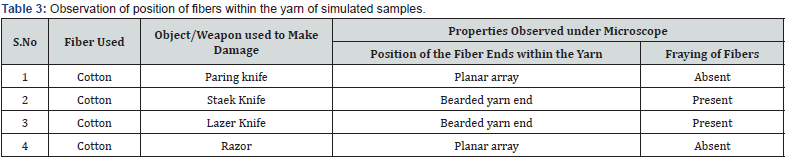
In the present study, two parameters are explored to differentiate the damage of clothing by different sharp objects. First parameter is observing the Position of the fiber ends within the yarn (planar array or bearded yarn ends). Planar array is where a clean-cut leaf all the fiber ends in the same plane of the yarn and a bearded yarn occurs during tearing where the fibers break at different positions in the yarn. Second parameter explored in this study was the fraying of the damaged fiber (Figure 1-4). (Fraying refers to the extent of the displacement of the yarn out of the weave). In case of damage due to paring knife, the fiber ends showed sharp cuts and margins were straight forming flat top with lip and the fibers were aligned in planar array with no fraying (Figure 5). In the case of damage due to razor, fiber end were found flat lip and loosely clustered and all ends of fibers terminating in similar plane, aligned in planar array. Fraying was absent in damage due to razor. (Figure 6). However, in damage due to steak knife and lazer knife, the damaged fiber ends exhibited “no clustering of fiber”. Fiber ends terminated in different planes with a random orientation and the fibers possessed bearded yarn end and significant fraying was also observed (Figure 7 & 8) Table 3.

Conclusion

Based on above observations, it is concluded that the SEM studies found to be a suitable technique to correlate weapon with the damage on clothing. However, the studies on the other types of clothing like rayon, polyester, silk, woolen etc. must be examined to establish the specificity of this technique.

To Know more about Forensic Sciences & Criminal Investigation


To Know more about our Juniper Publishers

Click here: https://juniperpublishers.com/index.php  

Planned Abortion Post COVID-19 - Juniper Publishers

  Public Health - Juniper Publishers Abstract Reproductive health policies, focused on the decriminalization of abortion, have effects on th...