Monday, May 10, 2021

After 180 Years, Is it Time for Something Better for Diagnosing UTI’s? - Juniper Publishers

 Urology & Nephrology - Juniper Publishers


Abstract

Urinary Tract Infections (UTI) are a common source of outpatient encounters for all ages of patients, along with emergency department visits and hospital admissions. Standard urine culture (SUC) has been the mainstay for diagnosis and treatment options for over 100 years, yet 25% of female patients develop recurrent, persistent urinary tract infections. New technologies are now available that quickly provide increased ability to detect organisms along with improved susceptibility information. We discuss the clinical validity and utility data available for Multiplex polymerase chain reaction (M-PCR) coupled with pooled antibiotic susceptibility testing (P-AST) for managing urinary tract infections.

Mini Review

Urinary Tract Infections (UTI) are a common source of outpatient encounters, emergency department visits, and hospital admissions [1,2]. Young and healthy people suffering from a UTI, while symptomatically aggravating, are unlikely to progress to serious complications [3]. However, older adults’ patients suffer from UTIs symptoms ranging from mild to severe, which may lead to delirium, sepsis, or mortality [4,5]. Effective treatment of UTI based on timely and accurate diagnosis is essential to keep elderly patients out of emergency rooms and hospitals. The Standard Urine Culture (SUC) has been the gold standard test for UTI diagnosis for over one hundred years, and has played a significant clinical role in managing patients with suspected UTI. Testing by SUC can often provide informative and actionable clinical information. However, SUC has limitations, particularly for providing complete information for the clinical management of older patients suffering from recurrent, persistent, or other complicated UTIs. These limitations include the inability to detect all relevant organisms causing the infection, producing results quickly enough to avoid empirical treatment and generating efficacious treatment recommendations [6-8]. Given the hospitalization and morbidity rates associated with UTIs in the elderly population, failure to identify a UTI, or adequately treat it, may have significant ramifications.

Novel advanced diagnostic testing methods such as multiplex polymerase chain reaction (M-PCR) coupled with pooled antibiotic sensitivity testing (P-AST) can provide clinically relevant microbiological data missed by SUC. Furthermore, the clinical need for these tests’ stems from the ability of these technologies to not only identify organisms but also provide optimal treatment options in a timely manner [9]. Four peer-reviewed published papers prospectively or retrospectively validated M-PCR/P-AST testing methods as a more useful diagnostic tool for managing complicated or recurrent UTI in the elderly [8,10-12]. A study by Wojno K et al. established the clinical value (or analytical validity) of using M-PCR to detect bacteria in urine. This study reported the results of 582 consecutive elderly patients with an average age of 77 years presenting to urologists with symptoms of a lower UTI. The authors compared the identification of bacterial organisms by M-PCR and SUC when tests were run in parallel on the same samples. The M-PCR detected uropathogens in 326 patients (56%, 326/582), while SUC detected uropathogens in 217 patients (37%, 217/582). M-PCR and SUC agreed in 74% of cases (431/582), and disagreed in 26% of cases (151/582): M-PCR was positive while SUC was negative in 22% of cases (130/582), and SUC was positive while PCR was negative in 4% of cases (21/582). The study identified polymicrobial infections, defined as 2 or more organisms present in a sample, in 175 patients (30%, 175/582), with M-PCR detecting 166 and SUC detecting only 39 (6.7%, 39/582). M-PCR identified polymicrobial infections in 67 cases (12%, 67/582) for which SUC results were negative. Additionally, M-PCR identified several microbes including Gram-positive bacterium, A. schaalii, A. omnicolens, C. riegelii, M. tuberculosis, M. hominis and Gram-negative bacterium, P. agglomerans, P. stuartii, and U. urealyticum missed by standard urine culture [8].

The publication concluded that SUC has limitations, including the inability to detect slow-growing, fastidious, or non-aerobic microorganisms, and SUC has a profound detection bias for fastgrowing Gram-negative aerobic organisms. Significantly, SUC had difficulty identifying most of the microorganisms that make up a polymicrobial infection. Similarly, the article by Vollstedt A. et al summarized results from a prospective trial comparing the detection levels of M-PCR and SUC of bacteria in UTI-symptomatic patients. The study enrolled 2,511 patients with UTI symptoms and an average age of 73 years from 37 urology clinics across the United States. M-PCR and SUC identified bacteria in 62.7% (1,575/2,511) and 43.7% (1,098/2,511) of cases, respectively. M-PCR detected 6 organisms which SUC failed to detect, including five Gram-positive bacteria A. schaalii, A. omnicolens, C. riegelii, M. tuberculosis, M. hominis and Gram-negative bacterium, P. agglomerans, P. stuartii, and U. urealyticum missed by standard urine culture and one Gram-negative bacterium (U. urealyticum), affecting 590 samples. Between the two testing methods, the study detected a total of 861 polymicrobial infections, with M-PCR detecting 834 (96.9%) and SUC detecting only 167 (19.5%). Polymicrobial detections made up 34.3% (861/2,511) of the total patients, and 53.0% of M-PCR positive cases (834/1575). SUC did not detect A. schaalii, which was the most common bacterium [53.0% (442/834)] detected in polymicrobial infections by M-PCR. The bacterial species detected by SUC but not detected by M-PCR, including Enterobacter species, the Enterococcus species, and several other rarely detected species, were detected in very small subsets of patients by SUC (0.9%, 0.2%, and 0.9%, of all patients, respectively). The M-PCR mix did not include primers for the missed species in the M-PCR assay at the time of the study [10].

This analysis has also showed the identity of microbes identified by M-PCR and the susceptibility results generated by P-AST takes an average of 29.7 hours (9 hours less than SUC) to provide physicians with urine pathogen and antibiotic susceptibility results. The difference in turnaround time improved a median of 19 hours (34.5 hours and 53.7 hours, for M-PCR/PAST and SUC, respectively) for patients with both positive pathogen identification and susceptibility results [10]. These two publications show the superior ability of M-PCR to quickly detect all relevant uropathogens in the sample, especially Gram-positive bacteria, along with more polymicrobial infections in patients with UTI symptoms [8,10].

A second paper by Vollstedt A. et al. focused on bacterial interactions in affecting susceptibility patterns. The study used a novel pooled antibiotic sensitivity testing method (P-AST) which assess the functional antibiotic sensitivity for a pooled sample. This assay measures optical density with a spectrophotometer, setting a threshold value to measure growth of organisms in a ‘pool’, or polymicrobial mixture. The benefit of the ‘pooled’ approach is that it allows real-world antibiotic sensitivity assessment of the polymicrobial community from the patient’s UTI. The average age of the patients were 74.9 years. This study analyzed 758 UTIsymptomatic patients with polymicrobial bacterial infections and antibiotic susceptibility results. By comparing results from these polymicrobial samples against monomicrobial bacterial samples from 594 UTI-symptomatic patients, the analyses revealed the odds of resistance to ampicillin (p = 0.005), amoxicillin/ clavulanate (p = 0.008), five different cephalosporins (p<0.05), vancomycin (p = <0.0001), and tetracycline (p = 0.010), increased with each additional bacterial species present. In contrast, the odds of resistance to piperacillin/tazobactam decreased by 75% for each additional species present (95% CI 0.61, 0.94, p = 0.010). Additionally, the comparison revealed 44 situations for which 13 pairs of bacterial species exhibited statistically significant interactions, which caused susceptibility patterns to change as measured by the Highest Single Agent Principle or Union Principle statistical analysis models [11]. These findings align with the results reported by De Vos et al, who examined the interactions between 72 bacterial isolates from elderly people with UTI symptoms. They measured the impact of species-to-species interactions on antibiotic efficacy. They assessed organism’s growth in response to two commonly used antibiotics for UTIs (trimethoprim-sulfamethoxazole and nitrofurantoin). Using media conditioned by donor isolates they observed that clinical isolates often protected each other from the antibiotics: 25% of tested species-to-species interactions showed greater than a 3.5- fold increase in tolerance for trimethoprim-sulfamethoxazole but decreases of the same magnitude only occurred in 12% of patient results [12].

Therefore, M-PCR is able to quickly detect all relevant uropathogens, especially Gram-positive bacteria, along with more polymicrobial infections in patients with UTI symptoms, [8,11] whereas the ‘pool’ approach used in the P-AST testing allows real-world antibiotic sensitivity assessment of the polymicrobial community from the patient’s UTI.

Does the improved performance of M-PCR/P-AST lead to better clinical outcomes? The study by Daly A. et al. addressed this question and shows the M-PCR/P-AST results are associated with better outcomes. The study used existing data from 66,381 patients seen for UTIs by primary care providers in the patient home or assisted living locations. The clinical outcomes measured in the study were numbers of hospital admission and/or emergency department utilization. Daly et al. divided patients into two non-overlapping cohorts. Physicians treated patients in cohort one (N=34,414) based upon the results from SUC. Physicians treated the other cohort (N=31,967) based upon the results from the M-PCR/P-AST assay. The patients in the two cohorts had similar demographics, comorbidities, Charlson/ Deyo Index Scores, number of provider visits, and enrollment locations. The analysis detected a 13.7% reduction in hospital admissions and/or emergency department utilization associated with the use of the M-PCR/P-AST assay compared with the use of traditional SUC. The 13.7% reduction in hospitalization when normalized to 34,414 patients in the SUC cohort would result in 156 fewer patients attending the ED/hospitalizations and/or ED utilization from a UTI [13]. Another study has shown that the savings of keeping patients out of the hospital for a UTI can be as high as $64,000 per patient when considering the dollars paid by the patient and insurance [14]. Thus, the cost avoidance for 156 patients is as high as $10,000,000. The cost of testing using M-PCR/P-AST for the target population suffering from UTI’s is well below the cost associated with adverse effects that result in ED or hospitalization.

For over a century, health care providers have accepted SUC as a tool to manage UTIs to identify both the bacteria and the appropriate treatment options. The diagnostic tool is well accepted, as is the notion that E. coli is the leading cause of UTI’s. Some may argue, therefore, that the pathogenic nature of bacteria uniquely identified by M-PCR is unknown as researchers have yet to perform Koch’s postulate studies on these species. Yet, though SUC has been accepted for over a century, 25% of UTI symptomatic women develop recurrent UTI’s despite SUC providing definitive bacterial identification and antibiotic recommendations [15- 17]. There is growing evidence of the pathogenic nature of the organisms uniquely identified by M-PCR. For example, A. schaalii, found to be involved in 53% of all M-PCR detected polymicrobial UTIs [10], was recently acknowledged as an uropathogen in older adults and young children [18]. Beyond missing critical pathogens, SUC fails to detect more than 2 organisms in an infection and does not consider bacterial interactions that impact susceptibility results. More than one in two patients (56.1%) who tested positive for UTIs were diagnosed with a polymicrobial infection based on M-PCR, and the odds of patients’ resistance to most antibiotics increased with each additional bacterial organism present, possibly due to bacterial interactions [11]. Additionally, specific combinations of bacteria either increase susceptibility or increase resistance, depending upon the specific pair of organisms and the antibiotic the pair is exposed to. Therefore, the P-AST methodology may detect the effects of these interactions, compared with the antibiotic susceptibility performed on isolates as in SUC [11].

What are the cumulative effects of SUC failure? Those with recurrent UTI’s are prophylactically prescribed antibiotics for recurrent UTI’s. As these patients age, the prophylactic use of antibiotic for UTI results in inappropriate antibiotic use, i.e. antibiotics being prescribed at a dose higher then recommended and for longer periods of time then recommended. In fact, some antibiotics are prescribed for life [19]. Indeed, studies show that 55% of antibiotics prescribed for UTI’s are inappropriate in the long-term home setting [20]. For older women, prophylactic antibiotic use was found to be associated with an increased risk of UTI related hospitalizations [21]. These patients may benefit from the M-PCR/P-AST test, which provide physicians with comprehensive and sensitive bacterial identification results from M-PCR along with susceptibility results from a pooled setting. As a result, therapeutic guidance may lead to more effective antibiotic selections. UTIs are a significant source of morbidity for the elderly. While conventional urine cultures remained the mainstay of diagnosing these patients for the last 180 years, evidence shows advanced diagnostic tests such as M-PCR/P-AST can identify pathogens not detected by SUCs, generates results more quickly, and provides antibiotic susceptibility results from real-world polymicrobial community [8,10-12]. Further evidence shows the detection and\treatment of these pathogens based on M-PCR/P-AST may lead to decreased hospital utilization as compared to patients who were managed using SUC [13].

To Know more about  Urology & Nephrology

To Know more about our Juniper Publishers


Friday, May 7, 2021

Evaluation of Biofield Energy Healing Treatment Based Proprietary Test Formulation on Gut Health Potential in Colon Cancer Cell Line (HT-29) - Juniper Publishers

Pharmacology & Clinical Research - Juniper Publishers

Abstract

The present study was aimed to evaluate the anti-inflammatory potential of Biofield Energy Healing (the Trivedi Effect®-Consciousness Energy Healing) on the test formulation in colon cancer cell line (HT-29). Each ingredient of the test formulation was divided into two parts, one part was denoted as the untreated test formulation and the other part was demarcated as the Biofield Energy Treated test formulation, which received Biofield Energy Healing Treatment by a renowned Biofield Energy Healer, Mr. Mahendra Kumar Trivedi. MTT assay showed that the test formulation was found safe and non-toxic upto 122 μg/mL in HT-29 cells with more than 78% cell viability. The level of interleukin-6 (IL-6) expression was significantly reduced by 34.09% and 59.41% (p≤0.001) at 3 and 15 μM, respectively compared to the vehicle control (VC) group under the stimulation of tumor necrosis factor - alpha (TNF-α). Moreover, IL-8 level was significantly suppressed in the Biofield Energy Treated test formulation by 57.09% and 42.88% at 0.1 and 3 μM, respectively compared with the VC group. However, the Biofield Energy Treated test formulation further substantial altered the level of interferon gamma compared to the VC group. The Trivedi Effect®-Consciousness Energy Healing significantly regulate the inflammatory condition after treatment with the test formulation in colon cancer cell line (HT-29). This experimental data suggested that the Biofield Treated test formulation can be utilized for many inflammatory disease conditions such as rheumatoid arthritis, multiple sclerosis, psoriasis, inflammatory bowel diseases, scleroderma, and type 1 diabetes mellitus.

Keywords: Biofield Energy Healing; The Trivedi Effect®; Inflammation; Colon cancer cell line (HT-29); Pro-inflammatory cytokines

Abbreviations: FBS: Fetal bovine serum; MTT: 3-(4,5-Dimethylthiazol-2-yl)-2,5-Diphenyltetrazolium Bromide; ELISA: Enzyme-linked immunosorbent assay; NCCAM: National Center for Complementary and Alternative Medicine; CAM: Complementary and Alternative Medicine

Introduction

The relation among of the gut health, micro biota, and cytokines have been well studies and reported in past two decades against various inflammatory bowel diseases (IBD) and associated mucosal inflammations. The cytokines play an important role in the mechanism in inflammation especially in Crohn’s disease and ulcerative colitis. The pathogenesis of IBD is not completely understood, but the role of cytokines in the intestinal immune system has significant impact in disruption of normal state of controlled gut inflammations [1,2]. Innate immune response is the major response in gut inflammation and its related diseases. Most of the cytokines are secreted by activated dendritic cells and the macrophages, which regulates the inflammatory response in gut inflammatory diseases. Once, these cytokines are secreted by antigen presenting cells, they triggers and differentiate various T cells by activating adaptive immune response. Gut inflammation dysregulates the T-cells, and manage the over-reactive and auto-reactive cells. T-cell regulation or its overproduction leads to the development of gut inflammatory diseases [3]. These cells along with various types of cytokines play a complex role in inflammatory gut diseases [4, 5]. Thus, there is the need of some novel formulation which alters the level of cytokines to improve the gut health. The present study was aimed to test the impact of the Biofield Energy Treated test formulation comprised of zinc chloride, ferrous sulphate, copper chloride (II-cupric), vitamin B6 (pyridoxine HCl), vitamin B12 (cyanocobalamin), magnesium (II) gluconate, and cholecalciferol (vit. D3) against the colon cytokines. The novel test formulation was treated with Biofield Energy Healing Treatment, as one of the best CAM approach with significant therapeutic outcomes. Biofield Energy Healing is one of the emerging frontiers aspect to CAM and various clinical approach has been used with significant results [6-9]. CAM therapies have been recommended by The National Center for Complementary/Alternative Medicine (NCCAM) and there therapies exist in various forms such as external qigong, Johrei, Reiki, therapeutic touch, yoga, Qi Gong, polarity therapy, Tai Chi, pranic healing, deep breathing, chiropractic/osteopathic manipulation, guided imagery, meditation, massage, homeopathy, hypnotherapy, progressive relaxation, acupressure, acupuncture, special diets, relaxation techniques, Rolfing structural integration, healing touch, movement therapy, pilates, mindfulness, Ayurvedic medicine, traditional Chinese herbs and medicines in biological systems both in vitro and in vivo. 

Human Biofield Energy has subtle energy that has the capacity to work in an effective manner [10] with its various clinical benefits [11]. This energy can be harness and transmit it into living and non-living things by the process of Biofield Energy Healing Treatment. Biofield Energy Treatment (the Trivedi Effect®- Consciousness Energy Healing Treatment) has been extensively studied with significant outcomes in the field of pharmaceuticals [12-14], nutraceuticals [15,16], metals and ceramics [17-19], microbiology [20-22], microbial genetics [23, 24], cancer research [25,26], livestock, agriculture science [27-29], improved bioavailability of many compounds [30-32], improved skin health [33, 34], improved properties of nutraceuticals [35, 36], improved overall bone health [37-39], human health and wellness. Thus, the study was planned on colon cytokines estimation that could significantly helped to improve the prevalence of gut inflammatory diseases using novel test formulation consisting of minerals such as Mg, Zn, Fe, Cu and vitamins including B6, B12, D3 in colon cancer cell line (HT-29).

Materials and Methods

Chemicals and Reagents

Antibiotics solution (Penicillin-Streptomycin) was purchased from HiMedia, India. 3-(4, 5-dimethyl-2-thiazolyl) 2, 5 diphenyl-2 H-tetrazolium) (MTT), Dulbecco’s Modified Eagle’s Medium (DMEM), NaHCO3, and EDTA were purchased from Sigma Chemical Corp. (St. Louis, MO), a subsidiary of Sigma-Aldrich Corporation. ELISA (enzyme-link immunosorbent assay) assay kits for all cytokines tumor necrosis factor alpha (TNF-α), macrophage inflammatory protein-1α (MIP-1α), and interleukin-1 beta (IL-1β) were purchased from R&D Systems, USA. Fetal bovine serum (FBS) was purchased from GIBCO, USA. Iron sulfate, copper chloride, and cholecalciferol (vitamin D3) were obtained from Sigma Chemical Co. (St. Louis, MO). Zinc chloride and magnesium (II) gluconate hydrate were obtained from TCI, Japan. Pyridoxine- HCL (vit-B6), cyanocobalamin (vit-B12) were procured from Alfa Aesar, USA. All other chemicals used in this study were analytical grade available in India.

Test Formulation and Reference Standard

The test formulation contained a combination of vitamins with minerals viz. iron sulfate, copper chloride, zinc chloride and magnesium (II) gluconate hydrate, cholecalciferol (vitamin D3), pyridoxine-HCL (Vit-B6), and cyanocobalamin (Vit-B12). Tumor necrosis factor alpha (TNF-α) was used as an inflammatory stimulant, while epigallocatechin-3-gallate (EGCG) was used as a reference standard (positive control) for immunomodulatory action in colon cancer cell line (HT-29).

Biofield Energy Healing Strategies

One part of each ingredient of the test formulation did not receive any sort of treatment and was defined as the untreated test formulation group, while another part received Biofield Energy Treatment known as Biofield Treated Test formulation by Mr. Mahendra Kumar Trivedi, a renowned Biofield Energy Healer under standard laboratory conditions for ~3 minutes. This treatment was provided through the Biofield Energy Healer unique Energy Transmission process (the Trivedi Effect®) to the test formulation. Further, the untreated test formulation was treated with a “sham” healer for comparison purposes. The “sham” healer did not have any knowledge about the Biofield Energy Treatment. After that, the Biofield Energy treated and untreated test formulations were kept in similar sealed conditions and used for the in vitro study on colon cancer cell line (HT-29) for cytokines estimation.

Experimental Design

The colon cancer cell line (HT-29) was divided into four different groups. Group 1 comprised of the HT-29 cells vehicle was denoted as the vehicle control, group 2 included cells with epigallocatechin-3-gallate (EGCG) as positive control at various concentrations. Group 3 and 4 included the cells with the untreated and Biofield Energy Treated test formulation, respectively at concentration range 0.1 to 15 μg/mL in presence of tumor necrosis factor - alpha (TNF-α).

Cytotoxicity by MTT Assay

The effect of the Biofield Energy Treated and untreated test formulations at a wide range concentration were tested for cell viability using 3-(4,5-dimethylthiazol-2-yl)-2,5-diphenyl tetrazolium bromide (MTT) assay. The number of viable cells were determined by the ability of mitochondria to convert MTT to formazan dye. The details procedure was followed as per Plikerd et al. 2017 [40]. The effect of the test formulation on cell viability of HT-29 cells was determined as:

%Cell viability=100-%cytotoxicity (1)

Where; % cytotoxicity = [(O.D. of control cells – O.D. of cells treated with the test formulation)/O.D. of control cells]*100.

The results of the concentrations that showed >75% viability were selected subsequently for cytokine estimation.

Determination of Cytokine levels by ELISA

The HT-29 cell suspension in DMEM medium containing 10% FBS was plated at a density of 0.5 X 10⁶ in 12-well cell culture plates. Cells were incubated at 37 °C for 24 hours. Cells were sera starved by replacing medium with DMEM containing 0% FBS and again incubated at 37 °C for another 24 hours. Cells were treated with proprietary test formulation at selected noncytotoxic concentrations and stimulated with Hu-TNF-α. Cells treated with TNF-α + EGCG were included as the positive control. After treatment, cells were incubated in a 5% CO2 incubator for 72 hours. After incubation, culture supernatants were collected from each well and stored at -20 °C until analysis. The level of cytokines (IL-6, IL-8, and IFN-γ) in culture supernatants of HT-29 cells were determined using ELISA as per manufacturer’s instructions.

Statistical Analysis

All the data were expressed as mean of three replicates ± SEM and were subjected to one-way analysis of variance (ANOVA) followed by Dunnett’s test and Student’s t-test for two groups comparison. Statistical significance was considered at p≤0.05.

Results & Discussion

MTT Assay

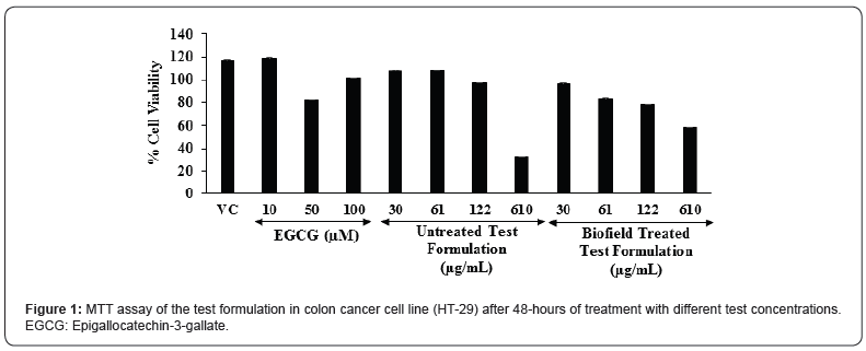
The cell viability results are summarized in the Figure 1. The percent cell viability in the vehicle control (VC) group was found as 117.5%. Moreover, the positive control, epigallocatechin-3- gallate (EGCG) showed 119.2%, 83%, and 101.2% cell viability at the concentration of 10, 50, and 100 μM, respectively. Further, the untreated test formulation showed more than 97% cell viability upto 122 μM; while the Biofield Treated test formulation showed more than 78% cell viability upto 122 μM. Based on the MTT cell viability assay the test formulation was found as safe and nontoxic upto the concentration of 122 μM. MTT assay is widely used for the cell toxicity against any test formulations. In addition, this assay was found as more rapid, less costly, less time consuming, and non-radioactive method as compared with the other assays. This assay display cell proliferation results on the basis of the cell growth and metabolic activity [41]. MTT assay suggest that the concentrations of the test formulation were found safe up to 122 μg/mL with respect to the viability in the colon cancer cell line (HT-29).

Estimation of Interleukin-6 (IL-6) Expression

The level of interleukin-6 (IL-6) expression in colon cancer (HT-29) cells is represented in the Figure 2. The positive control, epigallocatechin-3-gallate (EGCG) was significantly reduced the level of IL-6 by 38.71%, 92.43%, 91.74%, and 98.20% (p≤0.001) at the concentrations of 1, 10, 50, and 100 μM, respectively as compared to the vehicle control (VC) group. Moreover, the untreated test formulation showed significant reduction of IL-6 by 11.96%, 35.94%, and 52.3% (p≤0.001) at 0.1, 3, and 15 μM, respectively as compared to the VC group. Further, the Biofield Energy Treated test formulation group showed 14.19%, 34.09%, and 59.41% (p≤0.001) reduction of IL-6 at 0.1, 3, and 15 μM, respectively as compared to the VC group under the stimulation of TNF-α stimulation. Besides, the Biofield Treated test formulation also significantly reduced the level of IL-6 by 14.87% as compared to the untreated test formulation group. Overall, the minerals and vitamin-based Biofield Energy Treated test formulation showed an anti-inflammatory activity by reducing the level of IL-6 under the stimulation of TNF-α as compared with the vehicle control as well as untreated test formulation groups. Hence, the Biofield Energy Treated test formulation could be used a major role in immune-related disorders and also defined as controlling factor for many diseases [42]. Thus, it can be suggested that the Biofield Energy Treated test formulation can be used in many inflammatory disorders.

Estimation of IL-8 Expression

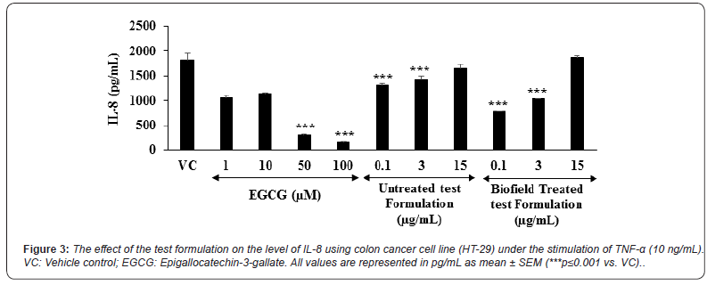
The level of interleukin-8 (IL-8) expression in colon cancer (HT-29) cells is represented in the Figure 3. The positive control, epigallocatechin-3-gallate (EGCG) was significantly reduced the level of IL-8 by 41.49%, 36.77%, 82.89% (p≤0.001), and 91.21% (p≤0.001) at the concentrations of 1,10,50, and 100 μM, respectively as compared to the vehicle control (VC) group. Moreover, the untreated test formulation showed significant reduction of IL-6 by 27.34% (p≤0.001), 21.03% ( ≤0.001), and 9.08% at 0.1, 3, and 15 μM, respectively as compared to the VC group. Further, the Biofield Energy Treated test formulation group showed significant (p≤0.001) reduction of IL-8 by 57.09% and 42.88% at 0.1 and 3 μM, respectively as compared to the VC group under TNF-α stimulation. Besides, the Biofield Treated test formulation also significantly reduced the level of IL-8 by 40.94% and 38.25% at 0.1 and 3 μg/mL, respectively as compared to the untreated test formulation group. Overall, the minerals and vitamin-based Biofield Energy Treated test formulation showed an anti-inflammatory activity by reducing the level of IL-8 under the stimulation of TNF-α as compared with the vehicle control as well as untreated test formulation group. Chronic inflammatory conditions leads to the massive production of proinflammatory factors such as chemokines. IL-8 is one of the chemokine in chronic inflammation and it is initially act as a neutrophil chemotactic and activating factor [43,44]. Overall, the experimental data suggested that Biofield Energy Healing Treatment has the significant capacity to reduce the level of IL-8 with respect to vehicle control and untreated test formulation.

Estimation of IFN-γ Expression

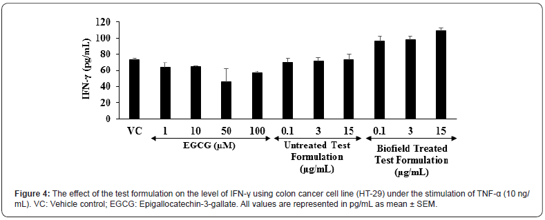
The level of interferon gamma (IFN-γ) expression in colon cancer (HT-29) cells is shown in the Figure 4. The positive control, epigallocatechin-3-gallate (EGCG) was significantly reduced the level of IFN-γ by 13.64%, 12.64%, 37.64%, and 22.99% at the concentrations of 1, 10, 50, and 100 μM, respectively as compared to the vehicle control (VC) group. Moreover, the untreated test formulation showed significant reduction of IFN-γ by 4.55% and 2.78% at 0.1 and 3 μM, respectively as compared to the VC group. Further, the Biofield Energy Treated test formulation significantly altered the level of IFN-γ as compared to the VC group under TNF-α stimulation. This suggests that the Biofield Energy Treated test formulation has significant immunomodulatory activity. Scientific reports suggest that various immunological and inflammatory functions of chemokines play significant role in controlling the immune response during infections. Overall, the immunomodulatory effect might be the result of specific modulation of NF-κB, a transcription factor involved in the activation of many inflammatory mediator genes [45,46].

Worldwide scope of alternative medicine and its outcomes have been increased significantly. However, an important phytoconstituents along with minerals and vitamins are reported to have beneficial role against many diseases such as diabetes, indigestion, inflammation of intestine, osteomalacia, blood disorders, infertility, potent revitalizer, etc. [47]. Due to high safety profile with the wide therapeutic action of alternative medicines, the scope has been increased worldwide [48]. Besides, the individual constituents of the novel proprietary test formulation has been reported to have substantial immunomodulatory action, and Biofield Energy Healing Treatment significantly alters the action of cytokines. Overall, the Biofield Energy Healing Treatment on the test formulation can be a novel approach in supports of the use of Biofield Treated test formulation for various types of autoimmune disorders in colon cancer cell line (HT-29).

Conclusion

On the basis of current study findings, it is concluded that the novel proprietary test formulation showed significant antiinflammatory action on the tested cytokines (IL-6, IL-8, and IFN-γ) in colon cancer cell line (HT-29) after administration of the Biofield Energy Treated formulation. MTT assay in the Biofield Energy Treated colon cancer cell line (HT-29) suggest that the test formulation showed more than 78% cell viability and found as safe and non-toxic. In addition, the levels of cytokine, interleukin-6 (IL-6) was significantly reduced by 34.09% and 59.41% (p≤0.001) at 3 and 15 μM, respectively as compared to the vehicle control (VC) group. Moreover, IL-8 level was reported to be significantly suppressed in the Biofield Energy Treated test formulation by 57.09% and 42.88% at 0.1 and 3 μM, respectively as compared with the VC group. On the basis of experimental results of various tested cytokines and their expression, significant anti-inflammatory activity in colon cancer cells was reported in the new test formulation after treated with the Trivedi Effect®- Biofield Energy Healing. Biofield Energy Treated test formulation can be used as a Complementary and Alternative Medicine (CAM) to prevent the immune-mediated diseases such as Irritable Bowel Syndrome, Rheumatoid arthritis, Ulcerative colitis and Crohn’s disease, Stress, Asthma, and many more with safe therapeutic index. Besides, it can also be utilized in organ transplants (for example kidney transplants, liver transplants and heart transplants), various autoimmune disorders such as Lupus, Addison Disease, Celiac Disease (gluten-sensitive enteropathy), Dermatomyositis, Graves’ Disease, Hashimoto Thyroiditis, Multiple Sclerosis, Myasthenia Gravis, Pernicious Anemia, Aplastic Anemia, Sjogren Syndrome, Systemic Lupus Erythematosus, Diabetes, Alopecia Areata, Fibromyalgia, Vitiligo, Psoriasis, Scleroderma, Chronic Fatigue Syndrome and Vasculitis, Type 1 to improve the overall health and quality of life.


To Know more about our Juniper Publishers


Thursday, May 6, 2021

A Brief Contribution to Understand the Structure of Knowledge and The Construction of Science - Juniper Publishers

Physical Fitness, Medicine & Treatment in Sports - Juniper Publishers

Abstract

Knowledge has rules. But there are rules imposed by the functionality of the process and rules that are imposed by rulers that have some power and try to impose it. Those are the good rules, these are “only” enforcement attempts. To understand the structure of knowledge and the construction of science is the beginning of the way to look for efficiency.

Keywords: Anthropology, Epistemology, Subsector level, Perceptions, Knowledge, Ambitions and desires, Reasoning abilities

Introduction

Knowledge is a tool to understand and explain phenomena and, in this way, allow to deal efficiently with the integration of man in the different contexts in which he takes part. A tool, and nothing more, which is certainly very colossal. The tools, which for a long time (naively) were thought that man was the only one to use, are fundamental to accomplish a work (conceptual or material) in an efficient way, we emphasize in an efficient way [Note: Einstein was nicknamed as the man who retained the laboratory - a laboratory is a tool - inside the hat, due to the conceptual experiences he carried out], they are so striking that many areas that have as object of study the evolution and development of man, (history, anthropology,...) base their work on the interpretation of the instruments and tools that have been used through the ages, as they allow not only to define the objectives that were aimed at, but also the perceptions, knowledge, ambitions and desires, reasoning abilities, etc..

Strategies, operating modes, such as tactics, technologies and even techniques, integration processes and established dialectics, all in short, count when we do not look at phenomena in an almost isolated way (such as events) but consider the processes in their integration (“internal” and “external”) with a concern to seek optimization of means and the efficiency of the whole. Knowledge, the primary object of science, must also be (perhaps even above all, being a multiplier) the target of these intentions and care. And yet, as we will try to show below, possibly because of its importance, it is one of the most fractionated and uncoordinated fields and tools that man uses. Not that knowledge is disrespected in a particular way. The same happens, for example, with the houses in which we often forget that have foundations, with the trees in which we do not attend to the roots that we do not see, with the sea that we consider as a water surface ignoring the kilometers of depth that it sometimes has and the currents and movements that develop there…

Epistemology, which some despise (or ignore?) classifying it as “mere philosophy” (in opposition, they defend, to a science that is “a solid thing”, but of which they must know little because they do not understand the importance of some of their foundations), also sometimes suffers from the same marginalization. Sciences themselves (classified as hard / soft, pure, experimental, natural, exact,...), also pass through this colander (also a mere instrument, which serves to “fix” the knowledge tools), a sieve that goes beyond its function as a mean to become an end in itself (abusively and at a cost because divert attention from what is fundamental, that is, literally, grounding, giving the support that it consolidates). Many forget that positivism (logical empiricism) had the purpose of, in addition to providing greater rigor (compared to empiricism) and thus facilitating the search for precision, defending its authors from the risk of expressing opinions (which sometimes went against the current with solidly established “truths”, and we don’t even have to go to the realm of the sacred, the inquisition or parallel things, such as “the heavier bodies that the air cannot fly”, or “the continents cannot move, Pangeia is idiocy”). The “crime of opinion” must be avoided (or disguised), the importance of the assumptions considered, corollaries, postulates, axioms, etc., does not make sense, “…from the data presented we conclude that…” what avoids more exposures, “these works should only be published after my death”, that avoids retaliation against the author…. The examples have no end, the harmful effects too and…, unfortunately, they are not past waters.

Alternatives? Certainly exist. The problem is in finding them, defining them (doing the necessary research and getting it to succeed) and, perhaps above all, implementing them. Next, we point out proposals for responding to the problems we have raised. However, we believe that it is an important and serious problem that justifies investments in terms of the enormous damage it causes to the development of science and, even more, to the fantastic effects that science can have on the lives of men and societies, so we will not only continue to engage in research on the topic, as well as we propose that there must be a coordinated action of the many positions that strive in this same direction.

A Restricted Vision: Advantages And Disadvantages

Parts help us to conceive the whole, given the limitations of the means (the tools) that we built in a process of millions of years in which, successively, we were responding to the problems we have faced and the adaptive capacities that we have (which were also being developed). The dialectics, fundamental in this whole process, cannot be overlooked, but the capacities we have compel us to constitute a conception of the global based on specific aspects (almost in an impressionistic context) in which, from specific stimuli, we compose an image of the whole, what passes, naturally, for sets of sensations, which give meaning to the multiple individual stimuli, for an interpretation strategy and for decision making that integrate a perception of the global (personalized, no doubt, but similar at least to those of us that are close… culturally) that have allowed us to survive and overcome difficulties for millions of years.

However, today we have undergone profound transformations in the context in which we are integrated and of which we are a part, transformations that are widely spoken, but little identified in their specifications and, even less, in the effects and implications that they have on our (individuals and societies) lives. Vision, one of the senses that most marked the development of man, is a good model of this whole process. Being a way of recording different stimuli, such as through rods (sensitive to light in general) and cones (sensitive to different rays that allow us to distinguish colors), which work in very specific ranges of stimuli, through a set of operations that optimize the detection of situations in different conditions (magnifications, low light, excess light, types of light, ...), giving rise to a perception that allows us a sense of the situation in which we are located, in order to be able to adapt our strategies to the solutions we need to find. It is, therefore, from the contribution of specific actions that we build the globality of our (particular) world, in a stratagem in which we start from punctual for the whole, the global, in the way we see it, in a process that is the opposite of what science did when looking to be more incisive in that it went from the whole to the specific, dividing into areas of knowledge, in analysis / synthesis processes, in deductions / inductions, etc.. Processes that can be complementary if, in science, we do not forget the final objective, which is globality, and that the isolations we make are useful as tools, instruments of an integration process that allows us to serve man... in an efficient way.

Resisting the temptation to stick to partial goals is, therefore, fundamental. This is because the search for truth is not the function of science (for a long time, although it is still ignored), what we are looking for are coherences. Coherences that seen at the local level may not be contradictory but at broader levels they become more evident, because the restrictions increase, and the dialectics gain lives that they would not otherwise have. The concretization, the execution of a work, is, therefore, a good way to test the consistency of a conjecture, because if in “laboratory” the isolation of variables can be done and can even be productive, when we move to application situations, models are confronted with the “globalities” of situations and validations that could otherwise be omitted (it is the leap that is taken when we move, in I, R & D, from a model to a prototype, before we even have to face the challenges of setting up industrial production lines…).

The Importance Of Understanding The Structure Of Knowledge

Knowledge, as we have argued above, faces the challenge of globality. Tasks can be distributed, roles assigned. We can (should we?) form teams, define strategies, set goals, make efforts, ... But all of this is justified in the sense that there is global coherence. Otherwise, the effort may be useless, or even counterproductive. The teams do not work, the strategies are meaningless, the tasks and functions are pointless. The tools that are essential for the realization of a work (conceptual or material) are no longer efficient. Knowledge, that is a tool, a tool and nothing more, to understand and explain phenomena, sense because the practical efficiency it offers has global. Everything that revolves around it, researchers, consumers of knowledge, distributors, and disseminators of knowledge, no longer makes sense. At least at the levels of the (huge) investment that exists. It is a whole space of development that disappears, unless, unguarded, we do not realize that the change must take place and we continue, automatically, to make the usual gestures, comply with habits and “live in second-hand”. And sometimes worsening everything. But there are solutions.

A Proposal

Assumptions

a) When water come to be some local events

b) Fountains

c) Streams and rivers

d) Home consumption

e) Small industry

f) …

g) To a notion of a WATER CYCLE:

h) Ocean evaporates

i) Clouds transport

j) Streams and rivers lead

k) Consumption

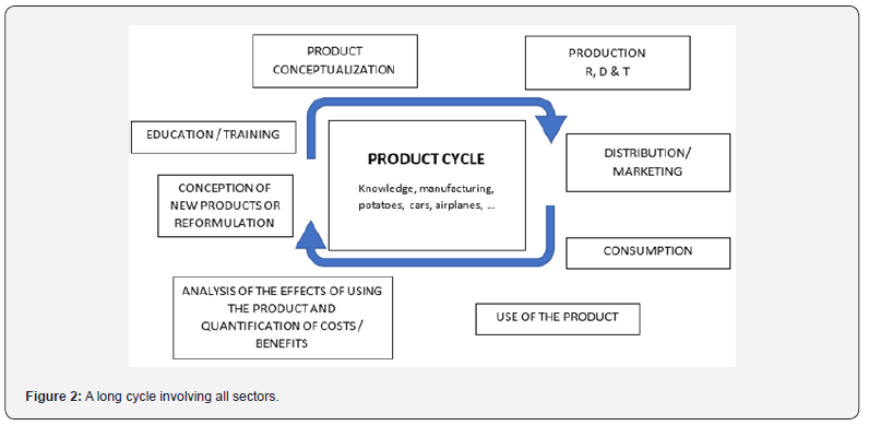
There was another conception not only of water and its availability, but of the world itself, which went from local and static to global and dynamic. Due to this change we have evolved and today we begin to ask ourselves about: groundwater; ocean and ocean dynamics; origin of water in space, in the terrestrial nucleus, in the formation of the Earth, ... - ANOTHER WORLD VISION IS PREPARED Figures 1&2.

The Proposal

Assumptions

a) With the integration of R, D & T (Research, Development & Technology), although still done imperfectly and partially, there was a great evolution and transformation in terms of resource profitability.

b) If the integration of the different fields of human production ceased to be seen in local terms and changed to a global view, would we not have similar gains? But much greater because the capital at stake increases and the resulting income has a much larger base.

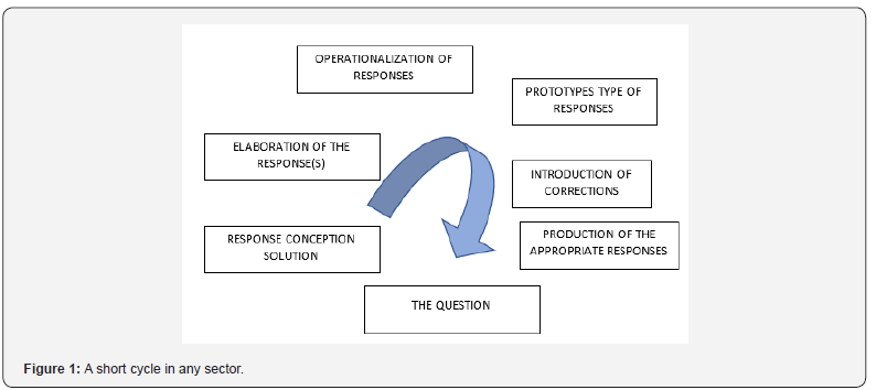
c) In a simplified and succinct way, we have in the development of any product Figures 1 & 2:

We assume that

a) The current trend is for the debate to be held at the SECTOR level (production, distribution, consumption, ...), if not at the SUBSECTOR level (research, development, technology, ...);

b) The big problems (big not only in size and effects, but mainly because they are ignored) occur in the articulation of the sectors;

Examples:

a) If education does not consider production, distribution and consumption, it can hardly be integrated understand, globally;

b) If production does not pay attention to distribution / marketing, and consumption no longer meets needs and has no meaning;

c) If planning does not consider the potential of education / training, as well as consumption, it will not be able to profit from the resources that will be available and the objectives that it should seek.

d) The debate that is taking place today in some thinkthanks, in some universities, in some political spheres and little else.

Knowledge is one of the great products (due to the dimensions it reaches, the multiplier effects it has, the investments (human, financial, time, etc.), which should be very concerned with the integration of the so, it different fields it seeks to serve, where it operates and of where it should (which it does not) gather the means it needs to develop and progress, it is certainly from the most unstructured fields and less coordinated with other areas of production, living closed in on itself and in a rapid process of increasing isolation (q.e.d).

Conclusion

The debate is urgent, and the identification of problems requires the collaboration of those interested in different areas of knowledge (basically all because knowledge all involves). Once the problem has been identified (as we know it well in research, in science) the solutions will be found and the articulations, in their own merit, will not fail to be established. Until then, ambiguities will prevail (which apparently may interest some, who certainly for lack of confidence in themselves to escape the dialogue). There will always be those who win with try any process, but distributing miseries does not favor anyone, especially when wealth is the alternative. To be an expert in this time of change is to be the one who knows best the flaws that can be committed and, therefore, is able to identify, evaluate and promote the safeguards to avoid them. It is not necessary to be in an ideal world, even utopian, to understand that the change will happen and, as any surfer will easily understand, it is necessary to know how to take advantage of the good waves.


To Know more about our Juniper Publishers

Wednesday, May 5, 2021

Analytical Method Development and Validation of UV-Visible Spectrophotometric Method for the Estimation of Saxagliptin in Gastric Medium - Juniper Publishers

Pharmacy & Pharmaceutical Sciences - Juniper Publishers  

Abstract

Aim: A simple, accurate, precise, cost effective, rapid and sensitive UV/visible spectrophotometric method was developed for the determination of Saxagliptin in active pharmaceutical dosage form. The developed method was validated as per ICH guidelines. Method: The purity of saxagliptin was characterized by solubility profile, melting point, Fourier Transform Infra-Red. The drug was analyzed using UV/visible spectrophotometric method was validated in terms of linearity, accuracy, precision, specificity, Limit of detection and Limit of quantitation. The solvent used was methanol: water (15:85, v/v) and the wavelength corresponding to maximum absorbance of the drug were found at 204 nm. Result: Melting point of drug was found 101°C nearly corresponds to its actual melting range. The linear response for concentration range of 2-10 μg/ml of saxagliptin was recorded with y = 0.1126x - 0.0103, regression coefficient r2 = 0.99068. The accuracy was found between 93.75- 104.16%. Precision for intra-day and inter-day was found to be within the limits. To establish the sensitivity of the method, limit of detection (LOD) and limit of quantification (LOQ) were determined which were found to be 6.77 μg /mL and 20.33 μg /mL respectively. Conclusion: The drug was confirmed by interpretation of UV spectra. Hence proposed method stands out validated and thus may be used for routine analysis of Saxagliptin in pharmaceutical dosage forms.

Keywords: Spectrophotometric method; Saxagliptin; Methanol; Water; Melting point

Abbreviations: UV spectroscopy: Ultraviolet Spectroscopy; ICH: International Conference on Harmonisation; DPP: Dipeptidyl Peptidase; FTIR: Fourier Transform Infra-Red; LOD: Limit of Detection; LOQ: Limit of Quantification

Introduction

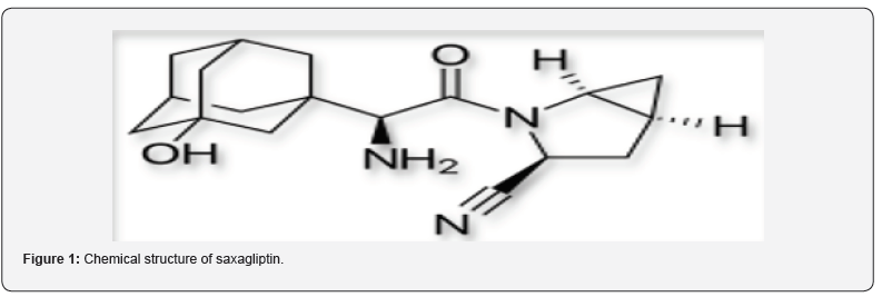
Saxagliptin (SXG) is chemically (1S, 3S, 5S)-2- [(2S)-2-Amino-2-(3 hydroxytricyclo [3.3.1.13,7] dec- 1-yl) acetyl]-2-azabicyclo [3.1.0] hexane-3-carbonitrile previously identified as BMS-477118 as shown in figure 1. The empirical formula is C18H25N3O2.H2O, and the molecular weight is 333.43 [1-4]. Saxagliptin is an oral hypoglycemic or anti-diabetic drug of the dipeptidyl peptidase-4 (DDP-4) inhibitor class. The inhibition of DPP-4 increases levels active of glucagon like peptide 1 (GLP-1), which inhibits glucagon production from pancreatic alpha cells and increases production of insulin from pancreatic beta cells. In 2009, U.S. Food and Drug Administration (FDA) approved saxagliptin and sold under the brand name Onglyza. In adults with type-2 diabetes mellitus, saxagliptin is suggested as an add-on to diet and exercise to improve glycemic control [5-11].

Literature survey reveals that the drug can be estimated only by LC-MS/MS, spectrophotometric method have been reported [12,13]. The aim of the present work was to develop a simple, sensitive, precise and accurate UV/Visible spectrophotometric method for the determination of saxagliptin in its pure form and pharmaceutical formulations further, to validate the developed method.

Materials and Methods

Chemical and reagents

All the chemicals used were of analytical grade. All the solutions were freshly prepared in Methanol and 0.1N HCl (15:85, v/v). Authentic of saxagliptin were obtained as gift samples from Mylan Laboratories Limited, Hyderabad

Methods

Saxagliptin was estimated using UV-Visible spectrophotometer (Shimadzu model 1800 double beam) at the wavelength of maximum absorption (204 nm) in acidic medium containing 0.1N HCl. The drug was characterized by solubility, melting point, and Fourier Transform Infra-Red (FTIR) techniques. The analysis of the drug was carried out by UV-Visible method which was validated analytical parameters like linearity, precision, and accuracy as per guidelines laid down by International Conference on Harmonization (ICH).

Physical characterization of saxagliptin

The physical characterization of procured drug sample of saxagliptin was determined on the basis of following parameters.

Organoleptic properties

The organoleptic properties have been determined for nature, colour, taste and odour of the pure sample of saxagliptin

Solubility

Excess amount of saxagliptin was dissolved in 10 ml distilled water till a saturated solution was obtained. The saturated solution of saxagliptin was stirred for 48 hrs on magnetic stirrer at 100rpm and room temperature (at 25 ± 1ºC). Then the sample was centrifuged for 10 min at 10,000 rpm. Clear supernant was collected using 0.22 μm syringe filter and analysed using UV spectrophotometer. The results were analysed and noted [14].

Melting point

Melting point of saxagliptin was determined using capillary melting point apparatus. In this method a small quantity of drug was filled in a capillary tube open both the ends and it was placed along with thermometer in melting point apparatus.

Fourier transform Infrared spectroscopy (FTIR)

FTIR spectrum was used as an analytical technique for identification of pure drug sample by KBr method using FTIR spectrometer (Agilent Technologies, USA). Peaks of individual pure drug were compared with reference FTIR peaks. Samples were previously prepared with KBr at 1: 5 (sample: KBr, w/w). KBr disks were prepared by compressing the powders at a pressure of 5 tons for 3 min in a hydraulic press and scanned against a blank KBr disk at wave numbers ranging from 500 to 4000 cm-1 with a resolution of 1.0 cm-1 [15].

Analytical method for drug concentration measurements (UV/Visible method)

The ultraviolet spectrophotometric method was selected for the estimation of saxagliptin in the range of 200 to 400 nm and the λmax was determined.

Diluent preparation

Methanol and 0.1N HCl (15:85, v/v) were used as a diluents.

Preparation of saxagliptin stock solution (20μg/ml) in 0.1N HCl

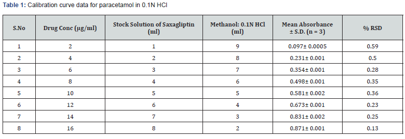
Saxagliptin was weighed equivalent to 100mg and transferred into 100ml volumetric flask then 15ml of methanol was added and shaked well to dissolve it after that the volume was made up to 100ml with 0.1N HCl. From that 2ml of solution was withdrawn and taken in 100ml volumetric flask. The volume was adjusted with diluent up to up to 100ml with 0.1N HCl [16,17] as shown in table 1.

Selection of detection wavelength

Drug solution was scanned over the range of 200- 400 nm. The wavelength of saxagliptin was determined to be 204 nm.

Preparation of working standard

Prepare a series of dilute solution from the above stock solution according to table 2 in 10 ml volumetric flask. Measure the absorbance of the solutions at 204 nm using distilled water as a blank. Plot a graph by taking concentration (μg/ml) on X-axis and absorbance on Y-axis. This plot gives a straight line and the linearity can be determined using y = mx + C formula. From the calibration curve calculate the Coefficient of determination R2 value, slope m and intercept C using the following formula, OR by excel sheet.

Construction of calibration curve

Pipette out 1,2,3,4,5,6,7, and 8 ml of working solution and transfer into separate 10 ml volumetric flasks. Dilute all of them to 10 ml with water to get solution of concentrations to 2, 4, 6,8,10,12,14,16 μg/ml respectively

UV Method Validation

The ultraviolet spectrophotometric method was validated for accuracy, precision, linearity, detection limit, quantitation limit and robustness [18-20].

Linearity

Appropriate aliquots of saxagliptin working standard solutions were taken in different 10 ml volumetric asks and diluted up to the mark with distilled water to obtain final concentrations of 2, 4, 6, 8, 10 μg/ml. Calibration curves were constructed by plotting absorbance versus concentrations and regression equations were calculated for both the drugs.

Range

The Range of the analytical method was decided from the interval between the upper and lower level of the calibration curve by plotting curve.

Precision

Intraday precision was determined by analyzing the drugs at concentrations (4μg/mL) and each concentration for three times, on the same day. Inter-day precision was determined similarly, but the analysis is carried out daily, for two consecutive days. Repeatability (intraday) of the method was determined by analyzing six samples of the same concentrations of the drug (4μg/mL). The absorbance of each was measured and reported in terms of relative standard deviation to obtain the variation.

Accuracy

The accuracy of the method was determined by calculating recoveries of saxagliptin by method of standard additions at three different levels 60, 100 and 140 %. Mean percentage recovery was determined. Recovery values were calculated and shown in table 1.

Detection limit

The Detection Limit of an individual analytical procedure is the lowest amount of analyte in a sample which can be detected but not necessarily quantitated as an exact value. The detection limit (LOD) may be expressed as. LOD= 3.3σ/S Where σ = Relative standard deviation of the response. S = the slope of the calibration curve (of the analyte).

Quantitation Limit

The Quantitation limit of an analytical procedure is the lowest amount of analyte in a sample, which can be quantitatively determined with suitable precision and accuracy.

Quantitation Limit (LOQ) may be expressed as: LOQ = 10σ/S Where

σ = Relative standard deviation of the response.

S = the slope of the calibration curve (of the analyte).

Results & Discussion

Organoleptic properties

Saxagliptin was found to be white colored, non-hygroscopic, crystalline powder.

Solubility

The solubility of saxagliptin in water was found to be 2.12 ± 0.28 mg/L corresponding to reference value of 2.9 mg/L.

Fourier transform infrared spectroscopy (FTIR)

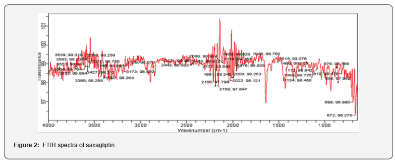
FTIR spectrum for identification of pure drug sample was done by FTIR spectrometer. Major absorption peaks of saxagliptin was found at 3450.12 cm-1 (N-H stretching), 2912.61 cm-1 (C-H stretching), 3301 cm-1 (OH stretching), 1614.47 cm-1 (C-N stretching), 1255.70 cm-1 (C-O stretching) as shown in figure 2 which corresponds to the literature peaks confirming the purity of drug sample.

Melting point

The melting point was determined by capillary melting point apparatus. The observed melting point of saxagliptin is 101°C

Analytical method for drug concentration measurements (UV/VIS Method)

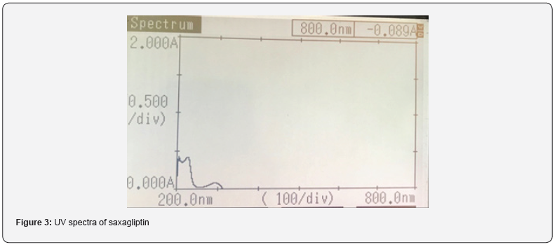
Selection of detection wavelength: The wavelength of saxagliptin was determined to be 204 nm as shown in figure 3.

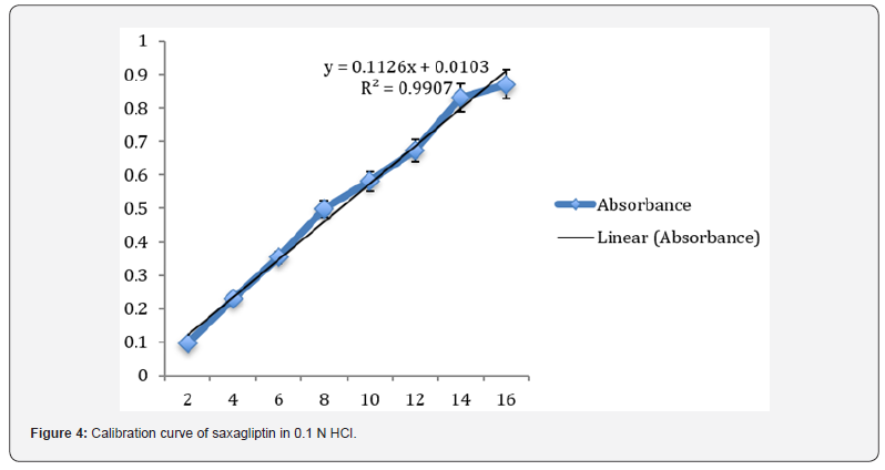
Preparation of standard plot for saxagliptin: Absorbance of the resultant solution was measured at 204 nm using blank. A graph was plotted between the concentrations and their respective absorbance. The response of the drug was found linear in the entire investigational range of 2 to 10μg/ml as shown in table 1. The calibration curve showed the linear equation as, y = 0.1126x - 0.0103, with a correlation coefficient, r2 = 0.99068, where y represents absorbance (optical density) and x represents the concentration (μg /ml) as shown in figure 4.

Method validation: The developed method was validated as per ICH guidelines for the following parameters:

Linearity: The linearity for Saxagliptin was found to be linear in the range of 2-10 μg/ml. The regression equation was found to be y = 0.1126x - 0.0103, r2 = 0.99068.

Range: The observed range of saxagliptin in test solution was observed from 0.097± 0.0005 to 0.871± 0.001.

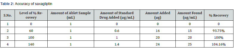
Accuracy: The accuracy of the analytical method for Saxagliptin was determined at 60%, 100% and 140% levels of standard solution. Absorbance was measured at 204 nm and results were expressed in terms of % recoveries in table 2.

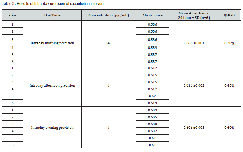
Precision: The Intra-day and Inter-day precision were carried out using same optimized conditions. The precision (measurement of inter-day, intra-day repeatability) results showed good reproducibility with the relative standard deviation (% RSD) below 2.0 % as shown in table 3 & 4 respectively. This indicated that the method was highly precise.

Limit of detection (LOD) and limit of quantification (LOQ): LOD and LOQ of method were determined to be 6.77 μg /mL and 20.33 μg /mL respectively. LOD and LOQ indicate that method was highly sensitive and fast.

Conclusion

The method was validated and found to be simple, sensitive, accurate and precise as per ICH guidelines [21]. The % RSD for the validation parameters was found to be less than 2%. Hence proposed method may be used for routine analysis of these drugs in pharmaceutical dosage forms. Accuracy of proposed method was confirmed by performing accuracy studies that showed the results within the range. Precision of proposed UV method was confirmed by performing intra-day and inter-day precision. Results were well within acceptance criteria that indicate excellent scope of the method for the determination of Saxagliptin in pharmaceutical dosage forms and bulk.


To Know more about our Juniper Publishers


Tuesday, May 4, 2021

Yoga Therapy for Treatment of Menstrual Disorders without Obvious Causes - Juniper Publishers

Yoga and Physiotherapy - Juniper Publishers  

Abstract

Introduction: Yoga is believed to help women of all ages lead healthier lives, by immediate remedy through relaxation, and deeper action through body mind regulation. It is believed to inspire system that influences one’s notions about wellness in life, even in presence of disorders. Menstrual abnormalities are one of most common disorders, women suffer. It may be less or more vaginal bleeding, for short or long duration, less or more interval between two menstrual phases, or continuous bleeding. Sometimes cause is obvious, can be treated. However, if it is not obvious, it may have linkage to notions, not easy to treat. Objective: To know about status of practice of Yoga for treatment of menstrual disorders in women who do not have obvious disorders. Material Methods: Studies, reviews in relation to effects of Yoga on menstrual disorders were searched. There was no inclusion, exclusion criteria of studies, reviews. Whatever could be accessed including recorded opinions were looked into. Results: Yoga is believed to be safe, cost effective therapeutic modality, for good health, wellbeing by regulating body weight, improving biochemical functions, obesity, and metabolic syndrome. Global Multidisciplinary group of researchers, who conducted large clinical trials to examine benefits of Yoga found positive effects of Yoga on Oligomenorrhea, infrequent menstruation, menorrhogia compared to exercise or no activity, in addition to taking an omega-3 supplement or placebo. Yoga might help by improving immune function of body, flexibility, endurance with added psychological benefits, directly and/or indirectly affecting menstrual disorders. However, after reviewing many studies about evidence of effects of Yoga on menstrual disorders, researchers concluded that more research with consistency in methods, measures, quality of studies was warranted. Conclusion: More research is needed about use effectiveness of Yoga practice to treat menstrual disorders.

Keywords: Menstrual disorders; No obvious cause; Yoga practice; Effects

Introduction

Menstrual abnormalities are one of the most common disorders encountered by women during reproductive life. Abnormal bleeding could occur beyond menopause too. Menstrual problems may be of less or more bleeding, during menstruation, for a short or long duration, with less or more interval between two menstrual phases, bleeding during interval or even continuous bleeding. In some women there are obvious disorders which are responsible for menstrual abnormalities but in others there is no cause. Such cases are usually given some or other hormones. However, hormones used for treating menstrual disorders, are not always effective in relieving symptoms and also, they do have side effects [1]. So search continues for knowing alternative therapies for menstrual disorders, specially those categorized as dysfunctional uterine haemorrhage, meaning there is no obvious pelvic or endocrinal or other such pathologies, but dysfunction in menstruation, difficult to treat.

Objective

To get information about effects of Yoga practice to treat menstrual dysfunction in women who do not have any obvious disorder.

Material and Methods

Literature search with available search engines was done to know about studies, and reviews, in relation to Yoga practices and effects on menstrual dysfunction. There were no inclusion or exclusion criteria of studies and reviews. Whatever could be accessed including opinions was looked into and self-observations were added.

Results

Yoga is believed to benefit health and wellbeing by regulating body weight and may be by improving body metabolism. So Yoga may be affecting menstrual function too. Menstrual disorders may result from physical disorders, emotional and hormonal disturbances, pathological development of reproductive organs, other organs or even psychological disturbances like depression, fear, or stress. All these are likely to get affected by Yoga practice. Prabhu et al. [2] did a study to know about effects of Yogasanas on menstrual cramps in young and adult women with primary dysmenorrhea and reported that both, Yogasans and Core exercises were useful in reducing menstrual pain. Rani [3] has reported that regular and proper practice of Yoga, Asanas and Pranayamas were beneficial to women who suffered from menstrual disorders, however rest, medicines and relaxation were also necessary. Yoga helped by correcting and balancing the functioning of the endocrine system, toning up the nervous system. It also relaxed the body and mind, which reduced the psychological problems. The Yogic postures helped in preserving the general health and reducing menstrual dysfunctions by improving the vigour and the strength of the body.

It seems that the roots of Yoga might have been in India but the perceived benefits of Yoga have led to its wide acceptability globally. It has been reported that around 15 million Americans practice Yoga [4]. Bullork [5] reported that a multidisciplinary group of researchers conducted large clinical trial to examine the benefits of Yoga compared to exercise or no activity, in addition to taking either an omega-3 supplement or placebo. Study participants included 355 peri- and postmenopausal women aged 40-62 years, who were randomized to one of the three groups, Yoga (107 participants), Exercise (106), and Usual Activity (177). These women were also randomized to receive either 1.8g/day of an Omega-3 supplement or an Olive oil placebo capsule. Also those in the Yoga attended weekly, 90-minute classes and were asked to complete a home practice daily for 20 minutes. Yoga practice entailed “doing cooling” pranayama, 11-13 Asans, and yoga nidra guided meditation. However researchers reported that no differences were found for vasomotor symptoms. However, women in the Yoga group reported modest improvement in sleep quality and a reduction in insomnia and depressive symptoms. Results of another pilot study revealed the benefits of Yoga therapy in lowering blood pressure, improving the immune function of the body which directly and indirectly affected menstrual disorders too by improving flexibility, endurance and added psychological benefits [6].

In various studies on the effects of Yoga on Amenorrhea, Oligomenorrhea, Dysmenorrhoea, Premenstrual Syndrome and Premenstrual Dysphonic Disorder enhanced mood, reduced pain, increased wellbeing and a heightened relaxation response were among the improved outcomes reported by women who practised Yoga [7]. Yeun et al. [8] looked into studies and reported that there was objective evidence for Yoga, as a useful complementary and alternative medicine on women with menstrual distress and primary dysmenorrhea. Vaghela et al. [9] reported that when compared the effects of Aerobic exercise and Yoga on premenstrual syndrome, both Aerobic exercise and Yoga practice were effective in treating PMS, however Yoga was more effective in relieving the symptoms of PMS than Aerobic exercise. Aggarwal et al. [10] did a study about effects of Yogasans and Pranayama on pain, severity and quality of life in primary dysmennorhea and reported that Yogasans along with Pranayama technique were more effective than using a single intervention protocol either of Yogasans or Pranayama in primary dysmennorhea.

Not much is known about menstrual irregularities in middle age with or without Polycystic ovarian syndrome (PCOS) and with or without Metabolic syndrome which commonly lead to menstrual abnormalities. Metabolic syndrome is becoming public health problem because it is believed to be a rising issue and probably 20 to 30 percent of women suffer with Metabolic syndrome [11]. 0ates [12]also looked at various studies for effects of Yoga on Amenorrhea, Oligomenorrhea, Dysmenorrhoea, Premenstrual Syndrome and Premenstrual dysphonic disorders reported by women who participated in Yoga practice and reported that menstrual dysfunctions were common and Metabolic disorders too were common. So it becomes essential to know more about the relationship between the two. There is a dramatic change in the way Metabolic syndrome is treated after a growing body of evidence has pointed to Insulin Resistance as an underlying cause of Metabolic Syndrome. Insulin resistance, believed to be a key pathogenic factor in both PCOS and the Metabolic syndrome, may be the thread that links the two conditions [13]. Research on the relationship between Yoga practice and menstrual disorders is warranted, but there must be both consistency in the methods, measures, and quality of studies and a shift towards research on Yoga practice that is replicable outside the clinical trial settings. Goel [14] also reported that various breathing techniques used in Yoga were to improve blood circulation which caused ease of the flow of blood during menstruation. This also relieves pain that is experienced during menstruation. Various positions of Yoga are useful to enhance uninterrupted down flow during menstruation and prevent dysmenorrhea. But they need to be performed with awareness.Yoga is believed to offer both immediate superficial remedy at the body and mind level in the form of relaxation practices and also deeper action by working on, notions about life.

Discussion

Menstrual disorders may result from physical disorders, emotional and hormonal disturbances, improper development with weak muscles of reproductive organs, imbalance of hormones, weak constitution, pathological and psychological disturbances like depression, fear, stress and mental tension. As Yoga is likely to benefit women with such health problems which have linkages to menstrual disorders, so it could help in relief of menstrual dysfunctions. Yoga seems to help by correcting and balancing the functioning of the endocrine system, toning up of the nervous system and also relaxation of the body and mind, thus reducing the psychological problems. Yoga is a correct approach to life, with right diet, exercise and hygiene, quite essential for the normal development and functioning of the reproductive system. The Yogic postures help in preserving the general health and reduce the complaints regarding menstruation by improving the vigour and strength of the body. There is a need for spreading the knowledge about benefits of Yoga practice to large section of the society. This is possible if practice of Yoga is validated through exhaustive research based on standard parameters. Scientific exploration of Yoga in women with interdisciplinary approach is needed. While treating menstrual disorders, the major goal is to treat the underlying cause and when cause is not obvious, it is usually difficult to treat. Hormonal therapies may or may not be effective in relieving the symptoms and they have their side effects too. In some women they are contraindicated too. So research continues to identify the efficacy of alternative therapies for menstrual dysfunction. Yoga seems to be an alternative or primary treatment, for maintaining health and wellbeing by regulating body weight, improving biochemical functions overcoming the obesity related complications as well as Metabolic syndrome which lead to relief from menstrual dysfunction.

So, Yoga is likely to be beneficial in treating Metabolic Syndrome and so menstrual disorders also [15]. More research is needed for identifying the efficacy of Yoga in menstrual dysfunction, to know about Yogasanas with various postures through which the physical revitalization, deep relaxation and mental calmness are achieved. Yoga recognizes the link between breathing and calmness of the mind, which could be beneficial in premenstrual syndrome too and also help relief from other menstrual disorders, like menopauserelated, hot flashes, night sweats, and fatigue negatively which impact the quality of life of millions of women. After looking at evidence of effects of Yoga on menstrual disorders by reviewing many papers Oates [12] also concluded that more research about relationship between Yoga practice and menstrual disorders was warranted. Also, there is need of a deeper understanding of the effects of Yoga on menopausal menstrual effects, especially those without any obvious organic disorders. Yoga is known to play an important role in reducing stress and sympathetic activity, increasing parasympathetic activity, improving one’s quality of life, and also by decreasing psychological symptoms. It also has beneficial effects on physical and mental health through down regulation of the hypothalamic- pituitary- adrenal axis and the sympathetic nervous system, which may also be useful for managing primary dysmenorrhea. Yoga asanas help in increasing the flow of vital energy to the reproductive organs and establishes a balance among hormones regulating menstruation. While practicing Yoga it should be kept in mind that the number and the sequences should always be modified according to the capacity and other conditions of the person. One must avoid practicing Yoga by just looking at the television or by reading a book on Yoga therapy. The practice should be learnt from a qualified Yoga therapist. This is cost effective and can be used as a exercise for menstrual well-being and help improve quality of life.

Conclusion

Available literature reveals that Yoga is helpful in management of menstrual disorders however more well-designed research is needed.

To Know more about Yoga and Physiotherapy

Click here: https://juniperpublishers.com/jyp/index.php


To Know more about our Juniper Publishers


Simultaneous Determination and Quantitation of Artemeter and Lumefantrine in Antimalarial Tablet Formulation using High Performance Liquid Chromatography with UV Detection - Juniper Publishers

  Pharmacology & Clinical Research - Juniper Publishers Abstract Artemether-lumefantrine (AL) combination therapy is now the most used a...