Tuesday, August 31, 2021

Significance of Culture in Shaping Societal Responses to Pandemic Assault - Juniper Publishers

 Annals of Reviews and Research - Juniper Publishers

Abstract

Influencing the general public response to pandemics is a public health priority. The preventive behaviours (e.g. wearing masks and reducing aggregation) that help in reducing the spread of pandemic found insufficiently followed across different regions, during recent pandemic influenza, 2009-10 H1N1 pandemic, including the current COVID-19 onslaught. Regional difference in public’s preventive intervention observed, owing to cultural values variables. Across the continents 209 COVID-19 pandemic affected countries reportedly adopted standard preventive protocol for COVID-19, but the pandemic spread, and onslaught showed different patterns in different countries. This human habit of maintaining or not maintaining physical distance is a result of a country’s culture which is rooted in its national heritage and traditions and the current pandemic scenario provides an opportunity to study the flexibility and adaptability of cultures between pandemic responses, on a governmental and societal level. In this study culture was defined using Hofstede's dimensions Individualism/Collectivism. An exploratory case-study methodology was taken after employing a post- positivist approach. The study findings indicated collectivism encourages faster and more effective COVID-19 responses and hence suggest cultural adjustments for the purpose of infectious disease preventive intervention.

Keywords: Culture; Individualism; Collectivism; Public health

Introduction

Influencing the general public response to pandemics is a public health priority. The preventive behaviours (e.g. wearing masks and reducing aggregation) that are effective in reducing the spread of pandemic found insufficiently followed across different regions, even during pandemic influenza, 2009-10 H1N1 pandemic, among others. Regional difference in public’s preventive intervention observed, owing to cultural values variables. Understanding the impact of social elements at the effect of pandemic assault across communities necessitates not just experimental observations, but also a conceptual framework for investigating and interpreting such observations. Societal individualism/ collectivism is one idea that could be useful as a guiding principle in this scenario; and the topic subsequently discussed with suitable references. Now to focus on the Coronavirus Disease 2019, as unfolded all over the world, and caused one of the most serious public health crises in recent times. Isolating actions as Social distancing, lockdowns, and other suggested and adopted as protective behavioral mechanisms to facilitate the avoidance of parasitic transmission along with other methods of managing local parasitic infections. However, in this pandemic, the enormous differences in transmission outcomes between countries and territories constitute a striking phenomenon. Social scientists suggest that the cultural difference of collectivism versus individualism could largely explain this divergence. Collectivist as opposed to individualistic values are important attributes of intercultural variation.

In this study culture was defined using Hofstede's dimensions Individualism/Collectivism. An exploratory case-study methodology was taken after employing a post- positivist approach, which was affected by Yin [1] and Noor [2]. This procedure is appropriate for the taking after reasons: i) the consider includes an observational examination of a developing marvel, which is the utilize of social media; ii) the consider accumulates realities to impartially degree event of certain design; and iv) the think about increases in value diverse elucidations of individuals on their encounters. Interestingly, the Parasite Stress Theory of Values [3-5] implies that the historical occurrence of infectious diseases may have played a role in the development of Individualist and Collectivist cultural differences. Societies with a high caliber of pathogenic stress were more liable to develop a collectivist culture that accommodates as a gregarious aegis against infectious disease spread, whereas societies with low caliber of pathogenic stress developed the individualistic value systems [3]. If this is true, modern societies with highly individualistic cultures may be more vulnerable to infectious diseases than more collectivistic societies. Another study [6] revealed that individualistic communities have had more infection disease outbreaks and zoonotic disease outbreaks in recent times but did not find a correlation for emerging infectious disease events.

The SARS CoV-2 situation reports of March 2021 brought out by world health organization (WHO) [7] indicate 209 pandemic affected countries across the continents adopted, by and large, uniform responses for pandemic control, but the spread and onslaught of COVID-19 has shown different patterns in different countries. Due to international variances in pandemic reactions on a governmental and societal level, the era of pandemic gives a chance to study the flexibility and adaptability of cultures. Due to the pathway of disease transmission through close patient contact, COVID-19 may be owing to close contact with the general population. This is where the culture of the susceptible population become evident and their behavior need detailed scrutiny. For instance, Italy was attributed to have a culture of close interaction (physical intimacy) as compared to Japan (culturally maintaining greater physical distance). Dissonantly the French saw social unification as a threat to the state, whereas Japanese culture intends to support the government’s response to COVID-19.

This human habit of maintaining or not maintaining physical distance is a result of a country’s culture which is rooted in its national heritage and traditions. Any attempt to abruptly change the behavior in order to prevent pandemic spread is impractical and will be met with public opposition, as has been proved in various countries.” The collective programming of the thoughts that separates the participants of one class of humans from another” is how culture is described [8]. As a result, population behavior and psychological elements that influence the said programming may be influenced in part by a country’s culture, which could be important in understanding Covid-19’s spread and mitigation. Indeed, a recent assessment found various social and behavioral traits, including cultural norms- that could help with the COVID-19 pandemic response, and encouraged researchers to cover gaps urgently [9]. There is also the pertinent issue of individualistic and collectivism cultural approaches of different countries that shape citizen’s cognitive processes. In general, individualism emphasizes non-public freedom and achievement. Individualist culture therefore gives social popularity to non-public accomplishments inclusive of critical discoveries, innovations, extremely good inventive or humanitarian achievements and all moves that project an individual. Collectivism, in contrast give importance to positioning individuals in bigger group. It whips up conformity and discourages individuals to balk and show up.

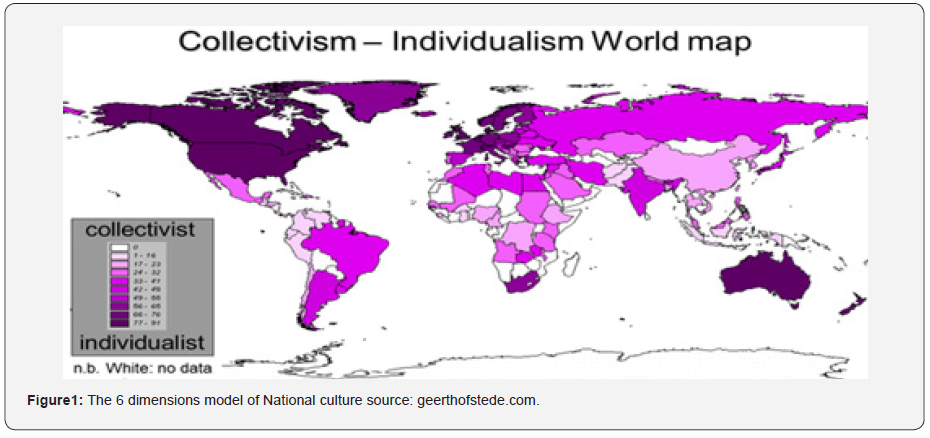
Individualism is said to hold sway in Western Europe and North America with their complex, stratified societies emphasizing independence and variety while collectivism is mean to thrive in Asia, Africa, Eastern Europe, and Latin America, in which agreeing on social norms is essential and jobs are interconnected [10]. For extra collectivistic societies like India, the self is described relative to others, is involved with belongingness, dependency, empathy, and reciprocity, and is centered on small selective coterie of fellow individuals in common business on the price of out-groups. Noted scientist Professor Geert Jan Hofstede put forward with six basic issues called dimensions of culture expressed on a scale that runs roughly from 0 to 100. His Collectivism-Individualism World Map provides a glimpse of nations’ cultural trait (Figure 1).
Hardin’s traditional article “The tragedy of the commons” [11] gives a prediction for the distinction among individualistic versus collectivistic societies dealing with the pandemic. Hardin defined a social ‘catch 22 situations’ wherein every decision-maker in a network is higher off performing egocentrically. Still, if others acted likewise without problem for the cumulative effect on society, “the commons” are in the end knocked down. Curiously, whilst not unusual place experience means that the virus will unfold extra fast in collectivistic societies because of their nearer and extra common social interactions, the mixture of way of life and Hardin’s principle endorse the opposite: the pandemic’s effect might be large in individualistic societies wherein human beings are less involved approximately in the greater good. COVID-19 has made it manner to almost every country within side the world and has but ended in very exclusive outcomes.

Epidemiologists have proposed lots of reasons for this variance, which includes variations in demographics, urbanization, excellent of health systems, the innate environment, and the rate of government responses [12]. Nevertheless, it is able to be argued strongly that way of life additionally matters. Because consensus is extra conveniently carried out in collectivist societies, their conditions are better for introducing speedy and effective motions to include disease containment. An investigation posted in February 2021 [13] researchers studied the connection among the individualism -collectivism size through the use of Hofstede’s cultural size version and the quantity of COVID-19 instances and associated deaths of all 69 countries, for which statistics became to be had in Hofstede’s national culture survey [14]. They conducted two simple correlations analysis and compared the variables among 36 Organization for Economic Cooperation and Development (OECD) countries and then performed identical correlations evaluation at the whole pattern at the same time as controlling eight applicable variables, i.e. days considering the facts that outbreak of the pandemic, percentage of population over 65 years of age, democracy index, Gini index, percent of finances for healthcare, human longevity, population density and general COVID-19 tests per million.

Taken together, those consequences counseled that the greater individualistic a society is, the greater it suffered from COVID-19 associated instances and deaths. As a result, it may be claimed that cultural differences between countries are equally as important in determining adherence to epidemic prevention measures and, as a result, a society’s susceptibility to COVID-19 outbreak. The virus has spread practically every country on the planet, yet the results have been wildly disparate. It is arguable that culture plays a considerable role. It has been observed that collectivist cultures are more likely to reach an agreement, they are better suited to putting in place swift and effective disease control measures. In collectivist societies, social networks are also more localized and directed around people’s intimate relationships (typically their extended family). This causes natural social bubbles, reduces social mixing and diversity, and hence inhibit virus’s transmission.

According to studies, having a more individualistic culture leads to more innovation and progress since those societies value social workforce more [15]. But there are also disadvantages, as Hofstede pointed out individualist societies are at a disadvantage in terms of swift collective action and coordination. This is because residents are encouraged to hold opposing viewpoints, express themselves, and challenge and debate decisions. As a result, achieving the necessary agreement for policies to succeed may take longer. Individually, cultural values can influence basic decision such as whether or not to wear face mask or maintain social distance. According to studies, persons in areas of the United States with a history of colonialism and more individualistic culture are less likely to wear face masks and keep physical distance in public venues.

It is possible to evaluate the country-wise COVID-19 incidences based on the national data publicly available. The discrepancies between individualistic and collectivist countries, as well as their differing response to pandemic containment, are most obvious when looking at data from the onset of the pandemic. There is a direct correlation between COVID-related mortality per capita and individualism scores in countries. Furthermore, studies and actual evidence demonstrate that collectivism promotes a faster and more effective COVID-19 response. Given the circumstances, individualist countries should make major attempts at cultural adaption in that direction, especially during outbreaks of diseases. The speculation of pathogen prevalence, or the parasite stress model [3-5], states that people living in areas with greater incidence of infections are more likely to become collectivist in the long run than people living in areas with fewer incidence. The aftermath of COVID-19 is likely to be something never witnessed to world citizens. A new social order is likely to emerge, a new structure of the society, a paradigm shift in human relationships.

To Know more about Annals of Reviews and Research


To Know more about our Juniper Publishers

Click here: https://juniperpublishers.com/index.php   


Monday, August 30, 2021

Calcineurin B Homologous Protein Regulation of the Sodium Hydrogen Exchanger Isoform 1: More than Competition, the Need for a CHP-Specific Therapy - Juniper Publishers

 Cell Science & Molecular Biology - Juniper Publishers

Abstract

The development of pathophysiologies including fibrosis and cancers involve the establishment of a tissue microenvironment characterized by hypoxia and low serum conditions. NHE1 directly influences cell proliferation, extracellular matrix composition, and cell migration in multiple disease states. Calcineurin homologous protein isoforms 1 and 2 (CHP1 & 2) are calcium-binding proteins whose function is not well understood. CHP1 is crucial for NHE1 membrane processing and basal transport activity. Additionally, CHP1 is involved in the regulation of gene expression. In contrast, CHP2 expression is limited to a small range of cells types its primary function is ascribed to the regulation of NHE1. Preferential binding of CHP2 to NHE1 under the tumor microenvironment supports cell survival by first activating the exchanger through the CHP2-NHE1 interaction and by freeing CHP1 to the nucleus where it also supports cell survival. The affinity of CHP2 is three-fold greater than CHP1, but endogenous CHP concentrations indicates that competition alone cannot explain the role of CHP2. Modelling of CHP binding identified two distinct approaches to establishing the CHP-NHE1 interaction; CHP1 binds in a two state-mechanism and CHP2 bound NHE1 in a one state-mechanism. Additional analysis of predicted and published phosphorylation sites of the CHPs suggest that phosphorylation can support a CHP specific interaction with NHE1. Together these data and models highlight a means to identify possible therapies that target CHP2 but not CHP1 to defeat cell survival in several disease states.

Keywords: Sodium Hydrogen Exchanger Isoform 1, NHE1, Calcineurin B homologous protein, CHP1, CHP2, Tumor metastasis, Protein-protein interaction, Tumor microenvironment, TME, targeted therapy, Intracellular pH

Overview

The Na+-H+ exchanger isoform 1 (NHE1) plays a critical role in directing cell functions including proliferation, motility and intracellular pH homeostasis. Poorly understood is how two closely related regulatory proteins, calcineurin B homologous protein 1 and 2 (CHP1 and CHP2), coordinate to control NHE1 related cellular events. Under normal conditions, CHP1 is bound to NHE1 maintaining basal activity. When CHP2 is expressed, it also binds NHE1 at overlapping residues with CHP1 and CHP2 regulates and enhances NHE1 transport. The physiological relevance and regulation of CHP2 has not been elucidated. As visualized in Figure 1, we hypothesize that in the tumour microenvironment, CHP2 displaces CHP1 from NHE1, altering aNHE1 function and freeing CHP1 to locate to the nucleus where it can drive expression of genes supporting cell survival.

NHE1 Regulation and Function

NHE1 belongs to the 13 member SLC9 gene family. Five isoforms, NHE1-5, are located on the plasma membrane and NHE1 is nearly universal in human tissues [1,2] NHE1 has two functions: (i) homeostasis of intra- and extracellular pH through Na+-H+ exchange and (ii) as a scaffold for a range of proteins through protein interactions at an extended cytosolic carboxyl tail. NHE1 regulation is complex and involves several phosphorylation sites, lipid binding domains and protein-protein interactions. How these regulators interact and whether certain forms have priority under different cellular conditions is unknown. Activation of NHE1 is one of the initial steps in the development of the transformed phenotype of cancer cells [3]. This activation leads to an alkalization of intracellular pH (pHi), which is a permissive requirement for cell growth. During cell migration, NHE1 localizes to the leading edge of cells where a combination of an elevated pHi and the binding of a range of proteins to NHE1 stimulates the formation of actin filaments that drive the forward movement of these migrating cells [4-8]. Abnormal regulation and function of NHE1 has been identified as playing a major role of a range of pathophysiological conditions. NHE1 directly influences cell proliferation and migration in multiple disease states. In pulmonary hypertension, pulmonary arterial smooth muscle cells increase proliferation and migration due to the activation of NHE1 in response to chronic hypoxia [9]. In dilated cardiomyopathy, elevated NHE1 activity induces cardiac hypertrophy in vivo driving disease progression [10]. In cancer, activation of NHE1 is an initial step in the development of the transformed phenotype of cancer cells [1-4]. Aberrant activation of NHE1 can also lead to changes in Na+ homeostasis which enhances disease progression. In both perinatal brain injury [11] and diabetic cardiomyopathy [12] anomalous activation is NHE1 drives an increase cytosolic Na+ level which alters Ca++ homeostasis leading to impaired energy metabolism and decreased cell survival. Each of the conditions listed share both the activation of NHE1 as a driving force in the initiation of a disease state and the fact that an underlying mechanism for that NHE1 activation has not been fully elucidated.

Role Of CHP 1 and CHP2

Both CHP isoforms bind and regulate NHE1 transport activity. CHP1 & 2 are calcium-binding proteins whose function is not well understood [13]. CHP1 is crucial for NHE1 membrane processing and basal transport activity. Additionally, CHP1 is involved in the regulation of gene expression [13-15]. No published data relates the distinct nuclear and membrane functions of CHP1 or the mechanisms by which these functions are regulated. In contrast, CHP2 expression is limited to a range of cells types including intestinal and skin cells (Figure 1) and its primary function is ascribed to the regulation of pHi via NHE1 [16]. In non-diseased tissue, CHP2 is expressed at low but detectable levels in most cell types including lung, muscle and other tissues ( Figure 2; RNAseq baseline Human Protein Atlas project: TMP 0.1-0.99), moderately found in kidney, adrenal, prostate, bladder and vagina (TMP 1-9.9) and highly expressed in intestinal track cells, some epidermal cells and spinal cord (TMP => 10.0). We have identified CHP2 expression in several cultured cell lines including CCL39 fibroblasts as well as other transformed lung epithelial and fibroblastic cell lines but not non-diseased cultured cells [17].


CHP1 has several identified functions:

A. at the membrane through binding to NHE1

B. in the cytoplasm engaging in the regulation of microtubule formation and vesicle trafficking

C. in the nucleus where it both positively and negatively regulates gene transcription and nuclear function.

Expression of CHP1 can inhibit, nuclear factor of activated T cells (NFAT) and its associated transcriptional activity by inhibition of calcineurin [18]. Both calcineurin and NFAT are expressed at 2-4 fold higher levels in non-small cell lung cancer (NSCLC) than in non-diseased lung tissue [19]. Hypoxic conditions enhance lung fibroblast proliferation and migration in lung fibrosis while increasing NFAT expression indicating a key role for CHP1 [20]. Both serum deprivation and hypoxia can lead to CHP1 translocation to the nucleus [21]. Nuclear CHP1 has been identified to stabilize HIF1a in hypoxic HepG2 cells inhibiting hypoxia-induced apoptosis [22]. Likewise, the deathassociated protein related apoptosis inducing kinase (DRAK2) binds to and is inhibited by CHP1 and may be involved in both nuclear translocation of CHP1 and inhibiting apoptosis [23]. CHP2 may have an important but unidentified role in several disorders. CHP2 expression was first identified in hepatoma, colon carcinoma, cervical carcinomas, and leukemia cells but not in healthy, non-diseased tissues [24]. Most recently, CHP2 expression was detected as a putative tumour marker in patients with acute leukemia, where it was not detected in paired, nondiseased tissue [25]. While most of the work investigating CHP2 has focused on its regulation of NHE1 in cancer, overexpression of CHP2 activates both calcineurin and NFAT, contrary to the effects of CHP1 [26]. Increased CHP2 expression affords cellular maintenance of an elevated pHi and enhanced cell survival in a low serum environment, as well as an increase in cell proliferation [17]. In addition, in nude mice knockdown of either NHE1 or CHP2 abrogates tumour formation [17]. Overexpression of CHP2 increased cell proliferation, cell adhesion, and cell invasion relative to cells lacking CHP2 expression [27]. Inversely, CHP2 expression knockdown in HepG2 cells inhibited cell proliferation, even in the presence of CHP1 [28]. These data suggest that CHP2 regulation of NHE1 allows a higher basal pHi which supports accelerated rate of proliferation [24].

NHE1-CHP1/2 Interactions As A Therapeutic Target

Current clinical trials using NHE1 inhibitors as therapeutic modalities for several disease have struggled for success, in part, because of the nature of small molecule inhibitors targeting a ubiquitously expressed protein resulting in negative impacts on non-diseased tissue [29,30]. Yet the optimism for an NHE1 related therapy remains high for several disorders as positive results have been observed [31,32]. Data suggests that both CHP1 and CHP2 play a critical role in cell survival, proliferation and motility in microenvironments found in both tumor and fibrotic tissues. Blocking the binding of both CHP isoforms would limit the gain of function that occurs with CHP2 expression but would concomitantly result in loss of CHP1 sequestering at the membrane where it could support diseased cell function in the nucleus. Thus a precision therapy targeting CHP2-NHE1 interactions while sparing CHP1-NHE1 interactions might be a promising approach. Unfortunately, there is currently not enough evidence to justify the development of such a therapy. We next provide evidence for such a CHP-specific approach. While both isoforms of CHP have a 75% amino acid homology with nearly identical binding sites on NHE1[31] there is a fundamental difference in the impact of each CHP-NHE1 interaction on transporter function. CHP1 does not activate NHE1 but its binding is required for agonist activation, while CHP2 increases both the Vmax and the H+ affinity of NHE1, thus serving to activate transport above and beyond CHP1. A single report indicates that CHP2 binds NHE1 with a five-fold greater affinity than CHP1 resulting in significantly increased NHE1 transport activity [33]. In cells stably transfected with CHP2, the resting pHi was 0.2 to 0.3 pH units higher than control cells [34]. No mechanism for the regulation of the CHP-NHE1 interactions has been reported.

CHP-NHE1 Binding

Using a semiquantitative competitive pulldown approach, [24] Pang et.al. demonstrated that CHP2 bound NHE1 with nearly a five times higher binding affinity than CHP1 [33]. These studies indicated that the two CHP isoforms could competitively bind to NHE1 and that concentration might be the driver for binding to NHE1. Recently, the impact of calcium on CHP-NHE1 binding was analysed using isothermal titration calorimetry (ITC; 34). In the absence of calcium, CHP1 binds to NHE1 with a 2.7 times greater affinity than CHP2. Yet, in the presence of calcium, CHP2 but not CHP1 affinity for NHE1 increases by nearly seven-fold, resulting with both isoforms bound to NHE1 with nearly the same affinity (KD = 7.7 nM for CHP1 and 3.2 nM for CHP2-NHE1).To qualitatively examine interactions between NHE1 and CHP, we bound GST or the GST- CHP binding domain of human NHE1 (aa 503-545) to a glutathione coated 96 well plate and determined binding of either RFP-CHP1 or GFP-CHP2 in the presence of 1 mM calcium chloride (Figure 3). Like the ITC generated data, the affinities were in the low molar range (CHP1-NHE1: 2.65 nM, CHP2-NHE1: 9.65 nM) with CHP2 binding NHE with 3.65 fold higher affinity than CHP1. Interestingly, while the KD for each binding interaction was modestly different, the maximal bound CHP significantly differed for each CHP isoform. The potential for an affinity tag to fold over and bind or block the NHE1 binding groove of CHP was suggested after examination of the structure of CHP1 and could be a potential explanation for the differences in bound between CHP1 and CHP2 [17,34]. Using recombinant His tagged purified CHP1/2 and the CHP binding domain of NHE1 after removal of the affinity tag by TEV proteolysis, we determined the melting point for each protein alone or in combination using circular dichroism spectroscopy during a thermal melt. The melting point shift for CHP1-NHE was 3.3 oC greater than for the CHP2-NHE complex (Tm CHP1-NHE1; 58.7 +/- 1.7oC and CHP2-NHE1: 55.4 +/- 1.2oC). The change in free energy (DG at 37oC) of binding for the two complexes was then calculated as -1.16 kJ/mol for CHP1-NHE and -3.91kJ/mol for CHP2-NHE complex. The 2.74kJ/mol difference in predicted binding energy at physiological temperature translates to an increase in affinity of CHP2 over CHP1 by a factor of 3.02. The increased affinity predicted here and identified by pull down assays [33] despite their high level of homology might suggest different binding mechanism for the association and protein recognition.

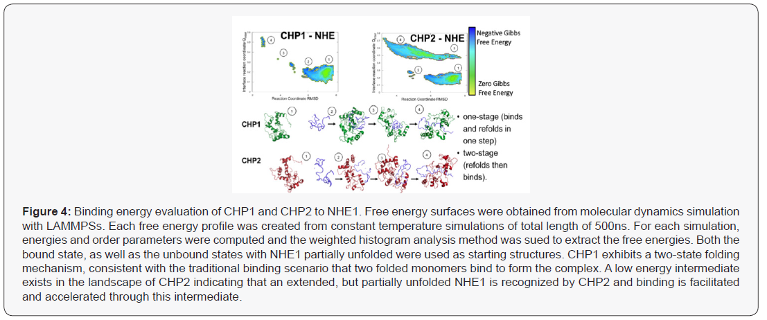
The thermodynamic predictions of binding were analysed using a Weighted Histogram Analysis Method (WHAM) and visualization software (LAMMPS) simulations to calculate binding energies to calculate predicted a binding mechanism (Figure 4). Protein binding landscapes obtained with molecular dynamics simulations show that both CHP1 and CHP2 binding is driven by the overall structure and binding site of the proteins. However, while the landscape of CHP1 exhibits a two-state binding of CHP1 to NHE1, CHP2, even though the monomer is stable on its own, shows binding through a partially unfolded, extended intermediate (Figure 5). Higher binding affinity and specificity through a partially disordered protein, although a bit counter-intuitive at first, has been documented in the literature. The intermediate in the CHP2 energy landscape suggest that the binding interface can be formed early during binding. Overall, the simulation study supports that the binding mechanism is different between CHP1 and CHP2 and CHP2 is accelerated due to the faster recognition of NHE1. Both of these modelled mechanisms are supported by the observed differences in binding between with the two florescent tagged CHP proteins (Figure 3). In this case, the flexible linker between the N-terminus of CHP and GFP/RFP could, as suggested, fold over and sterically block a folded binding site of CHP for NHE1. As modeled, CHP1, binding in a one-state mechanism would already be folded and thus binding events would be blocked but the affinity unchanged, a mechanism similar to a competitive inhibitor reducing Vmax but leaving Km unchanged. Our observation that CHP2 bound with the same affinity as CHP1 to NHE1, but with a higher binding maximum specific binding (Bmax), of 3.55nM for CHP2 and only 0.41 nM for CHP1 would support that the GFP portion of the fusion protein was distal to the unfolded CHP2 peptide which could then bind and fold in the one-state mechanism with less interference than a protein using a one-state binding mechanism.


CHP-NHE1 Interaction- Target For Drug Therapies

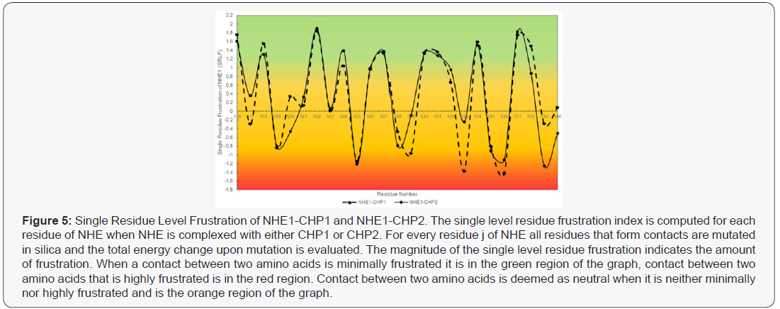
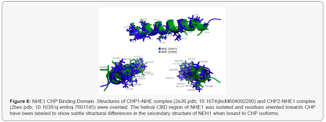
The structures of CHP1-NHE1 and CHP2-NHE1 have been solved [35,36]. CHP1 and CHP2 form a receiving groove for the NHE1 helix with the hydrophobic side chains of NHE1 aligned to one face making fourteen contact points with CHP. Several of the NHE1 residues are critical for binding CHP and mutation of these amino acids leave the transporter unable to bind either CHP isoform. While CHP isoform specific interactions with NHE1 were not identified, the mutation of NHE1 residues: Phe526, Leu527, Leu530, Leu531, Ile534, or Ile537 were all key for CHP binding and transport function [36,37]. Because CHP1 and CHP2 possess a divergent amino acid change at several of these NHE1 contact sites, it is likely that select NHE1 residues would be potential targets for defining CHP2-NHE1 interactions. Energy frustration computations using the Energy Landscape Theory were performed to quantitate the energy fit of each NHE1 residue making contact with CHP1 or CHP2 (Figure 5) [39,40]. Each NHE residue making contact with CHP1 and CHP2 was analyzed for the degree of local frustration using the freely available resource, EMBnet bioinformatic [41]. This tool scores the energies (local frustrations) of all possible amino acids vs the native amino acids involved in CHP-NHE binding. Additional evidence showing the unique binding of CHP for NHE1 is shown using an overlay of the NHE1 CBD domain bound to CHP1 and CHP2 with only the helix of NHE1 shown (masking CHP structure) (Figure 7) The shift between the two bound conformations of the NHE CBD indicates a subtle yet real difference in the structure of NHE1 as it binds each CHP (Figure 6) . It is possible that subtle contacts which render a specific interaction remain to be elucidated. Such analysis provides insight of biologically important regions of NHE1-CHP binding and identifies 11 sites of that may confer both the differences in binding affinities and highlight possible specific binding sites unique to CHP1 and CHP2.


Predictive modelling identifies that while both CHPs bind to nearly the same region of NHE1, there exists potential unique differences of binding of both CHP isoforms. Differences that could be exploited to design a CHP-specific inhibitor. However an argument remains that protein expression and competition alone could drive CHP1-CHP2 binding to NHE1. As seen in our and other published data, the affinity of CHP1 and CHP2 for NHE1 are similar showing a 3-5-fold higher affinity (KD) for CHP2-NHE1. This difference is also demonstrated by the binding models identified here. However, the difference in CHP affinity for NHE1 cannot explain a mechanism for the two CHP isoform binding. Several diseased tissues express higher levels of CHP2 [17,33]. We have shown that CHP2 expression increases in low serum [17] and other tumour microenvironment conditions (data not shown). As CHP2 expression increases due to the changes in the cellular microenvironment, CHP2 is thought to displace CHP1 bound to NHE1, increasing exchanger activity and altering cellular function. Thus at face value, it would seem that increases in protein expression and a slightly higher affinity for CHP2 could explain the phenomena. However, the estimated levels of protein in the highest CHP2 expressing cells favours CHP1 by 20-100 fold in cultured cells and in tissues ranged from 60 to 1000 fold in favor of CHP1 expression (Protein Atlas, GTX, Proteomics DB ; 17, 43). Therefore a model defined by competitive binding is not likely. More attractive is a combination of unique binding contacts and post translational modifications that could serve to regulate binding affinities.

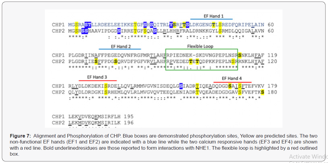
Interestingly, while NHE1 is phosphorylated at 22 putative and known phosphorylation sites [42], the CHP binding domain of NHE1 is not phosphorylated. The nearest modified site to the CBD domain is 50 residues distal to the carboxy-terminus. Both CHP isoforms are highly phosphorylated. Analysis of known phosphorylation sites identified in mass spectroscopy databases shows one site in common for both CHP1 and CHP2 while there are six unique phospho-sites of CHP1 and three unique sites identified for CHP2 [17]. Peptide sequence prediction analysis of NetPhos [43] highlights an additional six unique putative phosphorylation sites for both CHP1 and CHP2 (score, >0.5). There are seven additional sites predicted to be in common for both isoforms (Figure 7). CHP forms a hydrophobic receiving groove for the helical NHE CBD. Interestingly, the C terminal lobe of CHP which is key for binding to NHE1 is devoid of potential phosphorylation sites. The bulk of identified phosphorylation sites are on the N-terminus of the protein which forms the second lobe of CHP but is not a significant component of the NHE1 receiving pocket. Both CHP isoforms have similar C- terminal lobe domains with a unique linker domain at aa 81-101 (Figure 7). The structure of the CBD domain loops out between the N and C lobes when bound to NHE1, suggesting this is an important determination binding factor. Several putative CHP2 specific phosphor-sites are identified in the flexible loop that could be a site of specific regulation. The mechanisms for binding observed in the computer simulations might explain why the N-terminus is nevertheless very important. Despite the stark topological similarity of CHP1 and CHP2 as well as the high sequence homology, binding simulations clearly predict a different mechanism. The different recognition mechanisms suggest an important biological difference between CHP1 and CHP2 on a molecular level, and must be encoded in the proteinprotein interfaces. While the binding free energy landscape of CHP1 follows a more traditional lock-and-key two-state folding mechanism, CHP2 binding exhibits a clear intermediate in the binding pathway. These intermediates can often increase binding affinity by forming recognition-specific contacts. The intermediate found in the CHP2-NHE1 recognition has many of the proteinprotein interface contacts formed fairly early in the binding and recognition process (Figure 3). Interestingly, the intermediate consistently forms strong contacts to multiple residues found in the N-terminus that were predicted to be phosphorylation sites, namely series S3 and S37 (Figure 7) as well as threonine T7 (Figure 7). These residues play an important role in the binding but also in the regulation. A more detailed analysis of binding and regulation specific residues should shed further light on the binding affinity, and could provide a model system for designing therapeutic drugs.


Conclusion

The two proteins, CHP1 and CHP2 each bind to nearly the same site of NHE1 with subtly unique contacts. Endogenous levels of CHP1 are much higher than CHP2 even after expression is induced. While CHP2 has a 3-5 fold higher affinity for NHE1 than CHP2, the concentration of each CHP cannot explain how in particular disease states, CHP2 can displace CHP1 and bind to NHE1 supporting cell behaviour. While critical sites of contact for NHE and CHP have been evaluated, these experiments were all conducted investigating one CHP isoform or the other, never comparing the impact on both isoforms under the same conditions. It is unknown if these mutations would effect one or both CHP proteins. Thus a mechanism for CHP specific binding to NHE1 remains elusive. Because CHP1 has a second role in the nucleus supporting hypoxia and other environmental conditions, it could be possible that CHP2 not only activates NHE1 during disease progression, but CHP2 displacement of CHP1 from the membrane allows CHP1 translocation to the nucleus coordinating cell survival in hypoxic and other disease states. Thus a simple inhibitor non-specifically inhibiting CHP-NHE1 interactions could have the opposite effect by driving CHP1 to the nucleus. Thus what is needed is to further our understanding of how CHP1 and CHP2 each bind and compete for NHE1 binding. This work has identified and summarized possible mechanisms for the development of a new generation of inhibitors of NHE1 function in human disease. Thus, the generation of a functional precision therapy could come by identifying an entity that could block the unique binding interactions between CHP2 and NHE1 or by targeting the distinctive CHP2 phosphorylation sites.

To Know more about Cell Science & Molecular Biology


To Know more about our Juniper Publishers

Click here: https://juniperpublishers.com/index.php   


Friday, August 27, 2021

Evaluation of Biofield Energy Healing Treatment Based Proprietary Test Formulation on Gut Health Potential in Colon Cancer Cell Line (HT-29) - Juniper Publishers

Pharmacology & Clinical Research - Juniper Publishers


Abstract

The present study was aimed to evaluate the anti-inflammatory potential of Biofield Energy Healing (the Trivedi Effect®-Consciousness Energy Healing) on the test formulation in colon cancer cell line (HT-29). Each ingredient of the test formulation was divided into two parts, one part was denoted as the untreated test formulation and the other part was demarcated as the Biofield Energy Treated test formulation, which received Biofield Energy Healing Treatment by a renowned Biofield Energy Healer, Mr. Mahendra Kumar Trivedi. MTT assay showed that the test formulation was found safe and non-toxic upto 122 μg/mL in HT-29 cells with more than 78% cell viability. The level of interleukin-6 (IL-6) expression was significantly reduced by 34.09% and 59.41% (p≤0.001) at 3 and 15 μM, respectively compared to the vehicle control (VC) group under the stimulation of tumor necrosis factor - alpha (TNF-α). Moreover, IL-8 level was significantly suppressed in the Biofield Energy Treated test formulation by 57.09% and 42.88% at 0.1 and 3 μM, respectively compared with the VC group. However, the Biofield Energy Treated test formulation further substantial altered the level of interferon gamma compared to the VC group. The Trivedi Effect®-Consciousness Energy Healing significantly regulate the inflammatory condition after treatment with the test formulation in colon cancer cell line (HT-29). This experimental data suggested that the Biofield Treated test formulation can be utilized for many inflammatory disease conditions such as rheumatoid arthritis, multiple sclerosis, psoriasis, inflammatory bowel diseases, scleroderma, and type 1 diabetes mellitus.

Keywords: Biofield Energy Healing; The Trivedi Effect®; Inflammation; Colon cancer cell line (HT-29); Pro-inflammatory cytokines

Abbreviations: FBS: Fetal bovine serum; MTT: 3-(4,5-Dimethylthiazol-2-yl)-2,5-Diphenyltetrazolium Bromide; ELISA: Enzyme-linked immunosorbent assay; NCCAM: National Center for Complementary and Alternative Medicine; CAM: Complementary and Alternative Medicine

Introduction

The relation among of the gut health, micro biota, and cytokines have been well studies and reported in past two decades against various inflammatory bowel diseases (IBD) and associated mucosal inflammations. The cytokines play an important role in the mechanism in inflammation especially in Crohn’s disease and ulcerative colitis. The pathogenesis of IBD is not completely understood, but the role of cytokines in the intestinal immune system has significant impact in disruption of normal state of controlled gut inflammations [1,2]. Innate immune response is the major response in gut inflammation and its related diseases. Most of the cytokines are secreted by activated dendritic cells and the macrophages, which regulates the inflammatory response in gut inflammatory diseases. Once, these cytokines are secreted by antigen presenting cells, they triggers and differentiate various T cells by activating adaptive immune response. Gut inflammation dysregulates the T-cells, and manage the over-reactive and auto-reactive cells. T-cell regulation or its overproduction leads to the development of gut inflammatory diseases [3]. These cells along with various types of cytokines play a complex role in inflammatory gut diseases [4, 5]. Thus, there is the need of some novel formulation which alters the level of cytokines to improve the gut health. The present study was aimed to test the impact of the Biofield Energy Treated test formulation comprised of zinc chloride, ferrous sulphate, copper chloride (II-cupric), vitamin B6 (pyridoxine HCl), vitamin B12 (cyanocobalamin), magnesium (II) gluconate, and cholecalciferol (vit. D3) against the colon cytokines. The novel test formulation was treated with Biofield Energy Healing Treatment, as one of the best CAM approach with significant therapeutic outcomes. Biofield Energy Healing is one of the emerging frontiers aspect to CAM and various clinical approach has been used with significant results [6-9]. CAM therapies have been recommended by The National Center for Complementary/Alternative Medicine (NCCAM) and there therapies exist in various forms such as external qigong, Johrei, Reiki, therapeutic touch, yoga, Qi Gong, polarity therapy, Tai Chi, pranic healing, deep breathing, chiropractic/osteopathic manipulation, guided imagery, meditation, massage, homeopathy, hypnotherapy, progressive relaxation, acupressure, acupuncture, special diets, relaxation techniques, Rolfing structural integration, healing touch, movement therapy, pilates, mindfulness, Ayurvedic medicine, traditional Chinese herbs and medicines in biological systems both in vitro and in vivo. Human Biofield Energy has subtle energy that has the capacity to work in an effective manner [10] with its various clinical benefits [11]. This energy can be harness and transmit it into living and non-living things by the process of Biofield Energy Healing Treatment. Biofield Energy Treatment (the Trivedi Effect®- Consciousness Energy Healing Treatment) has been extensively studied with significant outcomes in the field of pharmaceuticals [12-14], nutraceuticals [15,16], metals and ceramics [17-19], microbiology [20-22], microbial genetics [23, 24], cancer research [25,26], livestock, agriculture science [27-29], improved bioavailability of many compounds [30-32], improved skin health [33, 34], improved properties of nutraceuticals [35, 36], improved overall bone health [37-39], human health and wellness. Thus, the study was planned on colon cytokines estimation that could significantly helped to improve the prevalence of gut inflammatory diseases using novel test formulation consisting of minerals such as Mg, Zn, Fe, Cu and vitamins including B6, B12, D3 in colon cancer cell line (HT-29).

Materials and Methods

Chemicals and Reagents

Antibiotics solution (Penicillin-Streptomycin) was purchased from HiMedia, India. 3-(4, 5-dimethyl-2-thiazolyl) 2, 5 diphenyl-2 H-tetrazolium) (MTT), Dulbecco’s Modified Eagle’s Medium (DMEM), NaHCO3, and EDTA were purchased from Sigma Chemical Corp. (St. Louis, MO), a subsidiary of Sigma-Aldrich Corporation. ELISA (enzyme-link immunosorbent assay) assay kits for all cytokines tumor necrosis factor alpha (TNF-α), macrophage inflammatory protein-1α (MIP-1α), and interleukin-1 beta (IL-1β) were purchased from R&D Systems, USA. Fetal bovine serum (FBS) was purchased from GIBCO, USA. Iron sulfate, copper chloride, and cholecalciferol (vitamin D3) were obtained from Sigma Chemical Co. (St. Louis, MO). Zinc chloride and magnesium (II) gluconate hydrate were obtained from TCI, Japan. Pyridoxine- HCL (vit-B6), cyanocobalamin (vit-B12) were procured from Alfa Aesar, USA. All other chemicals used in this study were analytical grade available in India.

Test Formulation and Reference Standard

The test formulation contained a combination of vitamins with minerals viz. iron sulfate, copper chloride, zinc chloride and magnesium (II) gluconate hydrate, cholecalciferol (vitamin D3), pyridoxine-HCL (Vit-B6), and cyanocobalamin (Vit-B12). Tumor necrosis factor alpha (TNF-α) was used as an inflammatory stimulant, while epigallocatechin-3-gallate (EGCG) was used as a reference standard (positive control) for immunomodulatory action in colon cancer cell line (HT-29).

Biofield Energy Healing Strategies

One part of each ingredient of the test formulation did not receive any sort of treatment and was defined as the untreated test formulation group, while another part received Biofield Energy Treatment known as Biofield Treated Test formulation by Mr. Mahendra Kumar Trivedi, a renowned Biofield Energy Healer under standard laboratory conditions for ~3 minutes. This treatment was provided through the Biofield Energy Healer unique Energy Transmission process (the Trivedi Effect®) to the test formulation. Further, the untreated test formulation was treated with a “sham” healer for comparison purposes. The “sham” healer did not have any knowledge about the Biofield Energy Treatment. After that, the Biofield Energy treated and untreated test formulations were kept in similar sealed conditions and used for the in vitro study on colon cancer cell line (HT-29) for cytokines estimation.

Experimental Design

The colon cancer cell line (HT-29) was divided into four different groups. Group 1 comprised of the HT-29 cells vehicle was denoted as the vehicle control, group 2 included cells with epigallocatechin-3-gallate (EGCG) as positive control at various concentrations. Group 3 and 4 included the cells with the untreated and Biofield Energy Treated test formulation, respectively at concentration range 0.1 to 15 μg/mL in presence of tumor necrosis factor - alpha (TNF-α).

Cytotoxicity by MTT Assay

The effect of the Biofield Energy Treated and untreated test formulations at a wide range concentration were tested for cell viability using 3-(4,5-dimethylthiazol-2-yl)-2,5-diphenyl tetrazolium bromide (MTT) assay. The number of viable cells were determined by the ability of mitochondria to convert MTT to formazan dye. The details procedure was followed as per Plikerd et al. 2017 [40]. The effect of the test formulation on cell viability of HT-29 cells was determined as:

%Cell viability=100-%cytotoxicity (1)

Where; % cytotoxicity = [(O.D. of control cells – O.D. of cells treated with the test formulation)/O.D. of control cells]*100.

The results of the concentrations that showed >75% viability were selected subsequently for cytokine estimation.

Determination of Cytokine levels by ELISA

The HT-29 cell suspension in DMEM medium containing 10% FBS was plated at a density of 0.5 X 10⁶ in 12-well cell culture plates. Cells were incubated at 37 °C for 24 hours. Cells were sera starved by replacing medium with DMEM containing 0% FBS and again incubated at 37 °C for another 24 hours. Cells were treated with proprietary test formulation at selected noncytotoxic concentrations and stimulated with Hu-TNF-α. Cells treated with TNF-α + EGCG were included as the positive control. After treatment, cells were incubated in a 5% CO2 incubator for 72 hours. After incubation, culture supernatants were collected from each well and stored at -20 °C until analysis. The level of cytokines (IL-6, IL-8, and IFN-γ) in culture supernatants of HT-29 cells were determined using ELISA as per manufacturer’s instructions.

Statistical Analysis

All the data were expressed as mean of three replicates ± SEM and were subjected to one-way analysis of variance (ANOVA) followed by Dunnett’s test and Student’s t-test for two groups comparison. Statistical significance was considered at p≤0.05.

Results & Discussion

MTT Assay

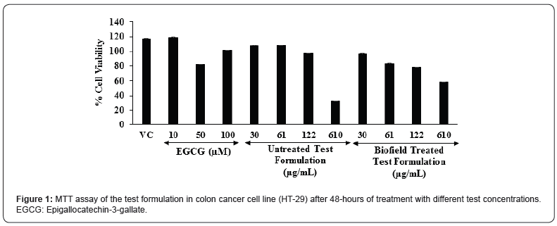
The cell viability results are summarized in the Figure 1. The percent cell viability in the vehicle control (VC) group was found as 117.5%. Moreover, the positive control, epigallocatechin-3- gallate (EGCG) showed 119.2%, 83%, and 101.2% cell viability at the concentration of 10, 50, and 100 μM, respectively. Further, the untreated test formulation showed more than 97% cell viability upto 122 μM; while the Biofield Treated test formulation showed more than 78% cell viability upto 122 μM. Based on the MTT cell viability assay the test formulation was found as safe and nontoxic upto the concentration of 122 μM. MTT assay is widely used for the cell toxicity against any test formulations. In addition, this assay was found as more rapid, less costly, less time consuming, and non-radioactive method as compared with the other assays. This assay display cell proliferation results on the basis of the cell growth and metabolic activity [41]. MTT assay suggest that the concentrations of the test formulation were found safe up to 122 μg/mL with respect to the viability in the colon cancer cell line (HT-29).

Estimation of Interleukin-6 (IL-6) Expression

The level of interleukin-6 (IL-6) expression in colon cancer (HT-29) cells is represented in the Figure 2. The positive control, epigallocatechin-3-gallate (EGCG) was significantly reduced the level of IL-6 by 38.71%, 92.43%, 91.74%, and 98.20% (p≤0.001) at the concentrations of 1, 10, 50, and 100 μM, respectively as compared to the vehicle control (VC) group. Moreover, the untreated test formulation showed significant reduction of IL-6 by 11.96%, 35.94%, and 52.3% (p≤0.001) at 0.1, 3, and 15 μM, respectively as compared to the VC group. Further, the Biofield Energy Treated test formulation group showed 14.19%, 34.09%, and 59.41% (p≤0.001) reduction of IL-6 at 0.1, 3, and 15 μM, respectively as compared to the VC group under the stimulation of TNF-α stimulation. Besides, the Biofield Treated test formulation also significantly reduced the level of IL-6 by 14.87% as compared to the untreated test formulation group. Overall, the minerals and vitamin-based Biofield Energy Treated test formulation showed an anti-inflammatory activity by reducing the level of IL-6 under the stimulation of TNF-α as compared with the vehicle control as well as untreated test formulation groups. Hence, the Biofield Energy Treated test formulation could be used a major role in immune-related disorders and also defined as controlling factor for many diseases [42]. Thus, it can be suggested that the Biofield Energy Treated test formulation can be used in many inflammatory disorders.

Estimation of IL-8 Expression

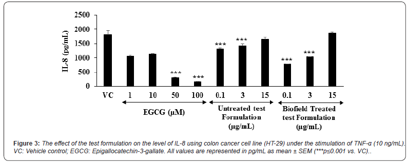
The level of interleukin-8 (IL-8) expression in colon cancer (HT-29) cells is represented in the Figure 3. The positive control, epigallocatechin-3-gallate (EGCG) was significantly reduced the level of IL-8 by 41.49%, 36.77%, 82.89% (p≤0.001), and 91.21% (p≤0.001) at the concentrations of 1,10,50, and 100 μM, respectively as compared to the vehicle control (VC) group. Moreover, the untreated test formulation showed significant reduction of IL-6 by 27.34% (p≤0.001), 21.03% ( ≤0.001), and 9.08% at 0.1, 3, and 15 μM, respectively as compared to the VC group. Further, the Biofield Energy Treated test formulation group showed significant (p≤0.001) reduction of IL-8 by 57.09% and 42.88% at 0.1 and 3 μM, respectively as compared to the VC group under TNF-α stimulation. Besides, the Biofield Treated test formulation also significantly reduced the level of IL-8 by 40.94% and 38.25% at 0.1 and 3 μg/mL, respectively as compared to the untreated test formulation group. Overall, the minerals and vitamin-based Biofield Energy Treated test formulation showed an anti-inflammatory activity by reducing the level of IL-8 under the stimulation of TNF-α as compared with the vehicle control as well as untreated test formulation group. Chronic inflammatory conditions leads to the massive production of proinflammatory factors such as chemokines. IL-8 is one of the chemokine in chronic inflammation and it is initially act as a neutrophil chemotactic and activating factor [43,44]. Overall, the experimental data suggested that Biofield Energy Healing Treatment has the significant capacity to reduce the level of IL-8 with respect to vehicle control and untreated test formulation.

Estimation of IFN-γ Expression

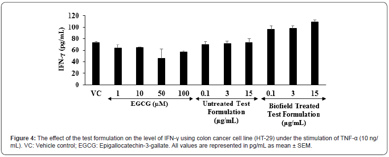
The level of interferon gamma (IFN-γ) expression in colon cancer (HT-29) cells is shown in the Figure 4. The positive control, epigallocatechin-3-gallate (EGCG) was significantly reduced the level of IFN-γ by 13.64%, 12.64%, 37.64%, and 22.99% at the concentrations of 1, 10, 50, and 100 μM, respectively as compared to the vehicle control (VC) group. Moreover, the untreated test formulation showed significant reduction of IFN-γ by 4.55% and 2.78% at 0.1 and 3 μM, respectively as compared to the VC group. Further, the Biofield Energy Treated test formulation significantly altered the level of IFN-γ as compared to the VC group under TNF-α stimulation. This suggests that the Biofield Energy Treated test formulation has significant immunomodulatory activity. Scientific reports suggest that various immunological and inflammatory functions of chemokines play significant role in controlling the immune response during infections. Overall, the immunomodulatory effect might be the result of specific modulation of NF-κB, a transcription factor involved in the activation of many inflammatory mediator genes [45,46].

Worldwide scope of alternative medicine and its outcomes have been increased significantly. However, an important phytoconstituents along with minerals and vitamins are reported to have beneficial role against many diseases such as diabetes, indigestion, inflammation of intestine, osteomalacia, blood disorders, infertility, potent revitalizer, etc. [47]. Due to high safety profile with the wide therapeutic action of alternative medicines, the scope has been increased worldwide [48]. Besides, the individual constituents of the novel proprietary test formulation has been reported to have substantial immunomodulatory action, and Biofield Energy Healing Treatment significantly alters the action of cytokines. Overall, the Biofield Energy Healing Treatment on the test formulation can be a novel approach in supports of the use of Biofield Treated test formulation for various types of autoimmune disorders in colon cancer cell line (HT-29).

Conclusion

On the basis of current study findings, it is concluded that the novel proprietary test formulation showed significant antiinflammatory action on the tested cytokines (IL-6, IL-8, and IFN-γ) in colon cancer cell line (HT-29) after administration of the Biofield Energy Treated formulation. MTT assay in the Biofield Energy Treated colon cancer cell line (HT-29) suggest that the test formulation showed more than 78% cell viability and found as safe and non-toxic. In addition, the levels of cytokine, interleukin-6 (IL-6) was significantly reduced by 34.09% and 59.41% (p≤0.001) at 3 and 15 μM, respectively as compared to the vehicle control (VC) group. Moreover, IL-8 level was reported to be significantly suppressed in the Biofield Energy Treated test formulation by 57.09% and 42.88% at 0.1 and 3 μM, respectively as compared with the VC group. On the basis of experimental results of various tested cytokines and their expression, significant anti-inflammatory activity in colon cancer cells was reported in the new test formulation after treated with the Trivedi Effect®- Biofield Energy Healing. Biofield Energy Treated test formulation can be used as a Complementary and Alternative Medicine (CAM) to prevent the immune-mediated diseases such as Irritable Bowel Syndrome, Rheumatoid arthritis, Ulcerative colitis and Crohn’s disease, Stress, Asthma, and many more with safe therapeutic index. Besides, it can also be utilized in organ transplants (for example kidney transplants, liver transplants and heart transplants), various autoimmune disorders such as Lupus, Addison Disease, Celiac Disease (gluten-sensitive enteropathy), Dermatomyositis, Graves’ Disease, Hashimoto Thyroiditis, Multiple Sclerosis, Myasthenia Gravis, Pernicious Anemia, Aplastic Anemia, Sjogren Syndrome, Systemic Lupus Erythematosus, Diabetes, Alopecia Areata, Fibromyalgia, Vitiligo, Psoriasis, Scleroderma, Chronic Fatigue Syndrome and Vasculitis, Type 1 to improve the overall health and quality of life.

To Know more about Pharmacology & Clinical Research


To Know more about our Juniper Publishers

Click here: https://juniperpublishers.com/index.php   

Thursday, August 26, 2021

Insulin Resistance in Hemodialysis Patients: How to Identify? - Juniper Publishers

 Urology & Nephrology - Juniper Publishers

Abstract

Insulin resistance is one of the common signs of end-stage renal failure. The onset of hemodialysis is accompanied by an increase in the sensitivity of peripheral tissues to insulin, but it does not eliminate insulin resistance. Clamp test in dialysis patients is difficult to apply. There is currently no consensus on how to replace it. It has been suggested that it is advisable to use a combination of HOMA-IR-1, HOMA2% B, Quantitative insulin sensitivity check index (QUICKI).

Opinion

Insulin resistance is identified as an impaired biologic response to insulin stimulation of target tissues (liver, muscle, and adipose tissue), resulting in a compensatory increase in beta-cell insulin production and hyperinsulinemia [1]. Over the past 20 years, the number of publications dealing with insulin resistance itself has sharply decreased. The emphasis shifted to metabolic syndrome. However, at present, there is no single understanding of metabolic syndrome. Some authors consider insulin resistance to be the trigger, while others consider central obesity. The gold standard for diagnosing insulin resistance is the euglycemic clamp test [2]. However, the technical features of its implementation do not allow its widespread use in hemodialysis patients.

However, the technical features of its implementation do not allow its widespread use in hemodialysis patients. It is proposed to use various indices as surrogate methods for studying insulin resistance: the Caro index [3], Fasting insulin resistance index [4], Fasting glucose-insulin ratio [5], Homeostasis Model Assessment of Insulin Resistance (HOMA-IR-1 and 2) [6,7], Quantitative insulin sensitivity check index (QUICKI) [8].

We conducted a comparative analysis of the use of these methods on a small group of 140 stable dialysis patients without diabetes and a fasting glucose level of not higher than 6 mmol / L. We concluded that for the diagnosis of insulin resistance, it is not enough to use any one formula. In our opinion, the optimal combination is the determination of the HOMA-IR-1 index, pancreatic β-beta cell activity index (HOMA2% B), and peripheral tissue insulin sensitivity index (QUICKI). The HOMA-IR-1 index was higher than 2.7 in 74 patients, the QUICKI index was less than 0.339 in 89 patients, the HOMA2% B index was more than 100% in 91 patients. Thus, in most clinically stable hemodialysis patients there is an increase in the activity of pancreatic β-beta cells and a decrease in the sensitivity of peripheral tissues to insulin. The HOMA-IR-1 index has been shown to be less sensitive. We believe that insulin resistance in dialysis patients develops in several stages: a decrease in the sensitivity of peripheral tissues to insulin - an increase in the activity of pancreatic β-cells in the pancreas - the development of true insulin resistance. The question of whether insulin resistance can be transformed into type 2 diabetes remains open. We believe that the cohort of patients on the kidney transplant waiting list is the most vulnerable in this regard. They are likely the risk group for the formation of steroid diabetes. However, this assumption needs to be clarified in the course of further research.

To Know more about Urology & Nephrology


To Know more about our Juniper Publishers

Click here: https://juniperpublishers.com/index.php   


Wednesday, August 25, 2021

Development and Validation of a Novel Analytical Method for the Determination of Dissolution Rate of Acetylsalicylic Acid in Enteric-Coated Tablet Dosage Form by Liquid Chromatography - Juniper Publishers

 Pharmacy & Pharmaceutical Sciences - Juniper Publishers 

Abstract

Objective: The aim of this study is to determine the applicability of UV detection method by the sensitive, simply, precise, specific, and low-cost High-Performance Liquid Chromatography (UHPLC) for acetylsalicylic acid in presence of its degradation product.

Methods: Method development and determination of dissolution rate for acetylsalicylic acid, were achieved by using reversed-phase liquid chromatography with PDA (Photodiode Array Detector) and TUV (Tunable UV Detector) detection by HCLASS-UHPLC. The new proposed method utilized by the Waters Acquity UHPLC® TUV and PDA systems using a UHPLC column Waters Acquity, BEH, C18, 2.1mm x 50mm, 1.7μm particle size with a mixture of heptane-1-sulphonic acid sodium salt solution which was prepared in acetonitrile: deionized water (15%: 85% v/v) in isocritic mode at a flow rate of 0.5ml/ min, at 25°C with a load of 5μL. The detection for all eluted compounds was carried out at 280nm.

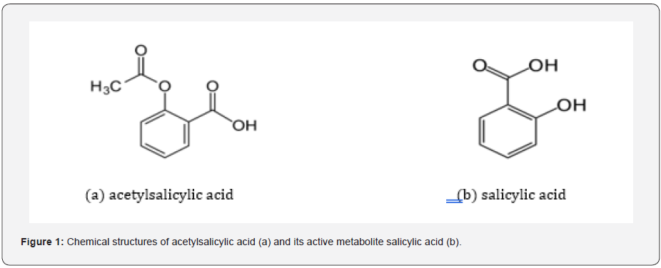
Results: The amount of acetylsalicylic acid’s active metabolite [1] salicylic acid (Figure 1) can also be determined in the same chromatographic conditions and resolution between Acetylsalicylic acid and salicylic acid is more than 2.0. The chromatographic parameters such as retention times, resolution, peak symmetry and theoretical plate count were determined. The validation studies of the developed method were conducted as to be perform the parameters specificity (resolution; 4.0, purity angle < purity threshold), linearity (r > 0.99), system precision, (RSD: 0.06% - 0.12%), repeatability (RSD: 1.69%), intermediate precision (RSD: 0.94%), accuracy and robustness (96.6% - 98.% for Acetylsalicylic Acid, 95.6% - 98.9% for Salicylic Acid) specified in the ICH (The International Conference on Harmonization) Q2(R1) [2]. Guideline and the results meet the acceptance criteria [3].

Conclusion: The developed method proved that the results obtained from the determination of dissolution rate in enteric-coated tablets containing acetylsalicylic acid are repeatable and sensitive. Also found suitable for the analysis of the stability samples of the drug product.

Keywords: Acetylsalicylic acid development; Dissolution; LC; Validation

Abbreviations: USP: United States Pharmacopoeia; LC: Liquid Chromatography; ACN: Acetonitrile; QSM: Quaternary Solvent Manager

Introduction

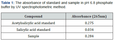
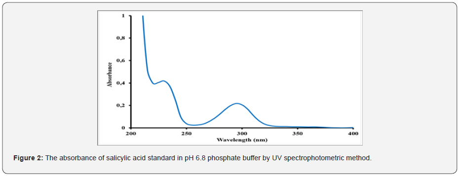
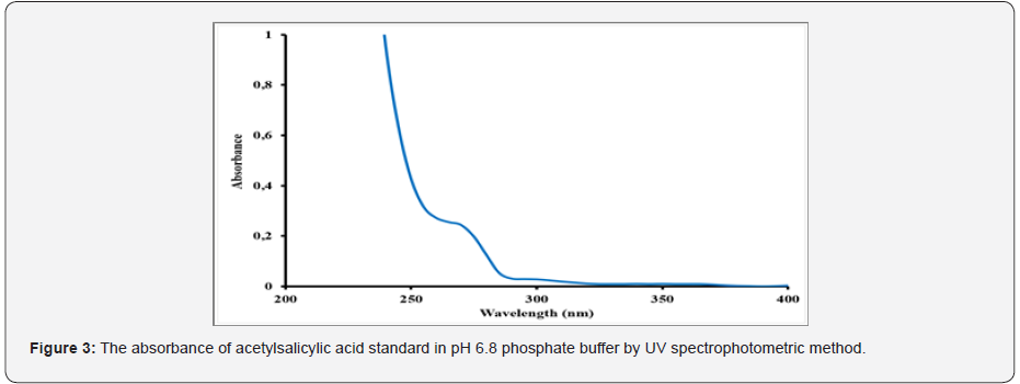
Acetylsalicylic acid, also known as an active substance of aspirin, is used an antipyretic analgesic for over a century. This widely known and accepted medicine can be used to treat wide range of diseases such as colds, fever, headaches, toothache, joint pain, and rheumatism. Aspirin can also inhibit platelet aggregation, prevent, and treat ischemic heart disease, cardiopulmonary infarction, cerebral thrombosis, and other diseases [4]. Moreover, it has shown that acetylsalicylic acid has the anti-cancer effect [5]. The US working group for preventive services has issued a guide that acetylsalicylic acid can be used as the primary prevention of cardiovascular disease and colorectal cancer. The guideline proposed for the first time that high-risk groups of non- colorectal cancer could use acetylsalicylic acid for the primary prevention of colorectal cancer [6]. It indicated that taking a certain dose of acetylsalicylic acid daily might effectively block the growth of breast cancer [7]. Additional studies are investigating the potential of acetylsalicylic acid to boost the immune system, treat cognitive decline and lower the risk of colon and ovarian cancer. A low daily dose, 75-81mg, is generally used in the preventive dose of acetylsalicylic acid therapy. Commonly, acetylsalicylic acid has been regarded as a potential gastric irritant [8] and studies have shown that the occurrence of gastric intestinal side effects might increase with regular use of the drug [9]. Enteric coating of the tablets is helpful for preventing stomach upset or irritation in those taking the daily dose of acetylsalicylic acid treatment. Due to the moisture-sensitive nature of aspirin drug, it can hydrolyze into acetic and salicylic acids when it is contacted to air with high humidity and elevated temperatures [10]. As the coating process will expose acetylsalicylic acid tablets to both high temperatures and humidity, it is significant that the formulation is resistant to moisture interaction during this process. The ideal enteric-coated tablets need to show perfect stability under accelerated storage conditions without the use of extra (and more expensive) packaging precautions such as desiccant packages or other specialized packaging materials [11]. Monographs of different forms of the official aspirin are available in USP, Polish Pharmacopoeia V, British Pharmacopoeia [12,13]. There are several analytical methods to assess aspirin product quality and batch to batch consistency of the production. One of the main methods is a dissolution performance test and salicylic acid determination. Dissolution test is used to obtain dissolution time and amount of dissolved active substances. Salicylic acid is an active metabolite of acetylsalicylic acid. The maximum allowed percentage for free salicylic acid in Aspirin Extended-Release Tablets and Aspirin Delayed-Release Tablets are 3% and in ‘Aspirin Effervescent Tablets for Oral Solutions’ is 8% [12]. Different methods have been reported for the determination of aspirin and salicylic acid [12-17] and USP 23 procedure includes HPLC analysis of the free salicylic acid. American Pharmacopoeia (USP) is often used as a reference in finished product testing applications. The specific dissolution technique employed is determined by the dosage form characteristics and the intended route of administration. For solid dosage forms, industry standard dissolution testing methodologies are the United States Pharmacopoeia (USP) Apparatus 1 (basket) and the USP Apparatus 2 (paddle). Floating capsules and tablets generally use USP 1 baskets [18]. According to monograph in the US Pharmacopoeia 39 (USP 39) [19] regarding tablet form containing the acetylsalicylic acid active substance, the method of dissolution rate determination is the UV spectrophotometric method. It was defined in the dissolution rate studies conducted according to this method that acetylsalicylic acid turns into its active metabolite salicylic acid in pH 6.8 phosphate buffer, Stage 2 and it could not be detected separately by applying UV spectrophotometric method (Table 1) and (Figure 2-4). The simultaneous evaluation of dissolution test of active substance acetylsalicylic acid and active metabolite salicylic acid is not possible by direct UV spectrophotometry. Therefore, it was decided to develop a more specific method by applying Liquid chromatography (LC) to determine both acetylsalicylic acid and its active metabolite salicylic acid in a short time. In this study, a new liquid chromatography (LC) method, which is more selective than UV spectrophotometric method used in determining the dissolution rate of enteric coated tablets containing acetylsalicylic acid was developed and validation study was performed. The significant number of method trials are performed to obtain the best results. The duration of the analysis was shortened but it was decided to switch to the faster response HPLC system to prevent the degradation of acetylsalicylic acid.

Material Science
Material Science
Material Science
Material Science
Material Science

Experimental

Chemicals and reagents

The chemicals that are used in the study were of analytical reagent grade. Acetonitrile (ACN), heptane-1 sulfonic acid sodium salt (C₇H₁₅NaO₃S), hydrochloric acid (HCl), methanol (MeOH), potassium dihydrogen phosphate (KH2PO4), sodium hydroxide (NaOH), the acetic acid anhydrous (CH3COOH) and phosphoric acid (H3PO4) were purchased from Merck (Darmstadt, Germany) and JT Baker (Center Valley, USA). Deionized and distilled water was used from PURELAB® Ultra Elga DV35 (High Wycombe, United Kingdom). Standards for acetylsalicylic acid [2-(acetyloxy) benzoic acid] was supplied by the Shandong Xinhua Pharmaceutical (P.R. China) and salicylic acid [2-Hydroxybenzoic acid] was supplied by the VWR Chemicals (United Kingdom).

Instrumentation

The HCLASS-UHPLC analysis was performed on both the Waters Acquity with column thermostat and the TUV and Waters Acquity PDA detector with column thermostat (Waters corp., Milford, MA, USA). ACQUITY H-Class System (Waters, Milford, USA) we used to consist of a Quaternary Solvent Manager (QSM), a Sample Manager with Flow Through Needle (SM-FTN) design. The system consisted of the data that were collected and evaluated by using Waters Empower 2 software. To separate acetylsalicylic acid and its active metabolite salicylic acid, a UHPLC column of Waters Acquity was used with BEH, C18, 2.1mm x 50mm with 1.7μm particle size, which was purchased from Waters (Torrance, CA, USA). The column temperature was set as 25οC. The injection port temperature was set at 25oC and the injection volume was 5μl. The total analysis time was 2.5 minutes. All the data for acetylsalicylic acid and its active metabolite salicylic acid were monitored at 280nm.

Samples

It was used Enteric Coated Tablets and its placebo were produced by Abdi İbrahim, Research and Development Center Department (Istanbul, Turkey).

Mobile phase

The mobile phase was prepared by Heptane-1-sulfonic acid sodium salt solution in Acetonitrile: Deionized water (15%:85% v/v) mixture. It was filtered through a 0.45μm membrane filter (Millipore, MA, USA) and degassed by sonication. Acetylsalicylic acid eluted at 1.5 minute in these conditions.

Standard preparations

Stage 1 (0.1 N HCl): A stock solution of acetylsalicylic acid (2.7mg mL-1) and salicylic acid (0.081mg mL-1) were prepared by dissolving an appropriate amount in stage 1 (0.1 N HCl). Working solutions containing 0.081mg mL-1 acetylsalicylic acid and 0.00243mg mL-1 salicylic acid were prepared from this stock solution. It was then filtered through a 0.45μm RC filter and transferred to an HPLC vial.

Stage 2 (pH 6.8 phosphate buffer): A stock solution of acetylsalicylic acid (0.081mg mL-1) and salicylic acid (0.00243mg mL-1) were prepared by dissolving an appropriate amount in stage 2 (pH 6.8 Phosphate Buffer). Working solutions containing 0.041mg mL-1 acetylsalicylic acid and 0.00122mg mL-1 salicylic acid were prepared from 0.1 N HCl. It was then filtered through a 0.45μm RC filter and transferred to an HPLC vial.

Sample preparations

Stage 1 (0.1 N HCI): Place 1000ml dissolution medium into each 6 vessel and warm up to 37οC ± 0.5οC. Drop one Tablet into each vessel and start the analysis at 100rpm paddle speed. At the end of 120 minutes taken sample solution from each vessel. It was then filtered through a 0.45μm RC filter and transferred to an HPLC vial. The drug substances in the final concentrations was 0.081mg mL-1 of acetylsalicylic acid in sample.

Stage 2 (pH 6.8 phosphate buffer): Place 1000ml dissolution medium into each 6 vessel and warm up to 37οC ± 0.5οC. Drop Tablets taken at the end of Stage 1 into each vessel and start the analysis at 100rpm paddle speed. At the end of 90 minutes transfer 5 ml of the solution from each vessel into 10ml volumetric flasks. Dissolve and dilute to volume with 0.1 N HCl. It was then filtered through a 0.45μm RC filter and transferred to an HPLC vial. The drug substances in the final concentrations was 0.041mg mL-1 of acetylsalicylic acid in sample.

Placebo solution

Stage 1 (0.1 N HCI): Place 1000ml dissolution medium into a vessel and warm up to 37 οC ± 0.5οC. An amount of 24.3mg placebo was weighed into a 1000mL dissolution vessel and start the analysis at 100rpm paddle speed. Then, the placebo solution was prepared in the same way as the sample solution.

Stage 2 (pH 6.8 phosphate buffer): Place 1000ml dissolution medium into a vessel and warm up to 37οC ± 0.5οC. An amount of 24.3mg placebo was weighed into a 1000mL dissolution vessel and start the analysis at 100rpm paddle speed. Then, the placebo solution was prepared in the same way as the sample solution.

Results and Discussion

Method optimization

The main purpose of the chromatographic method is the determination of Acetylsalicylic acid and active metabolite salicylic acid, along with maximum selectivity, resolution, retention properties, peak shape and convenient analysis time for the novel analytical method of the enteric-coated tablet dosage form by liquid chromatography according to the ICH guideline 2005 [2]. Different chromatographic conditions were utilized to achieve better efficiency of the chromatographic system. The mobile phase composition, column, detection wavelength, and column temperature were optimized by making necessary changes in the chromatographic conditions to obtain a well-established method. Several proportions of solvents and buffers were assessed to find a compatible composition of mobile phases. The development of the method was initiated by using the USP assay method that could separate Acetylsalicylic acid and active metabolite salicylic acid. This method includes parameters such as an isocratic system, mobile phase consisting of Heptane-1-sulfonic acid sodium salt solution in acetonitrile: deionized water (15%:85% v/v) mixture, the column with Phenomenex Gemini C18, 250mm x 4.6mm, 10μ and heated to 25oC at 280nm wavelength.

Development of the method

Acetylsalicylic acid and active metabolite salicylic acid show the same absorbance with UV spectrophotometric method. Therefore, it was decided to use and develop a more selective HPLC method to measure the amount of acetylsalicylic acid dissolved in enteric-coated tablets containing acetylsalicylic acid. First, dissolution analysis was performed using the assay method conditions in the USP monograph. Due to the conversion of acetylsalicylic acid into active metabolite salicylic acid after 12 minutes of analysis, column size was changed and Phenomenex Gemini, C18, 110A, 4.6 x 150mm, 5μm column was used instead of Phenomenex Gemini C18, 110A, 4.6 x 250mm, 5μm column. In this study, the duration of the analysis was shortened but it was decided to switch to the faster-responsive H-Class system to prevent the degradation of acetylsalicylic acid. As a result of the trial with column Waters, BEH, C18, 2.1 x 50mm with 1.7μm) analysis time decreased to 2.5 minutes and the conversion of acetylsalicylic acid into salicylic acid was prevented. However, the peak separation obtained from the standard injection which was prepared at 0.1N HCl and given to the system for stage 1 was convenient but the peaks obtained from the standard injection which were prepared at pH 6.8 phosphate buffer and given to the system for stage 1 were not convenient. For this reason, organic solvents ratios used in standard solutions have been changed but successful results have not been obtained in this study. Changes in the flow rate have been made by trying different columns. Despite these trials, appropriate peaks were not obtained. So, standard and sample solution prepared at pH 6.8 phosphate buffer at Stage 2 was diluted in an acid medium (0.1N HCl) and injected into the system. As a result of these studies, appropriate peaks were obtained.

Validation of the method

Specificity

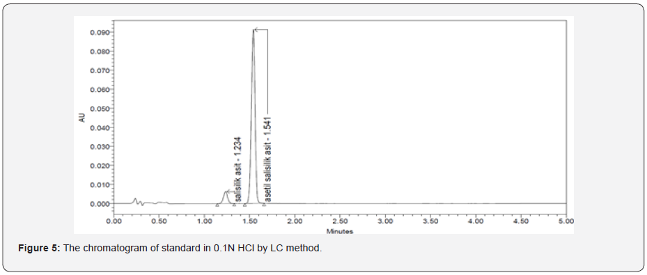
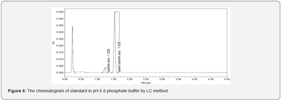
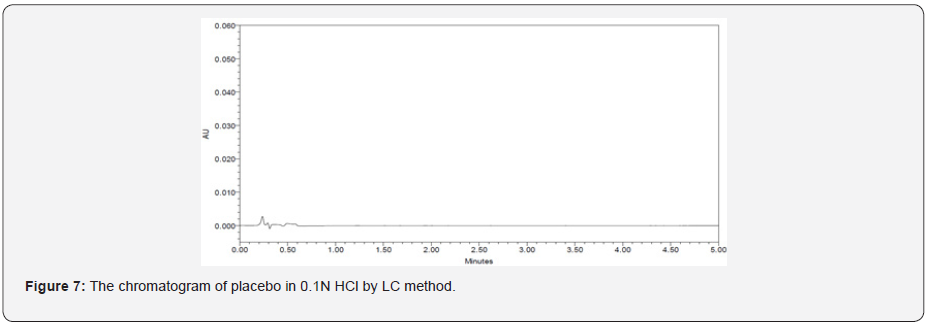
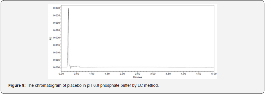
The specificity of a method is its suitability for analysis of a substance in the presence of potential impurities. It can also be used to validate the stability-indicating power of the analytical procedures used. The specificity of the LC method for acetylsalicylic acid has been determined in the presence of salicylic acid impurity (Figure 5 & 6). There was no peak found at the retention time of acetylsalicylic acid and salicylic acid active metabolite in placebo chromatograms proves no interference from placebo (Figure 7 & 8) These results confirmed the specificity of the method (Table 2 & 3).

Material Science
Material Science
Material Science
Material Science

Linearity

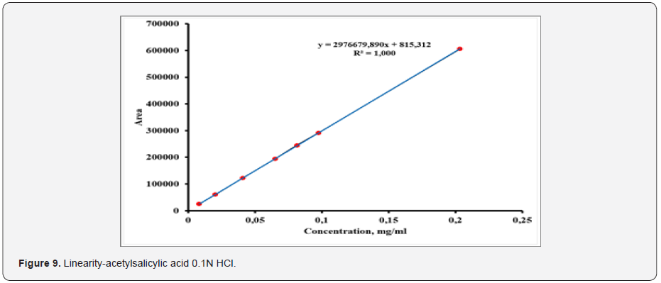
The linearity of the analytical procedure is its ability (within given range) to obtain test results which are directly proportional to the concentration in the sample. The linearity solutions were performed at concentrations levels; 0.0081mg mL-1 - 0.2025mg ml solution of acetylsalicylic acid and 0.0000243mg mL-1 0.00608mg mL-1 solution of salicylic acid corresponding to about 10% to 250%. A linear relationship was established by plotting the peak areas (on Y-axis) against the drug concentration in mg mL-1 (on x-axis). The data obtained in linearity experiments was subject to linear regression analysis. The coefficient of regression (R) was found to be 0.999 for acetylsalicylic acid and salicylic acid. The linearity of the method is proven (Figure 9 & 10).

Material Science
Material Science

Precision

The precision of the method verified by system precision, repeatability and by intermediate precision. System precision was checked by Waters Acquity H-Class system with UV detector, Milford, USA) injecting six preparations of standard solution. Repeatability was checked by (Waters Acquity H-Class system with UV detector, Milford, USA) injecting six individual preparations of the sample solution. The intermediate precision of the method was also evaluated using different analyst and different instrument (Waters Acquity H-Class system with UV detector, Milford, USA), and performing the analysis on different days. % RSD of the area for each result was calculated for both precision as well as intermediate precision and was found within 5%. System precision (RSD: 0.06% - 0.12%), repeatability (RSD:1.69%) intermediate precision (RSD: 0.94%).

Accuracy

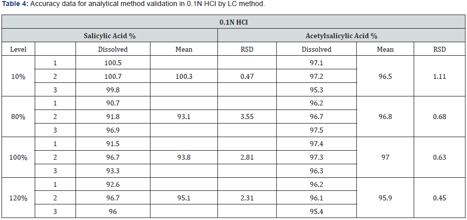
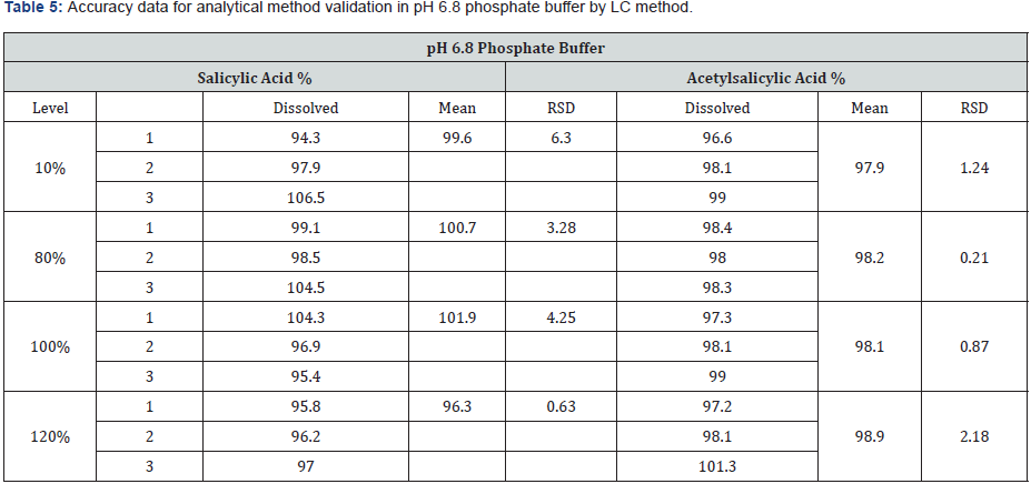
The accuracy was determined by preparation of three replicates of spiked sample solutions for each concentration level. The accuracy of the method was demonstrated. Recoveries for each level were found within the acceptance criteria. (95.3- 97.5%) for stage 1 (0.1 N HCl) and (96.6-101.3%) for stage 2 (pH 6.8 phosphate buffer) The mean recovery of acetylsalicylic acid enteric coated tablet should not be less than 95% and not more than 105%. The results from recovery data are depicted in the (Table 4 & 5).

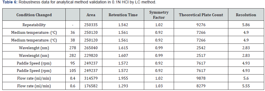
Robustness

To determine the robustness of the method the experimental conditions were deliberately changed, and the resolution of acetylsalicylic acid and salicylic acid were evaluated. To study the effect of flow rate on the resolution it was changed to 0.4 and 0.6- mL min-¹. The effect of paddle speed was studied at 95rpm and 105rpm. The effect of temperature of dissolution medium was studied at 36οC and 38οC. The effect of wavelength was studied at 278nm and 282nm. Under the different chromatographic conditions, the robustness of the method was proved. The results from the robustness study are depicted in (Table 6 & 7).

Material Science
Material Science

Stability in solution

Standard and sample solutions were used for the stability study. No significant changes in the amounts of the acetylsalicylic acid and its active metabolite salicylic acid were observed during solution stability experiments when performed using the dissolution method. The standard and sample solution were found to be stable for 6 hours during the determination of acetylsalicylic acid.

Conclusion

It was developed a more specific method by applying liquid chromatography (LC) to determine both acetylsalicylic acid and its active metabolite salicylic acid. The proposed analytical method has been proved to be simple, specific, precise, linear, robust, rugged, and accurate which fulfill all the parameters of the validation. All calculated statistical values, which were obtained throughout the development and validation of the UPLC method, were within acceptable limits. The method was successfully validated according to the current ICH Guidelines. The method is stability-indicating and can be used for routine analysis of production samples and to check the stability of acetylsalicylic acid enteric coated forms.

To Know more about Pharmacy & Pharmaceutical Sciences 


To Know more about our Juniper Publishers

Click here: https://juniperpublishers.com/index.php    
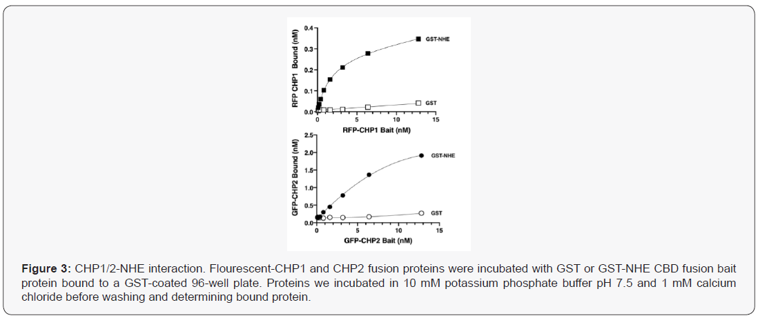

Artificial Intelligence System for Value Added Tax Collection via Self Organizing Map (SOM)- Juniper Publishers

  Forensic Sciences & Criminal Investigation - Juniper Publishers Abstract Findings:  Based on our experiments, our approach is an effec...