Journal of Surgery- Juniper Publishers
Abstract
Tension pneumothorax is an infrequently encountered
clinical condition in the emergency departments. It is a potential
life-threatening condition which can result in cardiopulmonary
deterioration and ultimately cardiac arrest if not promptly diagnosed
and managed. It has various etiologies with blunt chest injuries being
one of common causes. We report a rare case of a large traumatic tension
pneumothorax following a blunt injury which resolved without specific
intervention. The diagnosis was initially missed in the emergency
department and was identified on a retrospective radiology report after 2
days. A repeat CXR has surprisingly shown that the tension pneumothorax
resolved into “simple” pneumothorax without specific intervention.
Interestingly, the patient remained stable all through his hospital stay
for 7 days.
Conclusion: A spontaneous resolution of a
large traumatic tension pneumothorax in a previously healthy and
hemodynamically stable patient can occur. Nevertheless, the exact
mechanism of this observation still remains poorly understood.
Learning points
a. The possibility of the tension pneumothorax
should be considered in blunt chest trauma patients even when the
typical clinical features are not present.
b. A radiologically-diagnosed tension pneumothorax should be promptly managed.
c. An alert system should be activated if critical
radiological findings are detected and an effective response has to be
followed by the trauma management team
Keywords: Tension pneumothorax; Misdiagnosis; Critical radiological finding; Spontaneous resolution
Abbrevations: ATLS: Advanced Trauma Life Support; CXR: Chest X-Rays; ED: Emergency Department, GCS: Glasgow Coma Scale
background
Tension pneumothorax is an infrequently encountered
clinical occurrence with a variable incidence worldwide [1,2]. It merits
special attention as it is a potentially life-threatening condition due
to the progressive hemodynamic instability and cardiopulmonary
deterioration. A high index of clinical suspicion is therefore required
for a prompt diagnosis [1,2]. The recommendations regarding the
management of pneumothoraxes depend on size, underlying etiology, and
clinical stability of the patient [1]. The Advanced Trauma Life Support
(ATLS) guidelines recommend insertion of a chest drain in a patient with
a traumatic pneumothorax to prevent developing a tension pneumothorax
[3]. We report an unusual case of a large traumatic
tension pneumothorax in which the tension pneumothorax resolved
spontaneously without specific intervention. The patient was stable all
through his hospital stay; his oxygen saturation remained above 94% when
breathing room air. To the best of our knowledge this is only the
second reported case in the English medical literature in which
spontaneous resolution of a large traumatic tension pneumothorax
occurred [1].
Case Presentation
A previously healthy 23-year-old non-English speaking
male was brought to our Emergency Department (ED) four hours earlier
following an assault by a group of people. He stated that he had been
hit repeatedly by heavy objects on different parts of his body. He
complained of severe pain in both arms and right leg. There was no
history of loss of consciousness, headache or vomiting. He denied chest
pain or shortness of breath. His past
medical and surgical history was insignificant. He was alert but
in severe pain.
His vital signs on arrival were heart rate 102 beats per minute,
blood pressure 131/87 mmHg, respiratory rate 22 breaths per
minute and an oxygen saturation of 94% when breathing on room
air. His GCS was 15. Multiple superficial abrasions were noted on
his forehead and scalp, anterior aspect of the both forearms and
the right lower leg. Tenderness was elicited over the right lower
leg. Examination of the chest, abdomen and back was reported as
normal by the ED resident. Right leg X-ray showed a right distal
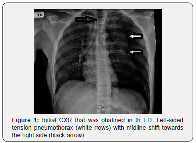
tibial fracture without deformity. His CXR revealed a large tension
left-sided tension pneumothorax (Figure 1).

However, this critical finding was missed by the ED resident
and the patient was admitted for neuro-observation and pain
management. He remained clinically stable with normal neuroobservations.
A retrospective radiology report received 2 days
later depicted those findings which were consistent with a leftsided
tension pneumothorax. On receipt this radiological report a
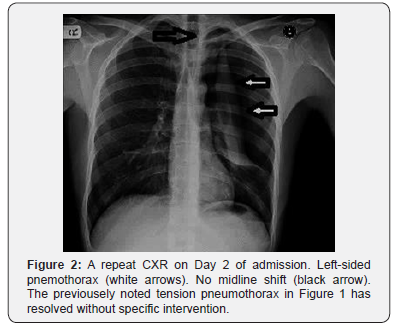
repeat CXR was taken which revealed a left-sided pneumothorax
with no midline mediastinal shift or tension features Figure
2, thus a spontaneous decompression of the previously noted
tension pneumothorax occurred without specific intervention, a
finding that was both a clinical as well as a radiological surprise.
The patient remained vitally stable all through his hospital
stay and his oxygen saturation was constantly maintained above
94% when breathing room air. He never complained of shortness
of breath or chest pain. Following this repeat CXR, size 32F chest
drainage was inserted by the surgical team after informed consent
for the “simple” large left-sided pneumothorax. A follow-up CXR
showed a satisfactory position of the chest drain. He was reviewed
at a Fracture Clinic on Day 5 for further management of the right
tibial fracture. The chest drain remained in-situ for 4 days and
was removed on Day 6 and the final CXR was relevant for a very
small (<5%) pneumothorax. He was discharged home on Day 7 in
a stable condition. The patient didn’t attend scheduled follow-up
visits.

Results
The importance of the prompt clinical diagnosis of tension
pneumothorax has been well-emphasized. The clinical features
suggesting a tension pneumothorax are chest pain and respiratory
distress (90%), tachycardia and ipsilateral decreased air entry
(50-70%), low oxygen saturation, hypotension and contra-lateral
tracheal deviation in less than 25% [2]. Nevertheless, these typical
features are not always present [1,2]. Moreover, these features
are poorly correlated with the diagnosis, and perhaps many
cases were missed before doing chest radiographs [4]. In fact,
there is increasing appreciation that tension pneumothoraces
are generally diagnosed radiologically rather than clinically [4].
We have only identified one case of spontaneous resolution of a
large traumatic tension pneumothorax in the reviewed English
literature [5].
The reported patient was a clinically stable 33-year-old female
who presented to ED following a fall. Her CXR showed a large
right-sided tension pneumothorax and rib fractures, however, the
diagnosis was missed by the ED resident and she was sent home.
The diagnosis was made on a retrospective basis after reviewing
her CXR and all attempts to contact her had failed. However,
she returned to the hospital after 50 days with intractable
vomiting due to methadone withdrawal and an admission CXR
showed a complete resolution of the previously noted tension
pneumothorax [5]. This reported case bears two similarities to
ours as the diagnosis was initially missed by the ED doctor and
the patient remained clinically stable through the course of the
disease. However, we re-evaluated the patient after 2 days and this
explains the resolution of tension pneumothorax with persistence
of pneumothorax, as the presumed re-absorption rate is 1.25%
per day on room air [6], and hence a complete lung re-expansion
will not be expected after 2 days only.
This case demonstrates that tension pneumothorax can
be a
radiological diagnosis in clinically stable patients, nevertheless it
should be managed promptly to prevent grave consequences that would
rapidly occur. An alert system should be activated if critical
radiological findings are detected and an effective response must
be followed by trauma management team.
To know more about Journal of Surgery
Click here: https://juniperpublishers.com/oajs/index.php
To know more about Juniper Publishers
Click here: https://juniperpublishers.com/index.php


No comments:
Post a Comment