Monday, October 16, 2023

Stress Among College Students is Linked to Headache, PCOS, Hypertension, and Depression: A Cross-Sectional Study - Juniper Publishers

 Psychology and Behavioral Science - Juniper Publishers


Abstract

Objective: In the present study, we aimed to find out the prevalence of stress among college students.

Methods: In this descriptive cross-sectional study, a simple random sampling approach was chosen for data collection from different colleges of Jammu & Kashmir (UT). Data analysis was done by using the chi-square test, and the t-test was utilized for discrete and continuous variables respectively. The logistic regression model using odds ratio was utilized to find out the association between the risk variable and the dependent variable.

Results: A total of 1405 college students were included in which stress was found in 60.49% of students with males representing 34% and females 65.88%. Educational stress was found to be the major contributor of stress-type faced by the students with 46.35%. Different disorders were also observed in students including hypertension 2.70%, PCOS (Poly-cystic ovarian syndrome) 2.34%, and depression 2.84%. Stress was found associated with the PCOS with OR: 3.62 [95% CI: 1.4850-8.84, p-value: 0.0047], headache OR: 3.67, hypertension OR: 5.31 [95% CI: 2.06-13.70, p-value: 0.0006] and depression OR: 7.24 [95% CI: 2.56-20.48, p-value: 0.0002].

Conclusion: Educational stress is found to be very common among college students which may alter the prosperous life of students. Female students were seen to be more stressed than male students. Stress has a strong detrimental impact on students’ health because it is considerably linked to conditions including hypertension, PCOS, and depression. Therefore, the educational system must need a better strategy that will ensure a better education system.

Keywords: Stress; Educational stress; Hypertension; PCOS; College students

Abbreviations: PCOS: Poly-cystic ovarian syndrome; SAA): Salivary Amylase Activity; FGD: Functional Gastrointestinal Disorders; IBD: Inflammatory Bowel Disease; PU: Peptic Ulcer; UC: Ulcerative Colitis; CRH: Corticotrophin Releasing Hormone; ACTH: Adrenocorticotropic hormone; OR: Odds Ratio; OECD: Organization for Economic Co-operation and Development

Introduction

In today’s world, many shortcomings need to be eliminated and education is the one that is often seen as a solution to many social problems. Education is an important instrument that has the power to make a person interactive, self-reliant, social, and disciplined. Education broadens the scope of learning by facilitating knowledge absorption and dissemination among people in order to advance society [1]. Every individual is bombarded with a multitude of perceptual information in their day-to-day life activities. Apart from such advantageous features of education, the ability to process information is limited and varies from person to person. These cognitive characteristics have the potential to disrupt students’ lives periodically, which might result in circumstances that resemble stress and anxietylike condition. According to physiological definition, stress may be described as “any form of change that results in bodily, emotional, or psychological strain or pressure”.

It could result from a traumatic, disappointment, or uneasy encounter or sensation [2]. It is not that stress is bad, “Eustress” describes stress as a “positive, motivating, and enhances functioning feature which leads to the excellent performance” while “distress” refers to bad and overwhelming stress full condition which leads to tiredness, despair, and a variety of other illnesses and thus impairs functioning [3]. Albeit intense or transient pressure can be helpful but ongoing pressure, which goes and remains for a more extended period, is very unfavorable to the body and causes hypertension, weight gain, sadness, and even coronary diseases [4]. The effects of anxiety on students’ academic performance were detrimental [5] and besides anxiety, another emotionally uncomfortable disorder that could interfere with a student’s life is depression.

Stress affects students’ life in a variety of ways, including how they perform academically, socially, physically, and emotionally [6]. Exam stress, a lack of interest in attending lectures, full schedules &responsibilities, lack of sleep, an inability to understand the topic, and academic pressure (pressure to achieve high marks and concerns about receiving poor grades) are some examples of stresses faced by students [7]. The cognitive system becomes overburdened under stressful settings, reducing the student’s attention and ability to process the perceived information [8]. Biologically, stress can lead to long-term health complications, chronic illness, and psychiatric conditions such as migraine, PCOS (poly Cystic Ovarian Syndrome), depression, and suicide [9-11].

The Organization for Economic Co-operation and Development (OECD) recently conducted a survey that included 540,000 students aged 15-16 years old from 72 nations which showed that education and academic achievement are major sources of stress for students [5]. Therefore, in the present study, we aim to find out the stress frequency among college students of Jammu division. To our knowledge, this is the first study that included 10 district colleges from our region. The remainder of the paper is laid out as follows: methods utilized in this study are depicted in Section 2, section 3 includes the result of the study, section 4 represents the discussion and the conclusion of the study is present in section 5.

Method

In this present descriptive cross-sectional study, a random sampling approach was chosen for data collection from different colleges in the Jammu division of Jammu & Kashmir (UT). The sample collection was done in two phases i.e., Phase-I (Face-face interview) and Phase-II (E-based sampling).

Sample Collection

A targeted survey was taken in the colleges of the Jammu division to gather data from the different college students. The information was gathered using two different phases, including face-to-face interviews (Phase-I) conducted by an experienced interviewer/ trained interviewer and online sampling/ E-Sampling using a google form (Phase-II). The questionnaire/ Google form was disseminated through emails and social networking sites. The present study design was duly approved by Animal and Human Experimentation Ethical Committee (AHEEC), University of Jammu vide notification number EC: DRS/22/4969.

Filtering and Data Cleaning

A large data set was obtained using the E-based sampling approach, and the data was cleaned and filtered using a variety of exclusion criteria to decrease redundancy and bias. Exclusion criteria include “students that did not belong from the district of Jammu division”, “non-college students”, “who don’t permit for use of their data (incomplete consent)”, and also the exclusion of subject data “who gave the partial information”.

Data analysis and statistics

For the descriptive data analysis, mean with Standard deviation and frequency distribution were used for the continuous and discrete variables respectively. The chi-square test and the t-test were utilized for the discrete and continuous variables, respectively, in the inferential statistics. The logistic regression model utilizing the odds ratio (OR) was used to determine whether the risk variable and the dependent variable i.e., diseases were associated or not. Online free statistical tools/calculators such as MedCalc’s Odds ratio calculator for calculation of Odds ratio, t-test were calculated by graph-pad Home-GraphPad and for chi-square statistics Social Science Statistics (socscistatistics.com) were used to draw out the inference.

Result

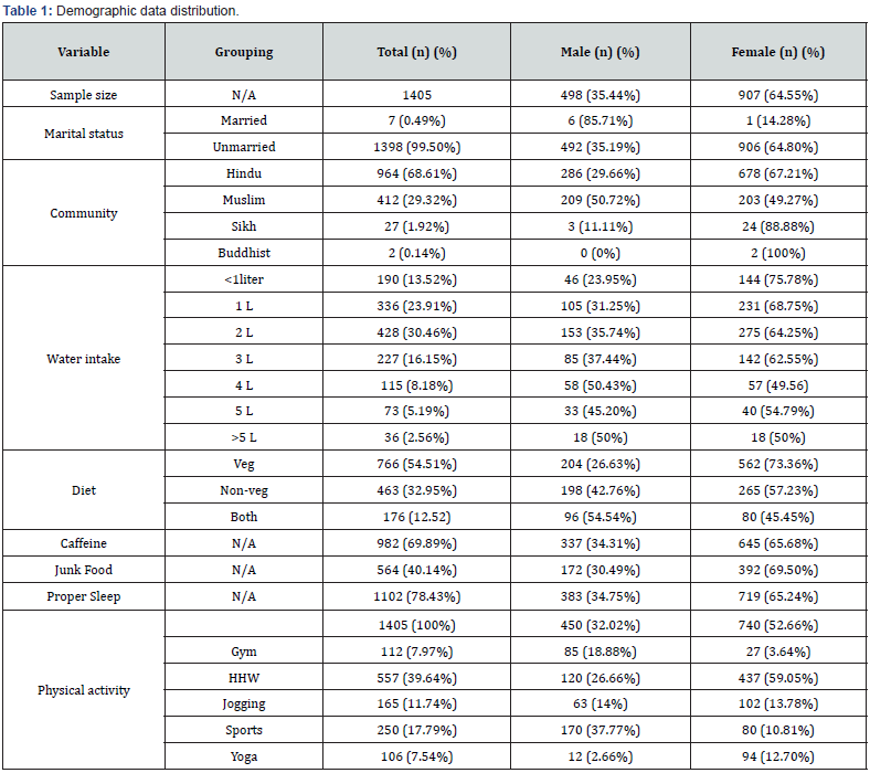
A total of 1405 college students were included with a mean age of 19.64±1.32, including 498 males (19.46±1.32) and 907 females (19.74±1.31). We found a highly statistical difference between the age of male and female students among college students (t-test: 3.8220: p-value: 0.0001). The majority of students were belonging to the Hindu community (n=964) in contrast to Muslims (n=412) and with minorities including Sikhs and Buddhists n=27 and n=2 respectively (Table 1) (Figure 2A). Regarding the martial estimates, the frequency of married students was not quite high (0.49%) (Table 1). Lifestyle activity of students has been also observed where it is found the lower frequency of alcohol usage 1.35% (n=19/1405) in which the male participants represent the dominancy 72.68% (n=14/19) over the female 26.31% (n=5/19).

Also, the increased smoking habit was observed in males 71.42% (n=15/21) in contrast to female students 28.57% (n=6/21). Around 1190 students (84.69%) were observed to do physical activities including n=450 males (37.81%) and n=740 female (62.18%) (Figure 2E) (Table 1). Regarding the dietary pattern, it was observed that 32.95% (n=463) were non-vegetarian (n=766) were vegetarian and 12.52% (n=176) take both (veg % and non veg.) and with respect to the caffeine 69.89% (n=982) were taken caffeine out of which32.29% (n=327) were occasional (Figure 2B) (Table 1). Stress was found in 60.49% students (n=850/1405) with the male representing 34% (n=290/850) and female 65.88% (n=560/850). We observed statistically significant difference (t-test = 3.3926, p-value: 0.0007) between the age difference between stressed males (19.46±1.33) and females (19.78±1.29).

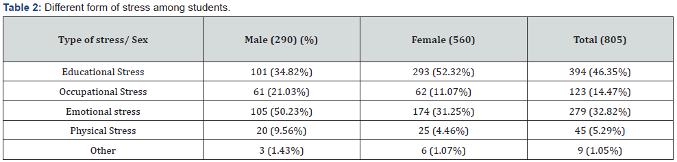
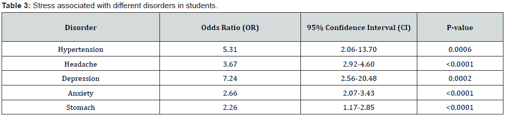
The frequency of the different types of stress was observed (Table 2) and the educational stress was at the top representing 46.35% including 34.82% (n=101/290) and females 52.32% (n=293/560) (Figure 2F). Different disorders were observed in students which include hypertension 2.70% (n=38/1405), PCOS (Poly-cystic ovarian syndrome) 2.34% (n=33/1405), and depression 2.84% (n=40/1045). To find out the risk attribute associated the condition Odds ratio (OR) were utilized. Stress was found associated with the headache OR: 3.67,95% CI: 2.92-4.60, (p value: <0.0001), PCOS with OR: 3.62 [95% CI: 1.4850-8.84, p-value: 0.0047], hypertension OR: 5.31 [95% CI: 2.06-13.70, p-value: 0.0006] and depression OR: 7.24 [95% CI: 2.56-20.48, p-value: 0.0002]. Also, caffeine risk on hypertension and PCOS were also observed including OR: 5.24 [95% CI: 1.60-17.17, p-value: 0.0061] and OR: 3.12 [95% CI: 1.13-9.34, p-value: 0.027] respectively.
We also observed the bidirectional association of headache and hypertension and found a significant increase of risk i.e., headache to hypertension OR: 14.65, 95% CI: [6.59-32.57], (p-value: 0.0001) and hypertension to headache OR: 3.61, 95% CI: [1.64-7.94] (p-value: 0.0014). Stress was also found significantly (p value <0.0001) associated with the risk of anxiety (OR: 2.66, 95% CI: [2.07-3.43] and risk of stomachache (OR: 2.26, 95% CI: [1.17-2.85]) (Table 3). Educational stress was found significant (p value: <0.0001) responsible for increasing the likely hood of headache by 3.8-fold (OR:3.84, 95% CI: 2.92-5.05). Emotional stress was found to be a high-risk variable associated with hypertension with an OR: 2.74 [95% CI: 1.35-5.54, p-value: 0.0050]. Also, the association between emotional stress and PCOS was observed with an OR: 1.14 [95% CI: 0.50-2.58] but did not reach at statistical significance (p-value: 0.74).
Discussion

Education has a crucial role in society since it may influence people’s attitudes, ways of thinking, and behaviors [12]. But stress which is an inevitable part of life has a detrimental effect on student’s physical and emotional health as well as their academic performance. In the present study, we observed that college female students were more stressed than the male participants, with the female group showing higher levels of educational stress (Figure 3F). Also, emotional stress was found to be more frequent in the female group as compared to the male students (Figure 3F). College students experience stress due to increased workload, new responsibilities, poor time management, and interpersonal relationships [13].

In comparison to our study, many research studies have provided significant data on stress and students and their relationship. According to Waghachavare and colleagues, 25.1% of medical students, 28.7% of dentistry students, and 19.7% of engineering students reported feeling stressed out. In comparison to the gender disparity, female students were found to be more stressed than the male participants [14-16]. According to published data, secondary school students had a medium degree of tension, with females being shown to be more stressed [17]. In the college institution, more than one-fifth of college students suffer from mental problems [18] and the academic domain was the most common source of stress, followed by the social activity area, and group activity domain [19]. Regarding the academic domains, science, and commerce domains students were shown to be more academically stressed than the students in the arts, management stream, and humanities [20]. The fact is that most of the female respondents feel stress in their college life because of fear of failure [21,22].

Reasons for the stress among the students were found the lack of appropriate support, a variety of personal and social issues, academic pressure, extracurricular activities, assignments overburden, and most importantly parents want their children to participate in the rat race and outperform their peers to improve their social status, the attitude of faculty members [16,23,24]. Rana and colleagues reviewed that stress can be either bad or positive for a person, depending on the severity and duration of the stress, the person’s personality, cognitive assessment of the stress, and social support. They must be raised in a positive environment and more attention is paid to the child’s growth as they enter adolescence [7]. Extreme stress can make it difficult to work effectively, as well as cause poor academic achievement and reported poor health and a lower quality of life [13] and there was no association between high-stress levels and students’ age [15].

The biological aspect of stress is complicated where stress has much negative feedback. One such example is the stress-induced stimulation of the hypothalamus to secrete CRH (Corticotrophin releasing hormone) which then further stimulates the anterior part of the pituitary gland to release ACTH (Adrenocorticotropic hormone) [25]. ACTH flows down from the brain to the kidney through the bloodstream (endocrine signaling) and binds to the specific receptor on the Adrenal gland (located on the kidney) and stimulus to secrete the adrenaline and cortisol. Adrenaline and cortisol alter the homeostatic mechanism of the different systems including heart rate, dilation of bronchioles, increasing blood pressure, increasing blood glucose level by converting glycogen, and decreasing digestive activity (Figure 4). Long-term and continuous stimulus results in a dysregulated mechanism which leads to different diseases such as heart diseases [26], asthma [27], Obesity, Diabetes [28], headaches including migraine [11], depression [29] and anxiety [30], gastrointestinal problems [31], Alzheimer’s disease [32] etc. (Figure 5).

In the present study, we have observed that stress negatively impacted the reproductive health of females where it significantly increases the chance of PCOS (Poly-cystic Ovarian Syndrome) OR: 3.62 [95% CI: 1.4850-8.84, p-value: 0.0047]. Also, structural changes in blood vessels i.e., hypertension found to be significantly associated with stress with OR: 5.31 [95% CI: 2.06- 13.70, p-value: 0.0006]. Mental status among students is the seed for progress but stress has created a distressing environment for the brain and alters its normal activity. We have also observed that stress increases 7 times more chance of depression among students with OR: 7.24 [95% CI: 2.56-20.48, p-value: 0.0002]. Emotional stress was found to be a high-risk variable associated with hypertension with an OR: 2.74 [95% CI: 1.35-5.54, p-value: 0.0050]. Also, the association between emotional stress and PCOS was observed with an OR: 1.14 [95% CI: 0.50-2.58] but was not reached statistical significance (p-value: 0.74). Students have a lot of stress related to their academics, where we found that educational stress is significantly associated with the increased likelihood of headache (OR:3.84).

PCOS results in low self-esteem which causes the majority of psychiatric conditions, such as depression and suicide [9]. Stressrelated factors such as Salivary Amylase Activity (SAA) and salivary cortisol levels were found to be higher in PCOS patients than in age-matched controls, implying an exaggerated response of the central stress stations in the affected women [33,34]. Early life stress such as abuse, and school bullying play a significant effect on the susceptibility to develop FGD (Functional Gastrointestinal Disorders) and IBD (Inflammatory Bowel Disease) later in life [35], gastrointestinal conditions like peptic ulcer disease (PU) and ulcerative colitis (UC). Psychological stress has been found to negatively impact the immune system which worsens the number of skin and hair conditions like psoriasis, alopecia areata, and atopic dermatitis [36].

Humensky and group have shown that self-reported depressive symptoms were associated with concentration difficulties and difficulty completing school tasks between 14 to 21 aged students in the United States were at risk for major depression [37]. One-fifth of college students had mental problems, indicating that mental health is a serious problem that increases with the children’s grade level [18]. These findings imply that there is a strong link between stress or stress-related factors and altered body composition. Also headache and hypertension has been found to be linked with each other (comorbid conditions) [38,39] and this might be due to the presence of diverse risk attributes such as environmental including stress and genetic factors [40].

To this end, the future of any nation lies in its students, who are blessed with incredible abilities; we simply need to discover them. Despite of advanced learning and teaching approach, enhancement of students is not at its peak. Stress is a major reason for the decreased development of students which negatively impacted their health, social status, future goals, their academic life, and many other important life aspects. Therefore, emphasizing the value of research, improving pupils’ growth may be accomplished by suggesting some coping mechanisms like college students should pay attention to their health and nutrition, take proper sleep, manage their time effectively, practice self-care, make connections with others, maintain healthy relations, focus on physical activities, use relaxation techniques like meditation, live organized life, practice positive thinking etc. All of these will help college students to increase their overall health and can reduce stress. Avoiding stress decreases and increases the likelihood of disease occurrence and happy and healthy existence respectively.

Conclusion

If a healthy solution cannot be found, stress can have a negative influence on academic performance and mental health. Therefore, there must be needed a system that will ensure a better education system. Colleges must develop coping strategies to reduce stress triggers and improve student experiences by identifying the causes of stress (financial struggles, academic pressure, studentteacher relationship, conflict with a roommate, family issues, relationship issues, career problems, post-graduation plans etc.) which disrupts student’s daily activities and how to manage it.

To Know more about  Psychology and Behavioral Science International Journal

To Know more about our Juniper Publishers
Click here: https://juniperpublishers.com/index.php

Friday, October 13, 2023

The use of Biomaterials in Infection Prevention in the Hospital Environment - Juniper Publishers

Biomedical Engineering & Biosciences Journal- Juniper Publishers


Abstract

Biomaterials are increasingly gaining space in the hospital environment because they provide researchers with a broad investigative aspect to compose new strategies for use in clinical practices. In this context, the present study aims to investigate the use of biocompounds with antimicrobial action as an alternative in the coating of surfaces in the hospital context. Thus, the guiding methodology of this study was a narrative review of the literature. To this end, the following databases were consulted: PubMed, Embase, Lilacs, using the PICo strategy and subjected to the following descriptors: (“Biomaterial” OR “Biopolymer” OR “Polymer”;) AND (“Nosocomial infections”) AND (“RCT OR randomized OR random OR case-control”). It was observed in this study a wide use of biomaterials, especially hydrogels related to natural compounds with the purpose of promoting antimicrobial action in the hospital environment, fighting pathogens, especially bacteria and fungi, which are responsible for most hospital infections. Furthermore, the use of copper and chitosan nanoparticles for coating dental and medical materials was verified, with bactericidal activity for the pathogens Staphylococcus aureus and Pseudomonas aeruginosa, affirming that the development of these surfaces is a promising tool against infections acquired in the hospital environment. Thus, the results obtained report a new perspective on combating antimicrobial agents responsible for aggravating patient life in the hospital setting.

Introduction

In the hospital environment, surfaces and objects such as syringes, scissors, catheters are more prone to the development of bacterial contamination, characterizing a potential mode of infection transmission in this environment [1]. Thus, microorganisms commonly acquired in this environment are characterized by presenting resistance to drugs, such as antibiotics and antimicrobial agents, especially strains of Staphylococcus aureus and Pseudomonas aeruginosa, which are the most responsible for the development of bacterial biofilms and transmission of infections [1,2].

Thus, the scarcity of drugs with effective action generates the need for measures that mitigate this problem. At the same time, antimicrobial coatings have presented themselves as a new aspect of research, in order to contribute positively to the progress of science in this field. In this context, biomaterials gain prominence, through sets of synthetic or natural substances, which aim to replace non-functional matter with biocompatibility, accessibility, biodegradability in certain situations [3]. Thus, the applicability of these materials in hospital environments in order to prevent the formation and proliferation of microorganisms is considered [2].

In this prism, it is coherent to emphasize the necessity of involving interaction of the living organism with the biocompounds, besides promoting biocompatibility in possible cases of surgery [4]. A vast number of these products are available on the market. One can mention biomaterials in the composition of intravenous catheters as prevention of phlebitis, such as the use of vialon, which demonstrates a lower rate of inflammation when compared to Teflon catheters. Or even, endotracheal tubes coated with biocompounds, which present themselves as a good proposition in preventing the formation of bacterial biofilms helping to prevent infections, are examples of strategies with good results in the hospital context [5,6]. In this conjuncture, the need to explore and develop research aiming at the promotion and application of these strategies as methods and treatment prophylaxis is exalted, as well as for exposing and reviewing data already available on the theme in the scientific literature. Thus, the study aims to describe the potential of the use of biomaterials in the hospital context aiming at preventing infections in patients.

Methodology

Characterization of the study

A narrative of the scientific literature was conducted as a methodological tool to conduct this study. It is a qualitative and descriptive study, which used the scientific bibliography to expose the theme. Thus, the analysis developed had the purpose of prioritizing the capture of the most relevant data in order to obtain a critical view of the subject.

The conception of the state of the art on the chosen theme occurred from a broad search, subsidized by the protocol mentioned by Almeida et al. [7] The synthesis of this article occurred from the elaboration of the guiding question. For this purpose, the acronym PICO, corresponding to population/ interest/context of the research in question, was used.

Conducting the investigation

The study in question raised the question: “What is the effect of hospital materials coated with biomaterials (P), as an antibiotic therapeutic strategy (I), considering cases of infections developed from the use of these objects (Co)?”. Moreover, the conduction of this investigation began during the months of January until March 2023 in online databases of scientific articles, such as PubMed, Embase, Lilacs. In this bias, descriptors were selected in the following search formula: (“Biomaterial” OR “Biopolymer” OR “Polymer”;) AND (“Nosocomial infections”) AND (“Biofilm” or “Antimicrobial” or “Antibiotic”) AND (“RCT OR randomized OR random OR case-control”). For the success of this review, a time interval of the last 5 years was prioritized, however, there were no restrictions when the relevance of the data was perceived. The chosen studies expose directly in their title and abstract that their research refers to a look directed to the issues of nosocomial infections after the use of medical objects.

Selection parameters

This research selected and prioritized works published as scientific articles, written in the languages: Portuguese, English and Spanish. Soon after the selection of findings, the works that did not meet or were not formulated to answer the central question of this publication were excluded. Thus, the studies approved to compose this article had their most relevant information highlighted in order to contemplate the purpose of the scientific development of this work.

Presentation of the results and synthesis of the information

Thus, after contemplating each paper individually, the review was drafted to match the scope of the investigation. Illustration 1 shows the methodological step followed. It is consistent to point out that submission to the Ethics Committee was not necessary, since the samples are in the public domain (Figure 1).

Results and Discussion

Currently, biocompounds have been gaining space in the biomedical and medical engineering area, because they present satisfactory results in the interaction in systems, such as coating objects, prostheses and implants and treatments of health problems [3]. Thus, the importance of such materials is perceived, because they present themselves as possibilities of innovation aiming to contribute with antimicrobial action against pathogens increasingly resistant to drugs.

That said, throughout the construction of this research, 11 studies were pre-selected, correlating natural or synthetic products with application in the hospital environment. Among these, 7 were correlated to the use of biomaterials in the hospital environment and 4 presented data and studies only on synthetic or natural compounds. After the selection, the chosen works were analyzed in their entirety, highlighting the main data for later achievement of a more critical analysis regarding this issue.

The characteristics of the selected studies were, as to the methodological approach adopted by the authors, literature review, exploratory experimental method, and randomized study. The languages of publication were Portuguese and English, and the publication ranged from the years 2013 to 2021, being selected articles published in journals, as well as thesis results. The synthesis of the main results observed is shown in (Figure 2).

In Heilman’s study [8] a catheter with a polyurethane coating with silver nanoparticles was developed, and it was observed that the use in various polymers, such as polyurethane and silicone, resulted in excellent antimicrobial activity against a broad spectrum of organisms in vitro, such as growth and adhesion of Staphylococcus aureus on the device surface.

Andrade [9] produced a hydrogel combined with chitosan nanoparticles to combat infections and burns infected by bacteria of the genus S. aureus, demonstrating that the compound encapsulated in Nps-H proved to be 2 to 3 times more active than the free compound (C-H) against the tested strains, thus being a possible application of the material in the treatment of infected burns.

Tavaria [10] studied chitosan as a biomaterial for coating dental objects, proving that the antioxidant, antimicrobial, healing power and anti-inflammatory action of this compound, inhibits the proliferation of biofilms potentiating the treatment of diseases associated with the oral cavity, especially Streptococcus mutans. Furthermore, it was observed that applying mouthwashes, toothpastes and dental materials based on chitosan can reduce and prevent the number of microorganisms present and accelerate the healing process.

Assunção [11] investigated copper-based coatings and their influence on the behavior of bacteria, and concluded that copper possesses bactericidal activity for the pathogens Staphylococcus aureus and Pseudomonas aeruginosa, stating that the development of copper surfaces are a promising tool against hospital-acquired infections.

Antezana [12] developed a biomaterial for coating dressings, with long-term antimicrobial effects from collagen directly related to hydrogel and silver nanoparticles against bacteria S. aureus and P. aeruginosa. Such compounds presented satisfactory results against infection caused by such pathogens and proved to be effective in wound healing application.

Gonçalves [13] performed a study testing the oil Melaleuca alternifolia Cheel, evaluating the antifungal activity of this compound against yeasts of the genus Candida spp. responsible for 80% of all hospital fungal infections. It was concluded that, the synthesized material showed a good fungicidal, stable and antioxidant action, revealing itself with great potential to combat these microorganisms.

From this, it is possible to demonstrate that biomaterials in the prevention of hospital infections have scientific support, but do not replace asepsis and sterilization strategies. Thus, it is proposed that a greater application of these compounds with antibiotic, antimicrobial, antioxidant potential and prevention of bacterial biofilm formation occur in the synthesis of such materials, in order to act together with the already recommended hygiene strategies to mitigate the number of infections acquired in these health institutions.

Conclusion

Given the tested and proven action in the most variable studies available in literature, biomaterials are revealed as a new potential alternative in the fight against multidrug-resistant pathogens. It is worth pointing out that these compounds have shown great relevance in several areas, from acting in the development of catheters, dental objects or wound coverings, proving to be a method of great versatility. Furthermore, it is of paramount importance to emphasize their high impact activities against pathogens that cause hospital infections, especially bacteria and fungi. Therefore, this study demonstrated the alternatives to combat the microorganisms that are in the hospital environment causing the worsening of the patient’s health through biomaterials. However, it is clear the need for a greater research framework in this area, such as the development of more clinical studies. It is recommended, therefore, the use of these strategies concomitant to the hygiene conducts present in the hospital environment, to achieve better results in fighting infection.


Thursday, October 12, 2023

Neuropsychiatric Lupus: A Review of Clinical Features and Treatment Strategies - Juniper Publishers

Orthopedics and Rheumatology - Juniper Publishers


Abstract

Neuropsychiatric lupus, also known as neuropsychiatric systemic lupus erythematosus (NPSLE), is a subset of systemic lupus erythematosus (SLE) that affects the central nervous system, leading to a range of neurological and psychiatric symptoms. It occurs in approximately 25-75% of SLE patients, varying prevalence across populations. NPSLE primarily affects women of childbearing age but can occur in individuals of any age or gender, with a higher prevalence among non-Caucasian populations and those with severe SLE symptoms. The exact cause of NPSLE is complex, involving immune dysregulation, autoantibody production, and central nervous system inflammation. Autoantibodies, such as anti-phospholipid and anti-NMDA receptor antibodies, may significantly affect neurological symptoms. Immune-complex deposition and proinflammatory cytokines within the brain contribute to the disease’s pathophysiology. NPSLE encompasses many symptoms, including cognitive impairment, mood disorders, psychosis, seizures, and headaches. Diagnosis relies on clinical evaluation, neuroimaging, cerebrospinal fluid analysis, and autoantibody testing. A multidisciplinary approach involving rheumatologists, neurologists, and psychiatrists is often necessary. Treatment for NPSLE is tailored to the specific symptoms and their severity. It typically involves immunosuppressive therapies like corticosteroids, disease-modifying antirheumatic drugs (DMARDs), and sometimes biological agents. Adjunctive therapies, including antiepileptic drugs, antipsychotics, and antidepressants, may be necessary for symptom management. Regular monitoring and follow-up are crucial to assess treatment response.

The article discusses the epidemiology of SLE and NPSLE, emphasizing the variability in prevalence across different populations. It highlights the challenges in diagnosing NPSLE due to its diverse and often nonspecific symptoms. Various diagnostic criteria and biomarkers are discussed, emphasizing the need for a personalized diagnostic approach. The article also covers the differential diagnosis of NPSLE, distinguishing it from other autoimmune diseases, infections, and medication-induced symptoms. Regarding prognosis, NPSLE is associated with a poorer quality of life and increased mortality compared to SLE without neuropsychiatric involvement. Specific factors, such as anti-ribosomal P antibodies and acute confusional states, are linked to worse outcomes. Mortality in NPSLE is often due to infections.

Neuropsychiatric lupus is a complex condition characterized by neurological and psychiatric symptoms. Early diagnosis and multidisciplinary management are essential to improve outcomes and the quality of life for patients with NPSLE. Further research is needed to understand its pathogenesis and improve diagnostic and treatment strategies.

Keywords: Neuropsychiatric Lupus; Systemic Lupus Erythematosus; Neuropsychiatric Systemic Lupus Erythematosus; Immune Dysregulation; Central Nervous System; Immune-complex deposition

Abbreviations: NPSLE: Neuropsychiatric Systemic Lupus Erythematosus; SLE: Systemic Lupus Erythematosus; CNS: Central Nervous System; DMARDs: Disease-Modifying Antirheumatic Drugs; IVIG: Intravenous Immunoglobulin; MRI: Magnetic Resonance Imaging; CSF: Cerebrospinal Fluid; MS: Multiple Sclerosis; APS: Antiphospholipid Syndrome; EULAR: European League Against Rheumatism; SLICC: Systemic Lupus International Collaborating Clinics

Introduction

Neuropsychiatric lupus, also known as neuropsychiatric systemic lupus erythematosus (NPSLE), is a subset of systemic lupus erythematosus (SLE) characterized by the involvement of the central nervous system. It encompasses a spectrum of neuropsychiatric symptoms and manifestations that can significantly impact a patient’s quality of life. NPSLE is relatively uncommon, occurring in approximately 25-75% of SLE patients, with variations depending on the population studied and the diagnostic criteria applied [1,2]. The disease predominantly affects women of childbearing age but can occur in individuals of any age or gender. The prevalence of NPSLE is higher among non-Caucasian populations and those with more severe SLE manifestations. The exact pathogenesis of NPSLE remains complex and multifactorial, involving immune dysregulation, autoantibody production, and inflammation within the central nervous system. Autoantibodies, such as anti-phospholipid, anti-NMDA receptor, and anti-ribosomal P antibodies, may play a pivotal role in developing neurological symptoms. Immune-complex deposition and proinflammatory cytokines within the brain parenchyma contribute to the pathophysiology of NPSLE [1-3].

NPSLE encompasses neurological and psychiatric symptoms, including cognitive impairment, mood disorders (e.g., depression, anxiety), psychosis, seizures, and headaches. Patients may present with focal neurological deficits, aseptic meningitis, or cerebrovascular events. Symptoms can be episodic and fluctuate, making diagnosis and management challenging [1,4]. The diagnosis of NPSLE is primarily clinical and relies on a combination of neurological and psychiatric assessments, neuroimaging studies, cerebrospinal fluid analysis, and excluding other possible causes of neuropsychiatric symptoms. Autoantibody testing, such as anti-dsDNA and anti-cardiolipin antibodies, can aid in diagnosis. A multidisciplinary approach involving rheumatologists, neurologists, and psychiatrists is often necessary for a comprehensive evaluation [5].

The management of NPSLE requires a tailored approach based on the specific neurological or psychiatric manifestations and their severity. Treatment typically involves immunosuppressive therapies, including corticosteroids, disease-modifying antirheumatic drugs (DMARDs), and, in some cases, biological agents. Adjunctive therapies such as antiepileptic drugs, antipsychotics, and antidepressants may be necessary to control specific symptoms. Regular monitoring and follow-up are essential to assess treatment response and adjust therapies as needed [3,6]. This narrative review aims to provide an overview of neuropsychiatric lupus’s clinical features and treatment strategies. Despite the complexity of this condition, early diagnosis and multidisciplinary management can help improve outcomes and the quality of life for patients with NPSLE.

Epidemiology

Patients diagnosed with systemic lupus erythematosus (SLE) may experience neuropsychiatric clinical manifestations due to abnormalities affecting the central, peripheral, and autonomic nervous systems. Considerable heterogeneity exists in the published estimates of SLE occurrence, even when examining data from within a single country or geographical area [7]. The available evidence from cohorts of patients diagnosed with SLE indicates that approximately 50% of these individuals will experience neuropsychiatric manifestations of the disease at some point during their illness [8]. A comprehensive analysis of several studies completed worldwide over the past 15 years shows that SLE’s prevalence varies between 9 and 241 cases per 100,000 person-years [7]. Similarly, the incidence of SLE ranges from 0.3 to 23.2 cases per 100,000 person-years. The observed variance may be attributable to genetic variation and exposure to environmental factors [7].

The American College of Rheumatology (ACR) categorization criteria for neuropsychiatric systemic lupus erythematosus (NPSLE) have gained significant acceptance in clinical research about NPSLE. Recent studies imply that SLE is responsible for 30 - 50% of all NP episodes in SLE patients, while the actual amount differs based on the type of NP event [8]. Some studies utilizing ACR criteria estimated the prevalence of NPSLE to be between 37% and 95%, though 90% of the estimate is pure CNS manifestations [7-9]. The prevalence of neuropsychiatric systemic lupus erythematosus (NPSLE) is estimated to be approximately 50 in 500,000 in the United States [8,9]. However, this statistic is challenging due to numerous study designs. The clinical manifestations of SLE can range from minor or organ-restricted disease to full-blown life-threatening illness. The most noted presentations of NPSLE were headache, mood disorders, cognitive dysfunction, seizures, cerebrovascular disease, and other minor nonspecific central nervous system (CNS) symptoms [7-9].

Pathogenesis

Two pathways have been proposed: an autoimmune or inflammatory pathway that causes damage to the bloodbrain barrier through the deposition of unclean complexes and inflammatory mediators. And the ischemic route through thrombosis that causes cerebral microangiopathy. Autoantibodies are considered a possible culprit in the pathogenesis of NPSLE [10].

The mechanisms by which inflammation contributes to neuropsychiatric symptoms are unclear. Some lupus autoantibodies cross-react with the N-methyl-D-aspartate (NMDA) receptor, a brain glutamate receptor. These antibodies are increased in acute confusional states in lupus, but there are no clear correlations between can cause mood alterations and fatigue and autoantibodies in SLE [11]. Most cerebrovascular disorders in NeuroPsychiatric Lupus (NPSLE) are due to antiphospholipid antibodies (aPL) associated with developing thrombosis. In addition, aPL has been reported to be associated with headaches, chorea, transverse myelitis, and epileptic seizures [12,13]. Anti- DNA/NR2 and anti-ribosomal P antibodies are considered to target specific parenchymal structures in the brain and underlie the onset of NP manifestations. Anti-Sm antibodies (anti-Sm) and anti-U1-ribonucleoprotein antibodies (anti-RNP) are frequently found in patients with NPSLE. In addition, anti-aquaporin 4 antibodies (anti-AQP4), diagnostic markers for neuromyelitis optica spectrum disorder (NMOSD), contribute to NPSLE [14].

Experimental studies in humans show that inflammatory cytokines released by the immune system can cause mood disorders and fatigue, and animal studies show that these cytokines can alter behavior by acting directly on the brain and brain endothelium. However, it is unclear to what extent immune activation and cytokine release contribute to altered mood and fatigue in SLE [15].

Clinical Features

Neuropsychiatric manifestation of systemic lupus erythematosus (NPSLE) is incredibly challenging. NPSLE has diverse and highly heterogeneous clinical phenotypes, including headaches, psychiatric symptoms, and peripheral neuropathy [16]. The symptoms of NPSLE have been classified into 19 neuropsychiatric (NP) manifestations by the American College of Rheumatology (ACR). The defined 19 NP manifestations consisted of two major classifications: central nervous system (CNS) and peripheral nervous system. CNS manifestations are further divided into focal and diffuse [17]. These manifestations are listed in Table 1.

The prevalence of NPSLE in patients with SLE is 30–40%. Additionally, 50–60% of these develop NPSLE within one year of the SLE onset [18]. The prevalence of each type of NPSLE significantly differs according to the study design followed. These discrepancies might be due to the ambiguity in diagnosing NP symptoms, especially those non-specific to SLE, such as headache or mood disorders. Cerebrovascular disorders and epileptic seizures are found in 5–15% of patients with NPSLE. Cognitive impairment, mood disorders, acute confusional state, or peripheral neuropathy are found in only 1–5% of patients. In contrast, psychosis, myelitis, involuntary movements of the limbs and facial muscles, and aseptic meningitis are extremely rare. In Systemic Lupus International Collaborating Clinic Criteria (SLICC) proposed in 2012 for the classification of SLE, epileptic seizures, psychosis, mononeuritis multiplex, myelitis, peripheral neuropathy, cranial nerve disorders, and acute confusional state are defined as the NP manifestations of SLE. In contrast, other types of NP symptoms, such as headaches, mood disorders, and cerebrovascular disorders, are excluded due to their low specificity [19]. In 2019, new classification criteria for SLE were proposed by the European League Against Rheumatism (EULAR)/ American College of Rheumatology (ACR) [20,21]. In the new criteria, only delirium, psychosis, and seizure are defined as the NP manifestations to improve specificity for diagnosis with SLE.

Diagnosis

The numerous clinical symptoms of neuropsychiatric lupus (NPSLE) can complicate obtaining an accurate diagnosis. Standard neuropsychiatric features of systemic lupus erythematosus (SLE) encompass cognitive dysfunction, mood disorders, seizures, and psychosis, which can manifest as focal or diffuse and vary in severity, making diagnosis complex [22]. Diagnosing neuropsychiatric lupus typically involves a comprehensive evaluation, including clinical assessment, neuroimaging, cerebrospinal fluid analysis, and laboratory tests. Physicians often rely on established criteria, such as the American College of Rheumatology (ACR) criteria and the Systemic Lupus International Collaborating Clinics (SLICC) criteria, to aid in diagnosis and classification [23].

Given the mild, nonspecific symptoms of NPSLE, absence of specific diagnostic markers, overlap with typical symptoms, comorbid conditions, potential medication side effects, and symptoms occurring independently from SLE activity, various biomarkers are employed in the diagnosis and treatment monitoring of NPSLE [24]. These include autoantibodies like antineuronal, anti-ribosomal P, anti-NR2 antibodies, chemokines, cytokines, and intrathecal levels of PAI-1 and MMP-9. It is crucial to rule out conditions that mimic NPSLE symptoms, such as infections or metabolic disorders. CSF analysis, including interleukin-6 levels, plays a role in differential diagnosis [25,26].

Assessment for neuropsychiatric lupus should be personalized. While neuroimaging is beneficial, it has limitations. Traditional MRI suits localized symptoms, whereas functional imaging is better for demonstrating perfusion and neurometabolic changes. However, more than half of NPSLE patients exhibit regular brain MRIs. Single photon emission computed tomography (SPECT), measuring cerebral blood flow, can be more sensitive than MRI for NPSLE, although research results vary. Functional MRI (fMRI) is also employed to assess cognitive function in SLE [25,26]. Various classification criteria, such as the American College of Rheumatology (ACR) criteria, have been developed to assist in NPSLE diagnosis based on specific clinical and laboratory features. Magro-Checa et al. proposed a diagnostic approach for NPSLE that aligns clinical symptoms with specific diagnostic tests or procedures. At the same time, attribution models have been devised to enhance diagnosis, considering their limitations [25,27].

Cognitive function and psychological well-being assessments can provide valuable information for diagnosis. Without more reliable diagnostic tools, identifying neuropsychiatric symptoms due to SLE depends on the clinician’s judgment, suspicion, and clinical analysis. A comprehensive approach involving neuropsychological, radiological, laboratory assessments, and rheumatology evaluation enhances the accuracy of diagnosing NPSLE [24,29]. Distinguishing whether neuropsychiatric symptoms are directly caused by SLE or result from other factors like corticosteroid treatment or psychological burden is crucial for treatment decisions. Various models have been developed to enhance the identification of SLE-related neuropsychiatric symptoms [24,28]. Individualizing the diagnostic approach for each NPSLE patient is necessary because there’s no one-size-fitsall method. The diverse signs and symptoms require a diagnostic algorithm that can be applied to a broad range of cases [24,29].

Differential diagnosis

Neuropsychiatric manifestations of systemic lupus erythematosus can vary, so its differential diagnosis becomes a challenge. The neuropsychiatric signs and symptoms of SLE are often relatively nonspecific. They may share many standard features with well-established psychiatric disorders, including paranoid and grandiose delusions as well as auditory and visual hallucinations, hyperactive and hypoactive delirium, anxiety, and depression.

Mood disorders and anxiety are prevalent among the general population, and their occurrence is higher in individuals with chronic illnesses. Unsurprisingly, a significant percentage of lupus patients, precisely 15%, experience mood disorders, while 5% develop anxiety disorders [30]. A meta-analysis conducted in 2017 on 59 studies involving a certain number of adult SLE patients showed that some studies, which used reliable clinical interviews such as DSM and/or ICD, found that 24% of SLE patients had major depression while 37% of them suffered from anxiety [31]. A proportion of psychotic events in lupus are temporally related to corticosteroid use. However, such observations are likely to be confounded by increases in systemic disease activity, which might precede increased steroid dose [32].

In addition to primary neuropsychiatric disorders, other autoimmune diseases should be ruled out, including multiple sclerosis (MS), antiphospholipid syndrome (APS), and autoimmune encephalitis. MS can present with focal neurological deficits and cognitive impairment, while APS may cause cerebrovascular events similar to those seen in NPSLE. Autoimmune encephalitis, characterized by autoantibodies targeting neuronal antigens, can manifest as seizures, psychosis, and cognitive dysfunction, closely mimicking NPSLE [33,34]. Furthermore, infectious causes of neuropsychiatric symptoms, such as neurosyphilis, HIV-associated neurocognitive disorders, and viral encephalitis, should be considered. Neurosyphilis can present with a wide range of neurological and psychiatric symptoms, while HIV-associated neurocognitive disorders can lead to cognitive impairment. Viral encephalitis can cause seizures and altered mental status, resembling certain NPSLE presentations [35,36,38].

Structural brain lesions, including brain tumors, stroke, and vascular diseases, can also produce neurological symptoms akin to NPSLE. Imaging studies, cerebrospinal fluid analysis, and thorough neurological assessments are vital in distinguishing these conditions from NPSLE [35,39]. Moreover, medicationinduced neuropsychiatric symptoms should be considered, as certain drugs, such as corticosteroids or antiepileptic drugs commonly used to manage lupus, can lead to mood disturbances and cognitive changes [37,40].

Treatment strategies

Treating neuropsychiatric lupus, a complex facet of systemic lupus impacting the nervous system and cognitive function, demands careful consideration. This condition presents a diverse range of neurological and psychiatric symptoms, encompassing memory impairment, mood disorders, seizures, and, occasionally, psychosis. Given its multifaceted nature, a collaborative approach involving specialists such as rheumatologists, neurologists, and psychiatrists is often warranted to devise a tailored treatment strategy aligned with the patient’s specific clinical presentation [41].

The foremost objective in managing neuropsychiatric lupus centers on mitigating the underlying inflammatory and autoimmune processes, the condition’s root causes [41]. Mild symptoms have been alleviated with immunosuppressive agents such as azathioprine and mycophenolate [42]. Treatment regimens may incorporate immunomodulator agents like high-dose corticosteroids, intravenous disease-modifying antirheumatic drugs (DMARDs)-cyclophosphamide, or biologic therapies to temper the hyperactive immune response and diminish inflammatory activity [42]. However, it is pertinent to acknowledge that these medications may entail side effects, necessitating vigilant surveillance and meticulous side effect management. In cases characterized by recalcitrance or severe manifestations, therapies such as intravenous immunoglobulin (IVIG), plasmapheresis, or rituximab might be contemplated to alleviate the burden of pathogenic antibodies [42,43]. Additionally, antiplatelet and anticoagulation agents have also been recommended by the European League Against Rheumatism (EULAR) committee in patients who carry antiphospholipid antibodies to try to prevent a thrombotic episode [41].

Concurrently, addressing the neuropsychiatric symptoms constitutes a vital aspect of treatment [42,43]. Pharmacotherapy may include antidepressants or antipsychotics for mood disturbances, acute confusional states and psychosis, and antiepileptic drugs for seizure control. Biofeedback-cognitive rehabilitation therapy, complemented by cognitive-enhancing strategies, proves beneficial for individuals grappling with cognitive dysfunction [41]. Routine follow-up with healthcare providers is essential for assessing the efficacy of treatment, managing potential side effects, and fine-tuning the therapeutic regimen to optimize outcomes for patients navigating the complexities of neuropsychiatric lupus.

Prognosis & Outcomes

Little is known about the outcome of neuropsychiatric (NP) involvement in systemic lupus erythematosus (SLE). However, it is generally associated with a poorer prognosis. Hanly JG et al. (2020) found that neuropsychiatric events significantly decrease the quality of life and increase mortality in SLE patients, with a 16% risk of death over 10 years for those with SLE-related events [44]. A study published in Modern Rheumatology (2019) emphasized the correlation between the anti-ribosomal P protein antibody and a heightened risk of death in patients with diffuse SLE NP involvement (NPSLE) [45]. A report from Seoul identified that NPSLE, especially of the focal type in the central nervous system, triples the mortality risk [46]. Zirkzee and his team pinpointed a high mortality rate in patients with acute confusional states and those diagnosed with NPSLE at older ages [47]. Jönsen et al., analyzing a Swedish cohort, observed that while mortality didn’t show significant discrepancies, there was a pronounced work disability and a notable clinical impact of the neuropsychiatric manifestations in SLE patients compared to those without these manifestations [48]. Finally, Monahan et al. (2020) highlighted an increase in mortality in NPSLE patients compared to the general population, with infections being the leading cause of death [49].

Xue Li et al. (2019) outlined an overall survival of 89%, 85%, and 84% at 1, 3, and 5 years respectively, with a higher mortality in females. They also deduced that acute confusional states are linked to a worse prognosis, with a survival rate of 64% and 52% at 1 and 3 years, respectively. The latter, combined with elevated intracranial pressure (>250cmH2O), were independent prognostic factors for death [50]. According to another study by Shangzhu Zhang et al., a SLEDAI score >15, proteinuria, raised serum creatinine, and hypocomplementemia are independent prognostic factors for death [51]. In summary, systemic lupus erythematosus with neuropsychiatric manifestations (NPSLE) poses a challenging prognosis associated with diminished quality of life and heightened mortality. Despite identifying specific indicators, the comprehensive prognosis landscape calls for more detailed research.

Conclusion

Neuropsychiatric lupus, or neuropsychiatric systemic lupus erythematosus (NPSLE), represents a complex subset of systemic lupus erythematosus (SLE) characterized by central nervous system involvement. It encompasses a broad spectrum of neuropsychiatric symptoms that can significantly impact a patient’s quality of life. While NPSLE is relatively uncommon, its prevalence varies depending on diagnostic criteria and the population studied, affecting mainly women of childbearing age and those with more severe SLE manifestations. The pathogenesis of NPSLE is multifactorial, involving immune dysregulation, autoantibody production, and inflammation within the central nervous system. Autoantibodies like anti-phospholipid, anti- NMDA receptor and anti-ribosomal P antibodies may be crucial in developing neurological symptoms. The clinical presentation of NPSLE is highly heterogeneous, including cognitive impairment, mood disorders, seizures, and more, often challenging diagnosis and management.

Diagnosing NPSLE relies on clinical assessments, neuroimaging, cerebrospinal fluid analysis, and excluding other potential causes of neuropsychiatric symptoms. Various autoantibodies and biomarkers can aid in diagnosis, but it remains a clinical challenge. A multidisciplinary approach involving rheumatologists, neurologists, and psychiatrists is crucial for accurate evaluation. The treatment of NPSLE necessitates a tailored approach based on the specific neurological or psychiatric manifestations and their severity. Immunosuppressive therapies aim to target the underlying autoimmune and inflammatory processes, including corticosteroids, disease-modifying antirheumatic drugs (DMARDs), and sometimes biologic agents. Adjunctive therapies, such as antiepileptic drugs, antipsychotics, and antidepressants, may be required to manage specific symptoms. Regular monitoring and follow-up are essential to assess treatment response and adjust therapies as needed.

It is important to note that NPSLE poses a challenging prognosis, potentially decreasing the quality of life and increasing mortality compared to SLE without neuropsychiatric involvement. The prognosis varies based on the specific manifestations and individual factors, and further research is needed to understand the long-term outcomes better and improve management strategies for this complex condition.

To Know more about  Orthopedics and Rheumatology Open Access Journal

Click here: https://juniperpublishers.com/oroaj/index.php

To Know more about our Juniper Publishers

Click here: https://juniperpublishers.com/index.php

Wednesday, October 11, 2023

Snapshot of International Radıotherapy Center: Single Center Experience - Juniper Publishers

Cancer Therapy & Oncology  - Juniper Publishers


Abstract

Cancer can become anywhere in the human body almost including trillions of cells. Cancerous tumors spread or invade into the tissues to form new tumors that named as the process of metastasis. Radiation therapy (RT) was first used to treat cancer over a hundred years ago. The progress has been made to improve the effectiveness of this awesome modality and minimize side effects. RT also has been used with surgery, chemotherapy or immunotherapy in combined modality approaches for numerous malignancies to maximize tumor control while minimizing toxicity and preserving the organs and tissues. We intend to present our patient population whom delivering radiotherapy in our radiotherapy center. The data includes the patients who still delivering radiotherapy, initial or last day of radiotherapy. This article is about a snapshot of a treated patient population in one day at our center. Mean KPS (Karnofsky Performance Score) was 90 (70-100). 2 patients were in stage 1 (2%), 25 (28%) were in stage 2, 35 (39%) wee in stage 3 and remaining 23 (26%) were in stage 4. 4 (5%) of all were benign.

Keywords: Cancer; Radiotherapy; Experience; Single center

Introduction

Cancer is a disease that some body’s cells grow uncontrollably and spread to other regions of the body. Cancer can become anywhere in the human body almost including trillions of cells. What human cells do is to grow and multiply to form baby cells as the body requires them. If cells grow old or damaged, new cells take their place of died ones. In case this process breaks down, abnormal or damaged cells grow and may form tumors. Tumors can be cancerous or benign. Cancerous tumors spread or invade into the tissues to form new tumors that named as the process of metastasis. Cancerous tumors may also be malignant tumors. Benign tumors do not spread or invade the tissues. If they are removed, benign ones often do not grow back. Benign tumors can rarely be quite large which can cause serious symptoms or life threatening such as in the brain.

Radiation therapy (RT) was first used to treat cancer over a hundred years ago. The progress has been made to improve the effectiveness of this awesome modality and minimize side effects. RT also has been used with surgery, chemotherapy or immunotherapy in combined modality approaches for numerous malignancies to maximize tumor control while minimizing toxicity and preserving the organs and tissues. Multidisciplinary team of clinicians, including surgeons, medical oncologists, radiation oncologists, and other specialists determine the best approach for the indivualized treatment [1,2]. RT may be the sole treatment or be given concurrently with chemotherapy, or may precede or follow surgery to minimize the microscopic disease after treatment. Additionally, RT might be used palliatively for incurable cancers. The duration of RT can vary from a single shot to eight weeks of daily irradiation.

Randomized clinical trials have established similar outcomes between surgery and organ-preservation treatment with an RT for selected patients with prostate cancer, oropharyngeal cancer, breast cancer, laryngeal cancer and sarcoma, Some trials have shown the appropriateness of organ preservation for patients with bladder cancer and early-stage lung cancer. Definitive RT is a very important option for treating selected patients with potentially curable malignancies. In this original article, we intend to share our real data of single radiotherapy center with characteristics, doses, cancer types and treatment approach of all patients.

Materials and Methods

We intend to present our patient population whom delivering radiotherapy in our radiotherapy center. The data includes the patients who still delivering radiotherapy, initial or last day of radiotherapy. This article is about a snapshot of a treated patient population in one day at our center. Age, gender, diagnosis, stage, treatment, fraction size, radiotherapy device and Karnofsky Performance Score were collected from files as the patient characteristics. The radiotherapy devices in our center are tomotherapy (HI-ART) and linear accelerator (ELEKTA, UK).

Statistical Analysis

Statistical Package for the Social Sciences, version 26.0 (IBM Corp. Released 2019. IBM SPSS Statistics for Windows, Version 26.0. Armonk, NY: IBM Corp) software was used for analysis and the level of significance was set at P < 0.05. Descriptive statistics and survival analysis were applied. Survival analysis and curves were done via the Kaplan–Meier method. Survival curves were analyzed due to the T stage. Logrank test was used for the analysis of T stage survival curves.

Results

Total treated patients number is 89. 15 patients (17%) were treated with tomotherapy and 74 (83%) were delivered radiotherapy with linac. Mean age of population was 57 (12-83) years. 48 (54%) of all were females whereas males were 41 (46%). Mean KPS (Karnofsky Performance Score) was 90 (70-100). 2 patients were in stage 1 (2%), 25 (28%) were in stage 2, 35 (39%) wee in stage 3 and remaining 23 (26%) were in stage 4. 4 (5%) of all were benign. 83 (93%) of patient population were treated with conventional fractionation and 6 (7%) of all were treated with SBRT (Stereotactic Body Radiotherapy). 16 (18%) patients were delivered palliative doses and 73 (82%) of all were given curative doses. The diagnosis of patients was seem to be a wide range of different type of cancers. 24 patients (27%) were breast cancer, 5 (6%) of all were prostate cancer, 15 (17%) patients were lung cancer.

Head and neck cancer patients were 6 (7%) of all. 4 (4%) of all group were lymphoma whereas 2 (2%) patients were gynecological cancer. Gastrointestinal cancer patients were 12 (13%) of all. 3 (3%) of all population were skin cancer. Central nervous system cancer were in 3 (3%) patients whereas 2 (2%) of all were pediatric tumors. Benign patiens were 4 (4%) and CUP (carcinoma of unknown primary) patient was just 1 (1%). 8 remaining (11%) of all were diagnosed with other type of cancers.

Discussion

According to the data of International Atomic Energy Association, there are 8403 centers delivering radiotherapy. 15276 mv therapy and only 107 light ion therapy are used in these centers. The number of kv therapy devices are 740. 3380 brachytherapy devices are used for treatment. In Turkey, there are 137 centers for radiotherapy. 281 mv therapy devices are used here and no light ion devices are there in our country. The number of brachytherapy devices are 31.

RT is a treatment approach that delivers energy to kill malignant tumor cells in the targeted area. What RT do is to damage the DNA of cancer cell by ionizing the atoms making the DNA chain. An optimal balance must be achieved between probability of a radiation-induced complication in a normal tissue and the probability of tumor control. The therapeutic ratio is achieved by minimizing the dose to normal tissues or organs while maximizing the dose to the target. At the same time, the total dose of radiation is often divided (fractionated) into smaller daily doses to allow normal tissues to repair the radiation damage between treatments.

External beam radiation therapy (EBRT) is the most common RT approach which delivers the radiation from a source outside the patient [3]. Electrons are accelerated to high energy and are allowed to either exit the machine as an electron beam or to strike a target that produces X-rays (also known as photons), which are directed at the tumor in linear accelerators. Photons are the most widely used radiation mode due to their ability to penetrate deeply and reach internal organs whereas electrons are specifically used for superficial targets such as the skin and breast to minimize radiation to deeper tissues and organs. Treatment planning include definition of tumor target, patient immobilization, imaging, delineation of the target volumes and critical organs, consideration of dose and schedule, set-up and finally treatment.

Intensity-modulated radiation therapy (IMRT) is an ultimate form of 3- dimensional conformal radiotherapy (3D-CRT) that shapes the intensity of radiation in different parts of a single radiation beam while the treatment is delivered [4]. Image Guided Radiotherapy (IGRT) is real-time imaging of the treatment target and normal tissues during each treatment allows for minimization of additional margins and the reduction of irradiated volumes which decreasing missing a target. Particle therapy is a special form of EBRT, with protons being often used [5]. Proton radiation reduces the dose to normal tissues by allowing for more precise dose delivery because of the unique physical specialities of heavy particles [6]. Neutron RT is believed to have an advantage in the treatment of certain tumors such as recurrent or inoperable salivary gland malignancies or incompletely resected sarcomas of soft tissue, cartilage and bone [7,8].

Stereotactic radiation therapy techniques administer the calculated dose of radiation in one or a very limited number of treatment fractions. Stereotactic techniques often utilize photons that are delivered by a linear accelerator or by a cobalt-60 source. Stereotactic radiosurgery (SRS) refers to a single-fraction treatment of intracranial or spinal targets whereas stereotactic body radiation therapy (SBRT) meaning multifractional (typically two to five fractions) treatment of intracranial, spinal, or extracranial sites such as prostate, liver, pancreas, head and neck and lung [9,10].

Total body irradiation (TBI) is widely used as a component of preparative cytoreductive regimens for hematopoietic cell transplantation. Brachytherapy is a form of RT in which a radiation source is located inside or close to the area requiring treatment. The radiation emitted is usually active over only a relatively short distance. Brachytherapy can be delivered with either high dose rate (HDR) or a low dose rate (LDR) system [11,12]. Intraoperative radiation therapy (IORT) that single-fraction treatment used is the delivery method of radiation at the time of surgery and dose is limited by critical structures [13].

To Know more about  Cancer Therapy & Oncology International Journal

Click here: https://juniperpublishers.com/ctoij/index.php

To Know more about our Juniper Publishers

Click here: https://juniperpublishers.com/index.php

Tuesday, October 10, 2023

The Effects of Oral Challenges on Antibiotic Prescribing Practices for Acute Bacterial Sinusitis - Juniper Publishers

Journal of Otolaryngology - Juniper Publishers


Keywords: Sinusitis; Antibiotics; Antibiotic allergies; Oral challenges; Bacterial rhinosinusitis

Abbreviations: CPT: Procedure Code Terminal; PPO: Preferred Provider Organization; HMO: Health Maintenance Organization; CDHP: Consumer Directed Health Plan; HDHP: High Deductible Health Plan; OR: Odds Ratios; CI: Confidence Intervals; EMR: Electronic Medical Record

Introduction

Beta-lactams, antibiotics that irreversibly inhibit the transpeptidation needed for bacterial cell wall synthesis, have been used for decades to treat a number of infections. Bacterial rhinosinusitis is most commonly caused by Streptococcus pneumoniae, Moraxella catarrhalis, and Haemophilus influenza [1]. A rhinosinusitis diagnosis requires purulent nasal discharge accompanied by facial pressure/pain/fullness and/or congestion of the nasal pathways [2]. Bacterial etiology is usually considered more likely over viral etiology if symptoms fail to improve within ten days or if there is an acute worsening of symptoms after prior improvement within the first ten days [1,2]. In the case of acute bacterial rhinosinusitis, the first-line treatment in adults is a five-to-seven-day course of high-dose amoxicillin-clavulanate [3].

Despite their broad utility for the treatment of rhinosinusitis among many other medical conditions, the use of beta-lactam antibiotics is often limited by the presence of penicillin allergy labels in patient medical records. In the United States, about 8% of the population report a penicillin allergy history; in practice, however, less than five percent of that group will acutely react to a full-dose oral challenge [4]. Several reasons may account for this over-estimated burden of penicillin allergy history. First, data shows that three-quarters of penicillin allergy labels are acquired before three years of age [5]. Many of these unnecessary labels are attributed to rashes, which are common features of many of the viral infections that affect the pediatric population [5]. Additionally, penicillin allergies tend to wane with time. Over ten years, for example, 80% of people with reaction histories to penicillin will no longer be allergic [6].

Given the prevalence of beta-lactam allergy labels and the well-defined detriments of carrying a penicillin allergy in one’s medical record, we sought to evaluate the impact of oral challenges on antibiotic prescribing practices for the treatment of acute bacterial sinusitis since first-line therapy is amoxicillin with clavulanic acid [3]. An oral challenge is a procedure commonly performed by an allergist in order to determine whether or not a patient is truly allergic to food, or medication in the case of this study. It specifically entails administering the substance in question in increasing amounts to patients while closely monitoring them for signs of an allergic reaction and is considered successful if the patient can tolerate it without difficulty. We hypothesized that drug oral challenges with subsequent antibiotic allergy delabeling increased the use of beta-lactam antibiotics for the treatment of acute bacterial sinusitis.

Methods

We performed a retrospective analysis using data from 2013 to 2018 from the Truven Health MarketScan Commercial Claims and Encounters database (hereby referred to as MarketScan). The database consists of reimbursed healthcare claims that include individual-level data of patients covered by diverse private insurance plans across all 50 US states and covers approximately 50 million privately insured individuals every year [7]. MarketScan (Cambridge, MA, USA) has information covering all insurance claims such as inpatient/outpatient claims, prescription drug claims, and other health care resource utilization claims. We included patients with no food allergy aged 18-64 who had at least one inpatient or outpatient medical claim for diagnosed acute sinusitis (using International Classification of Diseases, Ninth and Tenth Revision [ICD-9/ICD-10] code 461 and J01, respectively) from 2013 to 2018. Patients with food allergies were excluded because MarketScan is unable to differentiate food oral challenges from drug oral challenges.

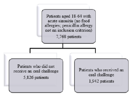
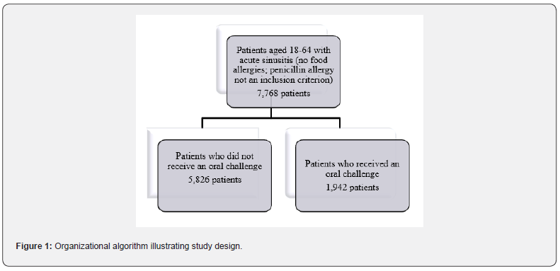
Furthermore, the presence of a penicillin allergy was not an inclusion criterion. The comparison groups were acute sinusitis patients who received oral challenge (Procedure code Terminal (CPT) 95076) compared with those who did not receive oral challenge. Each participant with oral challenge was matched to three participants without oral challenge, according to sex and year of birth (Figure 1). The main outcome of interest was the receipt of a first-line antibiotic (amoxicillin and amoxicillin/ clavulanic acid) within 30 days after the index date of an acute sinusitis diagnosis. Given the lack of statistical power caused by a small sample size, we were not able to examine other secondary outcomes such as infection with Clostridium difficile, multi-drug resistant organisms, and death. Demographic variables included age, sex, place of residence, and US regions. Another covariate of interest was insurance health plan types such as preferred provider organization (PPO), health maintenance organization (HMO), consumer directed health plan (CDHP), and high deductible health plan (HDHP).

SAS statistical software version 9.4 (SAS Institute, Cary, NC, USA) was used to conduct all statistical analysis using a two-tailed P value of < 0.05 as the significance level. We first examined the distribution of covariates between participants receiving oral challenges and those without. A multivariable analysis was conducted using conditional logistic regression models that accounted for matching variables, such as age and sex, and adjusting for place of residence (urban/rural) in US regions to estimate the adjusted odds ratios (aOR) and their 95% confidence intervals (CI). Crude OR was estimated using unadjusted conditional logistic regression that accounted for matching variables (age and sex). This study was approved by The Pennsylvania State University Institution Review Board.

Results

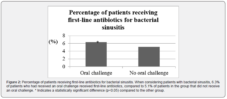
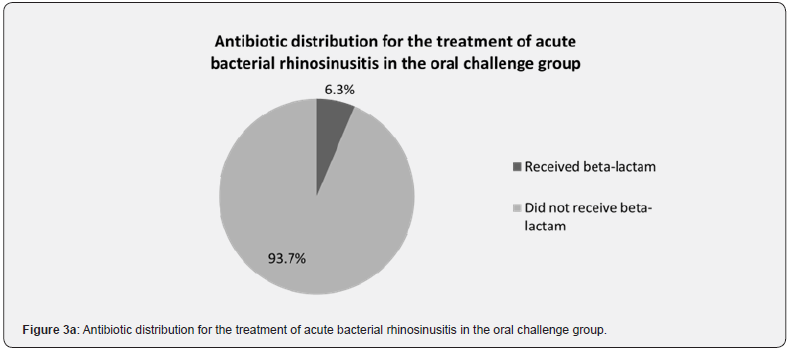
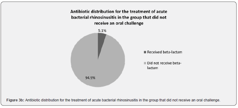
A total of 7,768 patients with bacterial sinusitis were included in the study (Table 1). Of these patients, 1,942 patients received an oral challenge, while 5,826 did not receive an oral challenge (Table 1). When considering patients who received the first-line beta-lactam-containing antibiotics, 6.3% (n=123) of the patients had received an oral challenge (Figure 2). This was significantly higher compared to the 5.1% (n=298) of patients who did not receive an oral challenge (Figure 2). When considering the converse, 93.7% (n=1,819) of patients who had received an oral challenge did not receive first-line antibiotics (Figure 3a). Additionally, 94.9% (n=5,528) of patients in the group without the oral challenge did not receive first-line antibiotics (Figure 3b).

Regardless of oral challenge status, however, an overwhelming majority of patients did not receive first-line antibiotics, which we assume is secondary to failure to remove the penicillin allergy designation versus inappropriate antibiotic prescribing practices.

Discussion

Data obtained from our research utilizing MarketScan demonstrate that patients who received an oral challenge were statistically more likely to receive the first-line beta-lactamcontaining antibiotics for the treatment of acute bacterial sinusitis compared to those who did not receive an oral challenge. Another key finding of this study was that, regardless of oral challenge status, first-line beta-lactam-containing antibiotics are not being frequently prescribed. While the data cannot provide any explanation for this finding, there may be a few reasons. First, the way a drug reaction is documented into the electronic medical record (EMR) can influence prescribing practices. A study conducted in Australia found that over 20% of reports of adverse drug reactions did not even include a description of the reaction [8]. Additionally, about 10% of the reports described as a penicillin allergy were actually more consistent with a drug intolerance, such as gastrointestinal symptoms [8].

Finally, a study conducted in a pediatric population in Saudi Arabia found that over 35% of antibiotic allergy descriptions in the EMR were “poor” [9]. From the perspective of antimicrobial stewardship, the presence of drug allergy labels, especially those that may be inaccurate, can be detrimental to both patients and to the healthcare system. Disadvantages of using alternate antibiotics include higher treatment costs, greater severity of side effects, and decreased treatment efficacy [5]. Additionally, a cohort study found that patients with a penicillin allergy label had significantly longer hospital stays and increased rates of infections with methicillin-resistant Staphylococcus aureus, Clostridium difficile, and vancomycin-resistant Enterococcus compared to those without a penicillin allergy label [10]. One study found that the presence of drug allergy labels was associated with increased use of quinolones and carbapenems and an overall increased rate of inappropriate use of antibiotics [11].

In the case of bacterial rhinosinusitis, second-line antibiotic treatments include doxycycline or respiratory fluoroquinolones (e.g., levofloxacin) [1,3]. The use of broad antibiotics such as levofloxacin can theoretically increase rates of antimicrobial resistance. Prior research found that S. pneumoniae-containing sputum samples had higher rates of antibiotic resistance (though not shown to be statistically significant) in patients with penicillin allergies compared to patients without penicillin allergies [12]. The presence of the penicillin drug allergy label in combination with a lack of meaningful reaction descriptions would likely deter clinicians from prescribing those antibiotics. The previously mentioned Australian study additionally found that the vast majority of the reaction descriptions could have warranted further evaluation and that about half of the documented histories appeared consistent with low-risk penicillin reactions [8]. Despite the demonstrated opportunities for further evaluation by allergists, the specialty is underutilized. For example, the study conducted in Saudi Arabia found that under 40% of the patients with concern for antibiotic allergies were referred for further evaluation [9].

Additional research showed that over 80% of research participants did not even know that penicillin drug allergy testing was available [13]. While these data show the potential impact of successful oral challenges, there are a number of limitations to the study. The most significant limitation was the small sample size of patients in the oral challenge group who received firstline antibiotics. A significant contributing factor to this limitation lies within the study design itself. As mentioned, the data was obtained through the use of MarketScan, which is limited to private insurance claims. As a result, the results are not directly generalizable to other kinds of insurance such as Medicare and Medicaid. Additionally, a diagnosis of food allergy was excluded from the patient population since MarketScan is unable to differentiate food oral challenges from drug oral challenges, subsequently reducing the generalizability of the provided data.

Finally, the CPT codes precluded us from being able to determine the identity of the drugs for which the oral challenges were intended or the outcomes of the oral challenges (e.g., whether or not the allergy label was removed from the medical record). Given how common beta-lactam allergies are, however, it is likely that the majority of the oral challenges were conducted for this particular class of medications. It should also be noted that MarketScan is unable to identify patients with a penicillin allergy label. Since the population studied is not definitively classified as having a penicillin allergy label, the reported difference in betalactam use for the treatment of acute bacterial rhinosinusitis between the group that has received an oral challenge versus the group that has not received an oral challenge could be an overestimation. While additional research is needed to better delineate the study population, the data interestingly highlights the lack with which these first-line antibiotics are used for acute bacterial rhinosinusitis.

Future work can focus on expanding the study population to include a broader array of insurance plans like Medicare and Medicaid. Given that most of the penicillin allergy labels are acquired during childhood, future studies might also be expanded to include the pediatric population. Such measures would likely increase sample size while broadening generalizability. Additionally, the types of antibiotics that are prescribed in place of these beta-lactams can also be examined. Finally, additional valuebased research can be conducted in order to further quantify the overall cost savings of drug oral challenges relative to the healthcare expenses associated with the use of alternative, less appropriate antibiotics.

While our study helps demonstrate the potential benefits of oral challenges, it appears that first-line antibiotics remain under-utilized even when allergy is excluded. In order to rectify this situation, further awareness (e.g., through the education of medical providers at all stages of training and practice) of the importance of penicillin allergy testing, penicillin challenges, education of the patient, and complete removal of a penicillin allergy label are crucial for promoting antimicrobial stewardship and improving health outcomes.

To Know more about  Global Journal of Otolaryngology 

Click here: https://juniperpublishers.com/gjo/index.php

To Know more about our Juniper Publishers

Click here: https://juniperpublishers.com/index.php
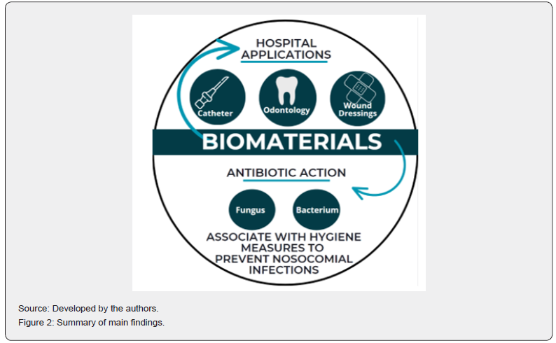
Monday, October 9, 2023

Female Sexual Dysfunction and a Plant-Based Diet - Juniper Publishers


Gynecology and Womens Health - Juniper Publishers


Abstract

Female Sexual Dysfunction (FSD) is a multi-causal and a multi–dimensional medical problem, comprising anatomical, physiological, psychological as well as social-interpersonal components, that adversely affects physical health and emotional well-being. A meta study showed that 41% of women had some level or form of Female Sexual Dysfunction. FSD is especially prevalent among women with chronic diseases such as metabolic syndrome, type 2 diabetes, hypertension, dyslipidemia and coronary artery disease, Hashimoto’s thyroiditis, Graves’ disease and Parkinson’s disease. A plant-based diet can lower the risk of Female Sexual Dysfunction (FSD) by substantially by lowering the risk of several pathologies that are risk factors for it.

Female sexual function, especially arousal, is significantly affected by genital vascular impairment which can lead to vaginal dryness and impaired genital engorgement mediated arousal. The plant-based diet is a safe and effective prophylaxis and treatment for hypercholesterolemia and atherosclerosis. The Mediterranean diet is a plant-strong diet that results in better female sexual function in a dose-dependent manner, regardless of menopausal and metabolic syndrome status. The plant-based diet has the potential to be at least as good a treatment of FSD. A plant-based diet has the significant advantage of having no contraindications or adverse reactions, and is an affordable prophylaxis for all patients over the long-term.

Keywords: Atherosclerosis; Clitoris; Dyslipidemia; Dyspareunia; Orgasmic disorder; Vaginal atrophy; Vaginismus; Vegan; Vegetarian

Abbreviations: FSD: Female Sexual Dysfunction; MD: Mediterranean Diet; NO: Nitric Oxide

Introduction

A healthy and satisfying experience of sexuality is often an important component of overall well-being for women (defined for the purposes of this article as individuals who have female genitalia), associated with increased life satisfaction, higher perceived and objective health status, and even increased longevity [1-4].

In addition, several significant health benefits have been identified for women related to regular sexual activity. These include postponement of natural menopause (and the subsequent hypoestrogenism) [5], reduced frequency of hot flushes during menopause [6], better vaginal health and less vaginal atrophy in post-menopausal women [7], better cognitive function in older adult women, [8] and a reduced risk of subsequent new severe disabilities in disabled women living with their spouse [9]. Some studies also showed that the thresholds for pain tolerance and pain detection were significantly increased when paired with genital self-stimulation, and considerably more so when achieving orgasm [10]. Female sexual function is a complex process coordinated by the neurological, vascular and endocrine systems [11]. Female Sexual Dysfunction (FSD) is a multi-causal and a multi–dimensional medical problem, comprising anatomical, psychological, physiological, as well as social-interpersonal components, that adversely affects physical health and emotional well being [12-16]. FSD is a general term comprising several sexual health concerns that can be distressing for patients, including female sexual interest/arousal disorder, female orgasmic disorder, and genito-pelvic pain/penetration disorders, including dyspareunia and vaginismus. [17,18].

Our understanding of female sexuality was only first formally addressed roughly 50 years ago. During this period and even today, the treatment of FSD has primarily focused on psychosocial/cultural therapy, and highlights that our limited knowledge is reflective of the inadequate treatment options available. Due to the complexity of FSD, a multifaceted approach, addressing neurobiological, vasoactive, hormonal as well as psychosocial/cultural aspects is necessary [19]. This article addresses only the physical etiological factors and symptoms.

Epidemiology

A 2016 systematic review and meta-analysis assessed the prevalence rate of female sexual dysfunction in 215,740 reproductive-age women worldwide and found the 41% of these women report some form of female sexual dysfunction [20]. Dysfunction is especially prevalent in women with chronic health problems, including metabolic syndrome, hypertension, dyslipidemia, coronary heart disease, diabetes, overweight and obese body mass indices (BMIs), anxiety, and depression [3,21-28]. In patients with overt cardiovascular disease, FSD is even more prevalent [29]. Studies have shown a positive association between cardiovascular diseases and sexual dysfunction in females [30]. Diabetes, heart disease, urinary tract disorders, and chronic illness are also significant risk factors for female sexual dysfunction [31].

Pathophysiology

The first phase of the female sexual response, associated with neurotransmitter-mediated vascular smooth muscle relaxation, results in increased vaginal lubrication, wall engorgement and luminal diameter as well as increased clitoral length and diameter. Physiologically, healthy pelvic blood flow is necessary for vaginal lubrication. Normal blood pressure pushes a transudative fluid through capillaries, which ultimately coalesces at the vaginal surface epithelium [32,33]. This process relies on vessels being both patent and able to dilate effectively. Therefore, low atherosclerotic burden and sufficient nitric oxide (NO) activity are protective against sexual dysfunction [33].

Evidence shows that female sexual function, especially arousal, is significantly affected by genital vascular impairment, which can lead to FSD. Specific physiologic impairments of vasculogenic female sexual dysfunction include vaginal engorgement and clitoral erectile insufficiency. Orgasmic female sexual dysfunction may be related in part to vasculogenic impairment of the hypogastric-vaginal/clitoral arterial bed [34]. These syndromes exist when during sexual stimulation, abnormal arterial circulation into the vagina or clitoris, usually from atherosclerotic vascular disease, interferes with normal vascular physiologic processes [35].

Clinical symptoms may include delayed vaginal engorgement, diminished vaginal lubrication, dyspareunia, diminished vaginal sensation, diminished vaginal orgasm, diminished clitoral sensation or diminished clitoral orgasm [35]. Some chronic illnesses, such as vascular disease, diabetes mellitus, neurologic disease, and malignancy, can directly or indirectly impact sexual function [36,37]. Type 2 diabetes, hypertension, dyslipidemia, chronic kidney disease, atherosclerosis, and traumatic injury are associated with diminished vaginal and clitoral blood flow and impaired sexual functioning [38,39]. For many cardiometabolic risk factors and diseases, such as hypertension, diabetes, dyslipidemia and metabolic syndrome, an adverse impact on women’s endothelial function, as well as an association with FSD, has been recognized [40].

A doppler ultrasound study found that the Clitoral Pulsatility Index - an index of vascular resistance in the clitoris - was positively correlated with body mass index, waist circumference, insulin, triglycerides, total cholesterol, and low density lipoprotein cholesterol [41]. Women who have neurologic diseases such as Parkinson's disease or multiple sclerosis may also have FSD [42]. Chronic diseases affecting multiple systems, such as thyroid disease, may also have an impact upon the female sexual function [42].

Nutrition

Nutrition plays a significant role in cardiometabolic disease, suggesting one pathway through which diet influences sexual health in females in the form of vaginal dryness and impaired genital engorgement-mediated arousal [43,44]. Atherosclerotic risk is minimized with a nutrient-dense diet whose foundation is plant-based foods [45]. Meanwhile, nitric oxide (NO) can function best when inflammation is minimal [33]. [46]. A high-quality diet can be a source of antioxidants, and nitrate-rich foods can directly increase NO stores [47,48]. Diet can also indirectly support NO availability through preventing and ameliorating conditions associated with inflammatory and pro-oxidant activity, such as metabolic syndrome, obesity, and atherosclerosis [33,48,49].

A plant-based diet can help prevent and treat diseases such as type II diabetes and cardiovascular disease, and can be very efficacious [50,51]. For instance in one study, a plant-based diet was found to be twice as efficacious in treating type 2 diabetes as Metformin [52]. A plant-based diet can also lower cholesterol as much as lovastatin [53]. In addition, a plant-based diet can reduce the risk of other chronic diseases that have been shown to promote FSD, such as Graves’ disease and Hashimoto’s disease, rheumatoid arthritis, chronic kidney disease, and Parkinson’s disease [54-57]. With regards to specific foods, consumption of soy is associated with increased vaginal blood flow, lubrication, and vaginal collagen content and decreased dyspareunia [58-60].

Fruits such as apples, watermelon, and cacao have been linked to enhanced vascular and sexual health. Daily apple consumption is associated with improved vaginal lubrication and general sexual function [61]. Apples are high in polyphenols, other antioxidants, and phytoestrogens, which together support an anti-inflammatory and anti-atherosclerogenic environment. It can be expected that other fruits would perform similarly, though further research is needed to confirm this. Watermelon in particular supports vascular health via an additional distinct mechanism. It is a rich source of citrulline, which the body readily converts to the NO precursor, arginine [62-64]. Chocolate, derived from the cacao bean, is rich in flavonoids and has been found to increase NO-mediated vasodilation, with promise for supporting sexual function [65,66]. Although more research is needed, these findings on individual foods offer support and further insight into how a plant-based dietary pattern can benefit female sexual health.

Intervention Studies

The Mediterranean diet (MD) is the most widely studied dietary pattern in this context. The Mediterranean diet is characterized by a high intake of plant foods, a high intake of olive oil, a moderate intake of dairy products, zero to four eggs a week, with fish and poultry consumed in low to moderate amounts and red meat consumed in low amounts [15]. Multiple randomized controlled trials and cross-sectional studies have analyzed the long-term effect of the MD on reported sexual function. They find that adherence to this diet results in better sexual function in a dose-dependent manner, regardless of menopausal and metabolic syndrome status [67-69]. ther lifestyle changes may help improve sexual function. These modifications include physical activity, nutrition counseling, and adequate sleep, in addition to a healthy plant-based diet. [70].

Clinical Considerations

Based on cultural norms and biases, conversations about sex are sometimes thought of as taboo in American society and in many other cultures worldwide. This is especially true for women, and particularly when sex is for pleasure rather than reproductive purposes. Failure to have informative discussions about sex often leads to misperceptions about sex and sexuality, including a sense that pain or lack of interest in sexual activity is inevitable and nonmodifiable, which can also lead to women not seeking the care they need. In addition, women sometimes assume that older people do not, or should not, engage in sexual activity [71].

Several U.S. and international surveys of women recently found that the majority of women surveyed did not discuss their sexual health-related symptoms with their health care provider, and discomfort and/or embarrassment with having this discussion was often cited as a reason for avoiding the conversation [72-75]. This finding was consistent for women across different demographics, including age, sexual orientation, race/ethnicity, educational level, and relationship status [76]. Given this situation, physicians need to develop clinical strategies when approaching their patient. For instance, a physician treating a female patient with type 2 diabetes should inquire as to her sexual function since their patient might not raise the issue. One study showed that 54% of women with type 2 diabetes had FSD [77]. Identification of concurrent comorbidities and implementation of lifestyle changes will help improve overall health and may improve sexual function [78,79].

A plant-based diet has the significant advantage of having no contraindications or adverse reactions, and is an affordable prophylaxis for all patients over the long-term. When treating a patient with a plant-based diet it is important to titrate relevant medications, for pathologies such as hypertension and type II diabetes in particular, as the effects of the diet become evident. Patient compliance on plant-based diets has been good in almost all studies. The degree of compliance has often been very high. For instance, one study obtained a 99% compliance [39]. In a 22-week study 94% of subjects on a vegan diet were compliant [40]. In a somewhat longer study, 84% of the participants in each group completed all 24 weeks [41]. In studies of patients placed on plant-based diets for coronary artery disease, high compliance has been noted even over several years. For instance, one study of patients placed on a plant-based diet showed 89% compliance for 3.7 years [42].

Evidence suggests that vitamin D and iron deficiencies are risk factors for sexual dysfunction, and resolution of these deficiencies may well be therapeutic [80-83]. Vitamin D receptors exist on the uterus and ovaries, where they can influence steroidogenesis and testosterone aromatization, with consequent effects on sex hormone levels [84-86]. In iron-deficiency anemia, fatigue is thought to mediate the relationship between deficiency and sexual dysfunction [82]. Testing for vitamin D and iron deficiency may be considered as a relatively simple and low-cost addition to the workup for female sexual dysfunction.

Discussion

Quality of life has become a large concern of patients with a wide variety of diseases. Patients with diseases ranging from several forms of cancer to rheumatoid arthritis want a satisfying sex life. Patient and nonprofit organizations are offering patients general advice on how they can have sex while they are being treated. While many patients may be reluctant to ask their physician about FSD, they may well be appreciative of the physician asking them about it.

While a plant-based diet can’t treat aspects of FSD such as psychological and social factors, it does deserve a place in its treatment. It can be used as a monotherapy in some cases, or as an adjunct to other treatments. It may be especially helpful in treating vascular causes of FSD. It can also help prevent and can efficaciously treat several risk factors for FSD including atherosclerosis, metabolic syndrome, type 2 diabetes, dyslipidemia and chronic kidney disease. It has no adverse reactions or contraindications and is affordable. This becomes even more important considering that many patients with FSD have comorbidities.

A plant-based diet, and specifically manufactured plant-based foods, are now considered mainstream and several studies have shown good patient compliance. Increasing numbers of physicians are prescribing a plant-based diet for their patients in order to prevent and treat disease. Asking a patient about their diet, and prescribing a plant-based diet, should be considered a new standard of care for FSD.


To Know more about Journal of Gynecology and Womens Health 

Click here:  https://juniperpublishers.com/jgwh/index.php

To Know more about our Juniper Publishers

Click here: https://juniperpublishers.com/index.php


How Does the Multi-Disciplinary Team Impact Chronic Kidney Disease Management? - Juniper Publishers

  Urology & Nephrology - Juniper Publishers Abstract Chronic kidney disease (CKD) is a condition where a gradual kidney function loss le...