Wednesday, October 7, 2026

How Does the Multi-Disciplinary Team Impact Chronic Kidney Disease Management? - Juniper Publishers


Urology & Nephrology - Juniper Publishers

Abstract

Chronic kidney disease (CKD) is a condition where a gradual kidney function loss leads to a build-up of toxic waste products in the blood, increasing morbidity and premature deaths from enhanced risks of cardiovascular disease, stroke and mineral bone diseases. The most common causes of CKD include type II diabetes and hypertension, both accounting for two-thirds of all cases. CKD is asymptomatic in early stages and is only diagnosed via routine screening tests unless severe symptoms arise from advanced CKD. Management of CKD via multidisciplinary team (MDT) consisting of professionals from various disciplines has been hailed as the best modality in treating such a multi-faceted chronic disease, leading to positive impacts on patients’ experience and clinical outcomes.

Introduction

More than 1.8 million people are diagnosed with CKD in England [1] where the advancing age is commonly associated with the higher incidence of CKD stage 3-5. Figure 1 is an illustration that on average, whilst there is only an estimation of 1.9% of the population having moderate to severe CKD, that figure is increased to 32.7% for those aged 75 and over. The latest guideline established that NHS England spent £1.45 billion in 2009-10 attributing to £1 in every £77 spent by NHS in managing CKD and associated illnesses stemmed from the condition [1]. Progression patterns and risk factors of CKD into end-stage kidney disease (ESKD) can be established well before the need to initiate renal replacement therapy (RRT). Therefore, proactive intervention strategies to prevent its progression form a basis of management of CKD which is mainly delivered via a multi-disciplinary team (MDT) to address all the various aspects and risk factors of CKD. This paper aims to explore and describe how effective this MDT approach is in treating CKD.


Chronic Kidney Disease (CKD)

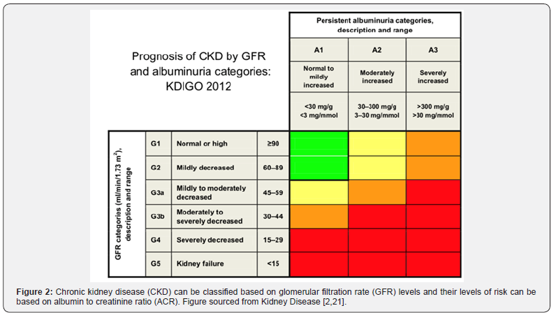
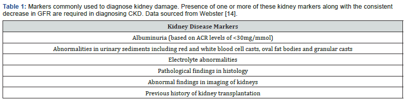
Chronic kidney disease (CKD) is a multi-factorial disease leading to progressive worsening of kidney functions. Kidney biopsies from most CKD patients will often show glomerulosclerosis, tubular atrophy and interstitial fibrosis. Moreover, at the end stage of kidney failure, they will feature a characteristic renal fibrosis where the kidney undergoes unsuccessful wound healing after chronic and sustained inflammation and injury to its tissues. CKD can be classified into 5 stages based on an estimated glomerular filtration rate (eGFR) which represents the amount of fluid each functioning unit (nephron) of kidneys filters through, per unit time. Figure 2 demonstrates the progression of CKD in stages using eGFR and albumin to creatinine (ACR) ratios [2]. A consistently high level of kidney disease markers (illustrated in Table 1) over a period of 3 months and a GFR at G3 stage are two criteria that need to be met in diagnosing CKD. G5 of GFR is termed as end-stage kidney disease (ESKD) where failure of kidney can only be managed with renal replacement therapy (RRT) including dialysis or kidney transplant unless the patient wishes to undergo conservative management.

Causes and Prevalence of CKD

Exact aetiology of CKD is not fully understood; however, older age, type II diabetes, and hypertension are associated with diabetic glomerulosclerosis and hypertensive nephrosclerosis both leading to CKD in Western world [3]. Main causes of CKD in developing countries include glomerular and tubulointerstitial diseases due to infections, drug and toxin exposure. A small percentage of CKDs also arises from congenital conditions such as polycystic kidney disease. Gender difference plays a role in CKD with males showing a more rapid and aggressive CKD progression, suggesting the potential roles of sex hormones in modulating synthesis of growth factors and chemical mediators leading to CKD progression [4]. Ethnicity and socioeconomic status are also key modifiers in CKD prevalence and progression. There is a 60% increased risk of worsening CKD in people of the lowest social quartile when compared with their richer counterparts. This progression is highest in people with ethnic minority backgrounds and can be attributed to having reduced medical awareness and access to specialist nephrologist care in both pre-dialysis and dialysis groups [5].

Clinical Presentation and Complications of CKD

CKD is non-symptomatic in the earlier stages and most diagnoses are made via routine tests unless patients present with severe symptoms from advanced CKD. As the kidney gradually loses its function from progressive disease, there is a rapid accumulation of uraemic retention solutes in the body which can then affect various parts of the body. Uraemic toxicity can lead to vascular damage increasing the risk of cardiovascular diseases and bleeding episodes, impaired inflammatory and immune responses, and altered microflora in the digestive system [6]. Impairment to normal kidney functions can lead to proteinuria, oedema, hypertension, hyperphosphatemia, hyperkalaemia, anaemia and bleeding diathesis, renal osteodystrophy, congestive heart failure, gastrointestinal disturbances and generalised myopathy [7].

Multi-Disciplinary Team in Management of CKD

With medication compliance and adequate lifestyle changes including keeping a normal weight, taking up exercise and following a renal diet, CKD patients can remain as healthy as possible. Management plan is aimed to treat primary pathological diagnosis along with an intervention based on eGFR stages and albuminuria to control hypertension, diabetes, dyslipidaemia and anaemia to prevent episodes of acute kidney injury (AKI) and reduce its complications. A referral to nephrology is to be initiated when patients with poorly controlled hypertension even after treatment with at least 4 anti-hypertensive drugs, reach either G4 or G5 stage with decreasing GFR of less than 30ml/min/1.73m2 and an ACR of 10mg/mmol or higher [8].

A typical renal department provides inpatient and outpatient services covering general nephrology, pre- and post-dialysis review, low clearance clinics and transplant follow-up. The department is made up of a team of professionals from various disciplines including consultant nephrologists, clinical nurse specialists (CNS) and dieticians. Joint clinics are typically run in parallel with endocrinologists for diabetic patients with CKD, with cardiologists to manage their cardiovascular health and lastly with rheumatologists for patients with hyperuricemia to manage their gout and joint pain associated with CKD.

In addition, a team of MDT managing CKD also includes surgeons working together with interventional radiologists to get an intravenous line in or to form fistulae for good vascular access for dialysis. Interventional radiologists are valuable for the team since they can also perform other key procedures such as image-guided percutaneous renal biopsy allowing the clinicians to treat the underlying causes. In addition, the maintenance of fistulae and veins which allows good vascular access for future dialysis is relied upon the great effort of vascular access CNS who work alongside surgeons and radiologists to support the patients regarding fistulae care.

Role of MDT in Management of CKD

Anti-Hypertensive Therapy

There is a 57% increased mortality from cardiovascular causes when patients reach a GFR of less than 60mL/min per 1.73m2 [9]. In addition, CKD patients have 5-10-fold increased risks of dying from other complications than progress into ESKD [10]. Keith [11], also published results from a longitudinal study which concluded that CKD patients are twice likely to die from CVD complications than develop ESKD. Therefore, one of the roles of the consultant nephrologist is to prescribe anti-hypertensives accordingly, and to monitor patients’ blood pressure and other side effects arising from these therapies. Antagonists of reninangiotensin- aldosterone system such as angiotensin convertingenzyme inhibitors (ACEIs) or angiotensin II receptor blockers (ARBs) are first-line choice of agents in CKD patients to reduce proteinuria which is the main culprit for disease progression [12]. However, ARBs and ACEIs can also lead to hyperkalaemia in CKD patients and therefore should be continuously monitored for their blood potassium levels.

Management of Anaemia

One of the main functions of kidney is to produce erythropoietin (EPO) which is a hormone stimulating red blood cell production. CKD patients typically present with hypo proliferative, normocytic, and normochromic anaemia due to impaired EPO production by the kidneys. CKD patients often have chronic anaemia which reduces patients’ quality of life and puts them at higher risks of deaths. There is a 29% increased chance of hospitalisation in patients with haemoglobin (Hb) of <10 g/dl than those with Hb between 11-12 g/dl [13]. Therefore, iron and derivatives of recombinant erythropoietin are commonly used in treatment of anaemia, decreasing the need to transfuse blood [14]. Chronic anaemia is normally managed by anaemia CNS who monitor patients’ iron levels and administer intravenous iron and erythrocyte-stimulating agents (ESAs) to the patients, as prescribed by the nephrologists. They are also important in providing counselling sessions for pre-dialysis and dialysis patients informing them of local and national treatment guidelines to help decide their future treatment plan.

Management of CKD Mineral Bone Disease and Gout

The kidneys regulate how calcium and phosphate are absorbed in the intestine by regulating vitamin-D metabolism which converts vitamin-D to calcitriol (activated vitamin D). CKD patients can have abnormal serum concentrations of calcium, phosphates and reduced calcitriol levels, leading to pain in the bone or bone fragility due to increased osteoclast activity. Management of CKD mineral bone diseases include renal diets restricting phosphates and prescription of calcium or non-calcium phosphate-binders and activated vitamin D in those with low serum calcitriol levels (>30 ng/mL) and normal parathyroid hormone levels [2]. In addition, CKD patients often have high uric acid level (hyperuricemia) which can lead to acute or chronic gout attacks for which uric acid lowering agents such as allopurinol and febuxostat are used to manage hyperuricemia. Therefore, it is the role of the renal dieticians to offer guidance on which food to avoid and how to prepare an adequate meal for good renal health, along with offering support and help to lose weight, exercise more and quit smoking, if need be.

Renal Replacement Therapy (RRT)

There is no universal definition or endpoint of CKD at which ESKD can be diagnosed and renal replacement therapy (RRT) is to be established since patients can have differing levels of comorbidities. When the patients’ eGFRs start to approach 15%, they are consulted to discuss and make timely arrangement for further treatment plans including starting dialysis (either peritoneal or haemodialysis), kidney transplant or undertaking conservative treatment. However, a study supported by USA National Kidney Foundation concluded that an acceptable surrogate endpoint should be when a decrease of eGFR of 30- 40% within 2-3 years excluding causes of acute kidney injury (AKI) [15]. Generally, GFR of less than 15mL/min/1.73m2 along with presentation of uraemic symptoms can warrant a consultation of RRT which involves either dialysis or kidney transplantation.

RRT takes up a huge portion of NHS kidney budget and there was an estimated total annual cost for both haemodialysis and peritoneal dialysis of £505 million and a total annual cost for all transplants was reported to be £225 million in 2009-2010 [1]. Having a strong MDT team behind the patients makes an important difference to ESKD patients regarding making lifechanging decisions when considering RRT. There is a huge burden for patients to consider whether to enlist for RRT or undergo conservative management. A randomised control study carried out by Cooper et. al., in Australia and New Zealand in 2010 (IDEAL study) did not find any difference or improvement in survival outcomes and mortality rates when stage V CKD patients were started on early or late dialytic treatments. Therefore, it is paramount that a patient needs to be educated and consulted on optimal timing of initiating dialysis, which modality of dialysis to choose, extent of time waiting for the matched donor kidney for transplantation and how to cope with immunosuppressants and post-transplant complications.

Overall Impact of MDT in CKD

Incorporation of MDT in CKD care has been supported widely due to its positive outcomes from several studies published over the years. A study by Lin [16], published in PLoS, found that MDT increased quality of life adjusted years (QALYs) by 0.23 per person, when compared with non-MDT care. Cost-effectiveness of MDT has also been reported where MDT care leads to a reduction of $1931 annually per patient from reduced RRT and emergent need for dialysis since MDT was shown to slow eGFR decline and infection-specific hospitalisation [17].

Furthermore, a prospective study led by Chen [18] in Taiwan, also reported that patients under MDT care had a 51% reduced mortality than patients under usual non-MDT care. Moreover, a holistic approach provided via MDT care ensures that one will be guaranteed with an individualised and targeted management plan including health education and awareness of nephrotoxic nonsteroidal anti-inflammatory drugs (NSAIDs) which are contraindicated in CKD patients since they can lead to severe renal injury and progression of CKD [19].

Summary

To conclude, CKD is a chronic disease associated with multiple co-morbidities. To manage such a multi-faceted disease accordingly, only a multi-disciplinary team with professionals working towards the same goals and acting in the best interests of patients, will be able to improve patients’ quality of life and bring positive clinical outcomes. As CKD progresses, there is a need for a more collaborative effort between members of the MDT to address the increasing needs of the patients as they present with more complications. However, integration of such collaborative works may also need enhanced communication between colleagues to provide optimal care for the patients. Although effectiveness of providing a more integrated care via MDT has been established, determination of one optimal professional in an MDT has not been elucidated yet, requiring more randomised studies to follow [20-22].

To Know more about   JOJ Urology & Nephrology

Click here:    https://juniperpublishers.com/jojun/index.php

To Know more about our Juniper Publishers

Click here: https://juniperpublishers.com/index.php


Tuesday, September 29, 2026

Efficacy of Prostaglandin Analogues for Induction of Labour and Associated Complications: A Retrospective Research Study conducted in Latifa Women and Children Hospital - Juniper Publishers


Reproductive Medicine - Juniper Publishers

Introduction

Induction of labour has become a more common worldwide medical intervention during the last few years [1]. The ideal cervical ripening agent must be effective, safe, easy to be administered and acceptable for the pregnant woman. Utilizing prostaglandins (PG) for cervical ripening during induction of labour (IOL) was first described in the 1960s [2]. Since that time various types of prostaglandins including PGF2α, PGE2 (Dinoprostone) and PGE1 (Misoprostol) were extensively studied to elicit the best prostaglandin pharmacological agent for pre-induction cervical ripening [2]. Dinoprostone was found to be superior to the others, as it increased the rates of successful vaginal delivery within 24 h without increasing the operative delivery rates. Vaginal route was found to be a safe and effective approach of bringing on labor.2

There are different pharmacological and mechanical methods that have been approved to ripen the unfavourable cervix [3]. Prostaglandins are the most effective drugs that cause cervical ripening by increasing inflammatory mediators in the cervix and inducing cervical changes. Prostaglandin E1 (PGE1) and prostaglandin E2 (PGE2) have different effects on these processes and on myometrium contractility [4]. The PGE2 is available for cervical ripening as a 3mg Dinoprostone vaginal pessary and also as a controlled release pessary (Propess®), which releases 10 mg of Dinoprostone over 24 hours. Prostin tablet is inserted into the vagina every 6 hours, with a maximum of 3 doses, as per our hospital protocol.

The effect of PGE2 has been investigated and there are many studies in the literature comparing the efficacy of the different formulations available in the market [5]. The incidence of Induction of labour is rising in the current era, with the advancement of technology viz. increasing frequency of ultrasound studies and CTG for fetal monitoring and assessment of fetal wellbeing by fetal medicine units. An appropriate and well tolerated pharmacological method of induction of labour cannot be decided without doing a detailed and in-depth analysis of the two commonly used drugs.

This research study was conducted to compare the efficacy of Propess and Prostin for induction of labor and their complications according to national and international standards.

Therefore, it will help us to update the current hospital guideline of Induction of Labour and eventually improve patient care.

Methodology

This is a retrospective study conducted over a period of 6 months from 1/10/2019 to 31/3/2020. The data was analysed from 597 patients in Latifa Hospital, Dubai, United Arab Emirates for the period of 12 months (1/10/2019 to 30/9/2020). The efficacy of two drugs Prostin and Propess, for induction of labour were compared with respect to their progression to labour and associated complications. The data was collected from the labour room records. Inclusion criteria included pregnant females that were 18-45 years old, gestational age of 28 weeks or more, singleton pregnancy, and cephalic presentation. Exclusion criteria included previous caesarean section/uterine surgery, any contraindication to vaginal delivery, suspected cephalo-pelvicdisproportion, multiple pregnancy, and unexplained antepartum haemorrhage. The data was collected using MS excel sheet.

Demographics

A total of 622 patients were enrolled in this study. Complete data was available for 597 out of them. These were distributed into two groups according to whether Prostin (56.8%) or Propess (43.2%) was used for induction of labour (IOL). Variations between the two groups were accounted for in terms of the following demographic factors: age, gestational age, parity, and indications for IOL. The mean age of women who received Prostin in our study was 31.6 (SD 0.32) and this was higher than that of Propess which was 29.6 (SD 0.33). The gestational ages between both groups were rather similar with the mean of Prostin being 38.4 weeks (SD 0.10) and Propess was 38.6 weeks (SD 0.12). Nulliparity was the most common parity amongst our study group with a valid percent of 37.4%. This was double the incidence of the next common parity which was 1 (18.6%). A varied range of indications for IOL existed amongst these patients ranging across Medical (HTN, DM, Cholestasis of pregnancy etc) and Obstetric (post successful ECV for unstable lie, IUGR, post term, foetal demise). The common indications were Diabetes Mellitus (31.5%) followed by post term (10.2%) and followed by intrauterine growth restriction (9.7%).

Statistical Analysis

Numerical data are presented as mean ± standard deviation or median (min/max) as appropriate and categorical data are presented as a percentage. Chi squared test or Fischer’s exact test was used to compare categorical variables (viz. complications) between the two groups of the study. T-test or Mann-Whitney test was used to compare numerical variables between the two study groups as appropriate.

All the tests are 2-sided tests and P value <0.05 indicates statistically significant results. SPSS 24 was used for data analysis.

Efficacy of drugs

The efficacy of these drugs was assessed by whether labour started or not after induction of labour. It was shown that a total of 79.1% of women from both categories collectively progressed into labour while 20.9% did not. 81.4% of those who were induced with Prostin progressed to labour while 76.0% of those who were given Propess progressed into labor. Using the Pearson Chi-Squared test this had a p-value of 0.105(not significant). Reinduction, which is defined as multiple doses of prostaglandin, was required in 1.84% of our patient population.

Complications

The most common complications following IOL were nonreassuring cardiotocography (CTG) and hyperstimulation/ tachysystole. These had an overall occurrence of 90.3% and 8.2% for Prostaglandins respectively. Other complications such as abruptio placenta and postpartum haemorrhage (PPH) were not seen as frequently with a percentage of 0.5%. While comparing the drug used for IOL, non-reassuring CTG was seen in 10.9% in women who received Prostin while it occurred in 6.2% in women who received Propess. Hyperstimulation/tachysystole was seen in 1.2% of Propess patients while there were none that experienced it in Prostin patients. The Pearson chi-squared test analysing this correlation had a p-value of 0.024 (significant). The incidence of no complications was significantly common too, with 92.6% of those on Prostin having no complications and 87.2% of those with Propess similarly. Fisher’s exact test was used to analyse the above correlation resulting in a p-value of 0.011(significant). The mean age of those who had complications was found to be 28.76 years for both Prostin and Propess and mean parity for the same was 0.91 (SD 0.251). Median parity of those who had complications is nulliparity. Test used for this comparison was Mann-Whitney test with p-value <0.001 (significant).

Mode of delivery

Those that had normal vaginal delivery (NVD) were 78.9% of the study population whilst the remaining 21.1% delivered via lower segment caesarean section. Progression to normal vaginal delivery was more common in women who received Prostin (81.4%) compared to those who received Propess (75.6%). Pearson chi-squared test revealed a p-value of 0.084 (not significant).

Discussion

The present study compared the induction of labour using Prostin vs Propess through a retrospective analysis in the hospital setting. Prostin 3mg was administered 6 hourly vaginally up to 3 doses, whereas Propess (10mg) controlled release single pessary was administered vaginally and left in place for up to 24 hours. The success rates, which was defined as onset of labour, of Prostin and Propess were 81.4% and 76% respectively. This was not statistically significant (p value= 0.105). Following induction of labor with both the agents, the incidence of “reinduction” was rather insignificant and did not bear weight on the outcomes of our study. Many studies that have investigated the efficacy of prostaglandin E2 in the induction of labour showed that Propess had a higher success rate than Prostin [6,7]. A most recent study evaluated the effect of Dinoprostine vaginal insert (Propess) compared to that of the vaginal tablet (Prostin) in primigravida specifically [8], and showed that Propess was the preferred and a better tolerated Prostaglandin E2 tablet for IOL because of the reduced need for vaginal examinations. On the other hand, our analysis shows an insignificant difference in efficacy between the two Prostaglandins E2, which was the primary outcome in this study. This is supported by multiple randomised control trials that also showed no significant difference between the two groups resulting in no preference of one drug over the other in terms of better efficacy [9-11].

The secondary outcome, being the complications resulting between the two drugs, proved to be a non-reassuring CTG and hyperstimulation/tachysystole combined. In our research, nonreassuring CTG was shown in 10.9% of the Prostin cohort while 6.2% of the Propess cohort which is insignificant (p value= 0.084). A study done in 1992, supports our results by showing that non-reassuring CTG was similar in both PGE2 pessary vs placebo groups [12]. Tachysystole was defined as more than 5 contractions/10mins minutes for two consecutive 10-minute periods. Hyperstimulation is defined as either > 5 contractions in ten minutes over a 30minute period, or contractions lasting more than 2 minutes in duration, or contractions of normal duration occurring within 60 seconds of each other as written by the NHS Wales protocol. But many authors define Hyperstimulation as exaggerated uterine response with late fetal heart rate decelerations or fetal tachycardia of more than 160 beats per minute or other worrisome fetal heart rate changes [13,14]. Our results indicate that the combination group of hyperstimulation/ tachysystole was higher in the Propess cohort while none of the Prostin cohort experienced this complication (p value=0.0024). Similarly, the same comparison done in Walsall, UK resulted in more cases of tachysystole in Propess rather than Prostin [11]. Walsall also had a higher rate of uterine hyperstimulation in both prostaglandins compared with the placebo. However, in all the cases, hyperstimulation resolved within 15 min after removal of the pessary indicating that the direct cause was from the effect of the prostaglandins.

The complications in IOL among nulliparous women is greater than that in multiparous as shown in a 2020 study done in Ireland. Many nullipara (32.63%) had undergone caesarean section compared to the multipara (4.37%). Therefore, it is in conjunction with our results that nulliparous women had a greater number of complications in comparison to other parities within our study (p value<0.001). Moreover, previous studies show that induction of labour in medically uncomplicated nulliparous women at term carries higher risk of emergency Cesarean section, compared to those who underwent spontaneous labour [13]. However, our study showed an insignificant difference between the rate of NVD versus caesarean section in the IOL with Propess and Prostin collectively (p=0.084).

A limitation of our study included the lack of a control/ placebo group which could have been used for a more effective comparison.

Conclusion

The results of our study concluded that the success of inducing labour between Prostin and Propess is not statistically significant and either can be used for a favourable outcome. Complications following their viz. non-reassuring CTG, failure of induction, reinduction of labor, higher risk of emergency Cesarean section and are quite similar with both agents. However, tachysystole/ hyperstimulation was more with Propess. These complications, however, are greater seen in nulliparous women compared to multiparous.

To Know more about   Global Journal of Reproductive Medicine

Click here:    https://juniperpublishers.com/gjorm/index.php

To Know more about our Juniper Publishers

Click here: https://juniperpublishers.com/index.php


Thursday, September 24, 2026

Simultaneous Determination and Quantitation of Artemeter and Lumefantrine in Antimalarial Tablet Formulation using High Performance Liquid Chromatography with UV Detection - Juniper Publishers


Pharmacology & Clinical Research - Juniper Publishers

Abstract

Artemether-lumefantrine (AL) combination therapy is now the most used anti-malarial treatment in the world. In Ghana, it has been used as a first line treatment for uncomplicated Plasmodium falciparum malaria since 2004. In this paper, a reliable High Performance Liquid Chromatography method method was developed and validated for the simultaneous determination of AL in commercial fixed-dose combination tablets in the Kintampo-North Municipality. The method employed a Jasco HPLC system equipped with C18 reverse phase column and a mobile phase of acidified methanol and triethylamine buffer (85:15) pH 2.7 as the mobile phase. The flow rate was 1ml/min and detection were by means of a UV detector set to 222nm. The isocratic mode of elution was employed. The retention time of lumefantrine was 5.22 ± 0.19 minutes and artemether, 4.19 ± 0.22 minutes. The method was validated by evaluation of different parameters such as accuracy, precision, linearity, ruggedness and robustness. The percentage recovery for artemether and lumefantrine ranged between 99.18-100.19 and 99.96-100.07 respectively. Six brands of artemether-lumefantrine fixed-dose combination tablets (two local and four foreign) from selected chemical shops and pharmaceutical shops in the Kintampo- North Municipality were analyzed. Of the six brands of artemether-lumefantrine fixed-dose combination tablets analyzed, all passed with respect to their artemether and lumefantrine content using the developed HPLC method. The percent recovery for the local brands ranges from 93.5 to 99.2% and from 91.3 to 97.2% for artemether and lumefantrine respectively. For the foreign brands, 92.05 to 105.0% and 95.8- 99.9% for artemether and lumefantrine respectively, which complies with the International Pharmacopoeia range of 90110. The optimized and validated RP-HPLC method is simple, sensitive, precise, accurate and reproducible. The developed method has been validated as per ICH guidelines and meets all the acceptance criteria given. Hence it can be used in routine analysis for the simultaneous determination of artemether and lumefantrine in bulk as well as in pharmaceutical preparations.

Keywords:Artemether; Lumefantrine; High; Performance; Liquid; Chromatography

Abbreviations:ACT: Artemisinin-based Combination Therapy; AM: Artemether; API: Active Pharmaceutical Ingredient; BP: British Pharmacopoeia; HPLC: High Performance Liquid Chromatography; ICH: International Conference on Harmonization ; IP: International Pharmacopoeia; LU: Lumefantrine; LOD: Limit of Detection; LOQ: Limit of Quantitation; ODS: Octadecylsilane; RP-HPLC: Reverse Phase HPLC; RSD: Relative Standard Deviation; SALMOUS: Standards for Articles Legally Marketed Outside the U.S; SD: Standard Deviation; USP: United States Pharmacopoeia; UV: Ultra-Violet; WHO: World Health Organizations

Introduction

Malaria continues to be one of the major public health problems in Africa, Asia and Latin America. About 219 million cases of malaria and an estimated 660 000 deaths were recorded, out of these, 90 % occurred in Africa [1]. In Ghana malaria accounts for more than 60% of under-five hospital admissions, and 8% of under-five mortality and 9.2% of maternal deaths. The use of antimalarial medications is the only practical means of preventing mortality and lowering morbidity brought on by the disease in many malaria-endemic locations, particularly in the African region [2]. Several routinely prescribed antimalarial medications, including chloroquine and sulfadoxine-pyrimethamine, no longer work on Plasmodium falciparum. The WHO has advised that all antimalarial medications should combine an artemisinin derivative with a co-drug as a result [3]. These treatments incorporate two active substances with various modes of action [1]. It is quite concerning because ACT-resistant P. falciparum has just been found along the Thailand/Cambodia border [4], There is evidence that drug-resistant falciparum malaria has migrated from Asia into Africa [5].

Artemisinin derivatives are incredibly important antimalarial medications, and because of their quick action and lack of adverse effects, they are in high demand in endemic areas, making them vulnerable to forgery and counterfeiting [6]. Checking the quality of antimalarials is necessary to prevent the emergence of resistance. For reliable and accurate results, High performance liquid chromatography is a very powerful tool for the quantification of these medications. There are currently HPLC methods for the analysis of lumefantrine and the assay of artemether in finished pharmaceutical products (FPP) [7] as well as for lumefantrine analysis [8]. However, only a few HPLC methods were reported for the quantitative determination of Artemether and lumefantrine in fixed combination anti-malarial products [8-11]. Hence this study seeks to add to the existing methods, a method that is rapid, economical, precise and accurate for the assay of artemether and lumefantrine.

Materials and Methods

Reagents and materials

Methanol, ethanol, Triflouroacetic acid, Hydrochloric acid, trielthylamine and acetonitrile used were HPLC grade from BDH, Europe. Sodium Phosphate was obtained from Sigma Aldrich, and Hydrochloric acid from Elitech Solutions, France. Artemether and lumefantrine standards were obtained as generous gift from Pharmanova, Accra, Ghana. AL tablets were sampled from selected chemical shops and three pharmaceutical shops in Kintampo.

Instrumental and analytical condition

The HPLC analyses were carried out on a JASCO HPLC system equipped with a stationary phase consist of an Ultracarb 3μ C_18, (20) 200*3.2mm and a UV detector. The injection volume was 20 μL. The separation of artemether and lumefantrine was evaluated in different proportions of different solvents or different proportions of the same solvents or same proportion with different conditions such as the flow rate and, for each condition, the retention times were noted. The optimized condition was achieved using a mobile phase comprising acidified Methanol and triethylamine buffer (85:15).

Preparation of standard solutions

Artemether–lumefantrine standard solution

4mg lumefantrine and 24mg artemether were accurately weighed and transferred into a 25mL volumetric flask, sonicated and diluted to volume with the mobile phase to give a solution of 160μg/mL of artemether and 960μg/mL of lumefantrine. The solution was then filtered using a sintered glass filter. 20μL of this solution was injected into the column and the chromatographs recorded.

Preparation and Analysis of Tablet Formulations

Twenty Tablets of artemether and lumefantrine were weighed and finely powdered. A quantity equivalent to 4mg of artemether and 24mg of lumefantrine was transferred into 25 mL volumetric flask and appropriate amount of diluent was added. The contents were sonicated to dissolve completely and the volume was made up to the mark with diluent and filtered through sintered glass filter. 1 mL of stock solution was transferred to a 10 Ml volumetric flask and made volume up to the mark with diluents to get final concentration of 16μg/mL and 96μg/mL for artemether and lumefantrine, respectively. 20μL of this solution was injected into the column and the chromatograph recorded. To calculate the artemether and lumefantrine content in the tablets, their respective peak areas were inserted into the linearity equation from the linearity graph to determine their contents.

Validation Linearity

Aliquot portions of standard stock solution 0.2, 0.4, 0.6, 0.8 and 1.0 mL were taken in separate 10 mL volumetric flasks. The volume was adjusted to the mark with diluent to obtain concentrations of 3.2, 6.4, 9.6, 12.8, 16.0μg/mL and 19.2, 38.4, 57.6, 76.8, 96.0, 115.2 μg/mL for Artemether and Lumefantrine, respectively. Calibration curve was plotted over a concentration range of 3.2-16μg/mL for artemether and 19.2-115.2μg/mL for lumefantrine. Calibration curve was constructed by plotting peak area v/s concentration, the graph must be linear and the regression equation was calculated.

Precision

One set of three different concentrations of mixed standard solutions of artemether and lumefantrine were prepared. All the solutions were analyzed in triplicates, in order to record any intraday variations in the results. For inter-day variations study, three different concentrations of the mixed standard solutions in linearity range were analyzed on three consecutive days. The peak areas were recorded and the Relative Standard deviation (RSD) was calculated for both series of analyses.

Robustness

In the robustness study, the influence of small, deliberate variations of the analytical parameters on retention time of the drugs was examined. The following factors were selected:
1. Flow rate of the mobile phase (2.7±0.02ml/min)
2. Wavelength at which the drugs were recorded (222±1nm).

Accuracy

The accuracy of the method was determined by calculating the recovery of the analyte of interest by the standard addition method: Known amounts of working standard of artemether (1.6μg) and lumefantrine (9.6μg) were added to solutions of various concentrations like: 3.2μg, 6.4μg and 9.6μg of artemether and 19.2μg, 38.4μg and 57.6μg of lumefantrine. Each sample was prepared in triplicate and injected. Sensitivity

Limit of detection (LOD) and quantification (LOQ) were estimated from the signal-to-noise ratio. The LOD and LOQ were calculated by the use of the equations:
LOD = 3 σ/s.
LOQ = 10 σ/s

Where σ is the standard deviation of intercept of calibration plot and sis the average of the slope of the corresponding calibration plot.

Ruggedness

Sample solutions of artemether (16μg/mL) and lumefantrine (96μg/mL) were prepared and analyzed using slightly different operational and environmental conditions.

Results and Discussion

Chromatographic method development

Optimization of chromatographic mode, wavelength: Artemether and lumefantrine, both the API’s are non-polar in nature, hence either reversed phase or ion-pair or non-aqueous chromatography was used. Detection wavelength was selected by scanning reference standards over a wide range of wavelength from 200nm to 400nm. A fixed concentration of AM-LU (10μg/ml) was analyzed at different wavelengths. From the responses, the λ max value was found to be 209nm and 335nm for artemether and lumefantrine, respectively. Using this data 222nm was selected as a detection wavelength at which the components showed well resolved peaks. The standard solution of artemether and lumefantrine was prepared and run through the system and different combinations of mobile phase and column were tried for isocratic mode to get well resolved, symmetric peaks. The method employed acidified methanol/triethylamine phosphate buffer (85:15), which is economical but have well resolved peaks.

Validation

Linearity: The method demonstrates linearity over a concentration range of 10-100ug/ml for lumefantrine and 3-20ug/ml for artemether. From the calibration curve the R2 value over this range was found to be 0.9991 and 0.9995 for artemether and lumefantrine respectively. This indicates a linear relationship between the concentrations of the two analytes.

Robustness: When some conditions of the mobile phase such as pH and column used for the method were varied, there was no statistically significant difference between results of the varied and old conditions of mobile and stationary phases using the student t-test. The flow rate and wavelength of detection were varied, with no significant difference in the peak areas. This indicates that the method is robust under varying condition of both flow rate and wavelength of detection (USP SALMOUS edition 2008).

Precision: Precision is reflected by percentage RSD values less than 2. These low values suggest high sensitivity of the developed method (USP SALMOUS edition 2008).

Sensitivity: With the developed method, the LOD and LOQ for LU were 338 and 1129ug/ml (0.333 and 1.129mg/L) respectively. AM had 90 and 300ug/ml (0.090 and 0.033mg/L as LOD and LOQ respectively.

Accuracy: The percent recoveries for Accuracy was within range of (97.6 -99.86) % for AM and 97.5-99.2 for LU which indicates that the method was accurate.

Assay of Tablets

According to the USP SALMOUS standard, lumefantrine tablets should contain not less than 90.0 percent and not more than 110.0% of the labeled amounts of artemether and of Lumefantrine. (USP SALMOUS edition 2009). Six commercial brands of (two local and four foreign) AM-LU tablets were analyzed for active substances using the developed method. Triplicate determinations were carried out. The respective contents of AM and LU were 93.5/91.3, 99.2/97.2, 96.62/97.0, 95.82/98.5, 105.2/99.9, and 92.05/95.8 percentage of the declared contents for Malar2DS, DANMETHER, Malafantrine, Coartem, Lonart and Artemos plus. The formulations complied with the (90-110%) of the label claim for the IP.

Application

Analysis of Marketed formulation

The validated method was used for the simultaneous estimation of artemether and lumefantrine in fixed dose combination tablets. Six brands were procured and analyzed with the proposed method and the results are presented in Table 5. The content (mg) and percentages of each of artemether and lumefantrine in the tablet sample was computed using peak areas and the regression equations from the calibration curves. The mean contents obtained for artemether and lumefantrine in the formulated products were very close to the labeled amount. The results show that the method is accurate in determining the content of the two active ingredients in fixed dose combination tablets.

Conclusion

Considering the increasing use of ACT to treat malaria in endemic areas, the availability of simple and rapid analytical method is essential to evaluate the quality of formulations being used currently. From the present study it can be concluded that the optimized and validated RP-HPLC method is simple, sensitive, precise, accurate and reproducible. The developed method has been validated as per the ICH guidelines and it meets all the acceptance criteria given in ICH guidelines. Hence the method can be used in routine analysis for the simultaneous determination of Artemether and Lumefantrine in bulk as well as in pharmaceutical preparations.

To Know more about   Journal of Pharmacology & Clinical Research

Click here:    https://juniperpublishers.com/jpcr/index.php

To Know more about our Juniper Publishers

Click here: https://juniperpublishers.com/index.php


How Does the Multi-Disciplinary Team Impact Chronic Kidney Disease Management? - Juniper Publishers

  Urology & Nephrology - Juniper Publishers Abstract Chronic kidney disease (CKD) is a condition where a gradual kidney function loss le...