Wednesday, November 30, 2022

Tumor-Related Epilepsy and Post-Surgical Outcomes: Tertiary Hospital Experience in Saudi Arabia - Juniper Publishers

 Neurology & Neurosurgery - Juniper Publishers

Abstract

Objectives: Recent studies reported that tumor histopathology plays a significant role in predicting freedom from seizures after epilepsy surgery; however, no consensus among researchers regarding this issue exists. This study retrospectively examined different types of tumor-related epilepsy and post-epilepsy surgical outcomes and the relationship between various tumor Histopathology and these outcomes.

Methods: In this hospital-based retrospective study, patients with brain tumors and drug-resistant epilepsy, which is defined as the failure of two tolerated and appropriately chosen antiepileptic drugs (whether as monotherapies or in combination) to achieve and alleviate sustained seizure freedom, were recruited. These patients underwent a thorough pre-surgical evaluation in an Epilepsy Monitoring Unit (EMU) before deciding to undergo surgical intervention according to an epilepsy case management conference.

Results: One-hundred patients (including 45 children) with brain tumors were included in this study (male: female = 3:2). Most of the patients (93) had G/GNT. No significant differences in outcome were observed among sex, age, or histopathological categories. However, during the first year after epilepsy surgery, most of the low-grade G/GNT cases showed favorable outcomes based on ILAE classes 1 and 2 (61.3% and 9.7%, respectively), whereas high-grade gliomas and meningothelial tumors showed outcomes of ILAE class 1 (40% and 100%, respectively).

Conclusion: Post-epilepsy surgical outcomes of different brain tumors have been achieved with favorable outcomes in children and adults with low-grade gliomas and meningiomas. Thus, pre-surgical evaluation in EMU is highly recommended to enhance better post-epilepsy surgical outcomes.

Keywords: Epilepsy; Brain tumor; Histopathology; Surgery; Saudi Arabia; ILAE

Introduction

Approximately 30%–50% of patients with brain tumors have epilepsy as an initial presentation [1]. However, 6%–45% of patients with brain tumors develop seizures later in life [2,3]. Although the exact mechanism of seizure development in patients with brain tumors is not clearly understood, recent studies have identified that some changes in the peritumoral regions affect the release of neurotransmitters that lead to seizure development [4]. Many studies have reported a relationship between tumor type and seizure frequency. For example, low-grade gliomas and glioneuronal tumors (G/GNT) are associated with a high rate of seizure incidence (85%–92%) [5-7]. In contrast, glioblastomas, which are high-grade tumors, are associated with a low rate of seizure incidence (20%–50%) [8,9]. The incidence rates of seizures in meningiomas, especially in atypical and malignant subtypes, remain understudied [10]. Despite the abundance of knowledge regarding surgical management of resistant forms of epilepsy, including those associated with brain tumors, especially of the low-grade type, prospective studies regarding the medical treatment of epilepsy in this type of patient are scarce [11]. Reportedly, resection of the epileptogenic zone due to the development of brain tumors lead to freedom from or significant control of seizures in 70-90% of patients [12,13]. Furthermore, tumor type, seizure severity, early surgical intervention, frequency during the pre-operative stage, histopathology of the tumors, and the extension of surgical resection to include peritumoral tissues are reportedly the factors that increase the likelihood of freedom from seizures post-operatively [14]. Two of the best predictors of freedom from post-operative seizures include a duration of less than one year since the onset of epilepsy and gross total surgical resection [15,16]. Tumor histopathology plays a significant role in predicting freedom from seizures after epilepsy surgery; however, there is no consensus among researchers regarding this issue [17,18]. This study retrospectively investigated different types of tumor-related epilepsy and their outcomes in the first year after epilepsy surgery.

Materials and Methods

Study design

A hospital-based retrospective study was conducted using secondary data from the epilepsy registry at King Faisal Specialist Hospital and Research Center (KFSH&RC) between 1998 and 2017.

Study population

The included patients underwent surgery for drug-resistant epilepsy. The patients were admitted to the Epilepsy Monitoring Unit (EMU) for long-term monitoring; they underwent presurgical evaluations such as surface electroencephalography (EEG), 3-tesla magnetic resonance imaging (MRI) of the brain, and fluoro-deoxy-glucose positron emission tomography (PET) brain scans. Additionally, a qualified neuropsychologist was present during the evaluation of the enrolled patients with epilepsy. In some patients, intracranial subdural recordings, intracarotid amobarbital procedure (Wada test) and electrocorticography (ECoG), and motor, sensory, and language mapping were performed.

Detailed information about the patients was collected. This information included demographic characteristics (age, gender, handedness, age at onset of the disease) and history and clinical data (type of seizure and frequency, seizure observed at EMU, MRI findings, PET scan, ictal EEG (IEEG) location and type, subdural EEG recording, and inter-ictal EEG [IIEEG] location and type). Moreover, final diagnosis, surgical procedures, and pathology were recorded as the primary outcome. All tumor cases enrolled in this study were reviewed and graded independently by a neuropathologist according to the World Health Organization (WHO) classification [19]. Epilepsy data were discussed in an epilepsy surgery conference with epileptologists, epilepsy surgeons, neuroradiologists, and neuropsychologists to determine the status and surgical candidacy of the patient.

According to the International League Against Epilepsy (ILAE) commission report (1997–2001), six outcomes of interest were proposed [20]. However, the categories 4, 5, and 6 are difficult to measure and implement in daily practice, particularly when parameters related to the quality of life are included. Hence, we suggested simplifying the ILAE classification of epilepsy surgical outcomes with only four categories rather than six to facilitate its application for a new modification that shares the ILAE classification for classes 1, 2, and 3, while classes 4, 5, and 6 would be merged into only one class, called class 4. Thus, the definitions of the classes were divided into four groups:

1. class 1, patients who were completely seizure-free with no auras;

2. class 2, patients with auras but no seizures;

3. class 3, patients experiencing one to three seizure days per year ±auras; and

4. class 4, ranging from four or more seizure days per year to those experiencing ≥ 50% reduction of baseline seizure days ±auras to ≥ 100% increase in baseline seizure days; ±auras. Furthermore, outcomes of classes 1 and 2 were considered favorable, while those of classes 3 and 4 were considered unfavorable. These four outcomes were investigated during the first post-surgical year.

Data analysis

Statistical analysis through cross-tabulation of the tumor groups, pathologies, and progressive outcomes was performed using SAS software (ver. 9.4). Due to the small sample size, some subgroups, pathologies, and outcomes were collapsed. Proportional statistics, and chi-square and Fisher’s exact test were used to explain the findings within a 95% confidence interval (CI). A P-value of < 0.05 was considered statistically significant.

Results

Among the 100 patients with brain tumors related epilepsy included in this study, 59 (59%) were males, and 41 (41%) were females. The incidence of brain tumor-related epilepsy (BTRE) in our center was lower among children than adults (45% versus 55%). However, the differences among sex, age, and the histopathology categories were not statistically significant (P = 0.111 and 0.878, respectively) as depicted in Table 1.

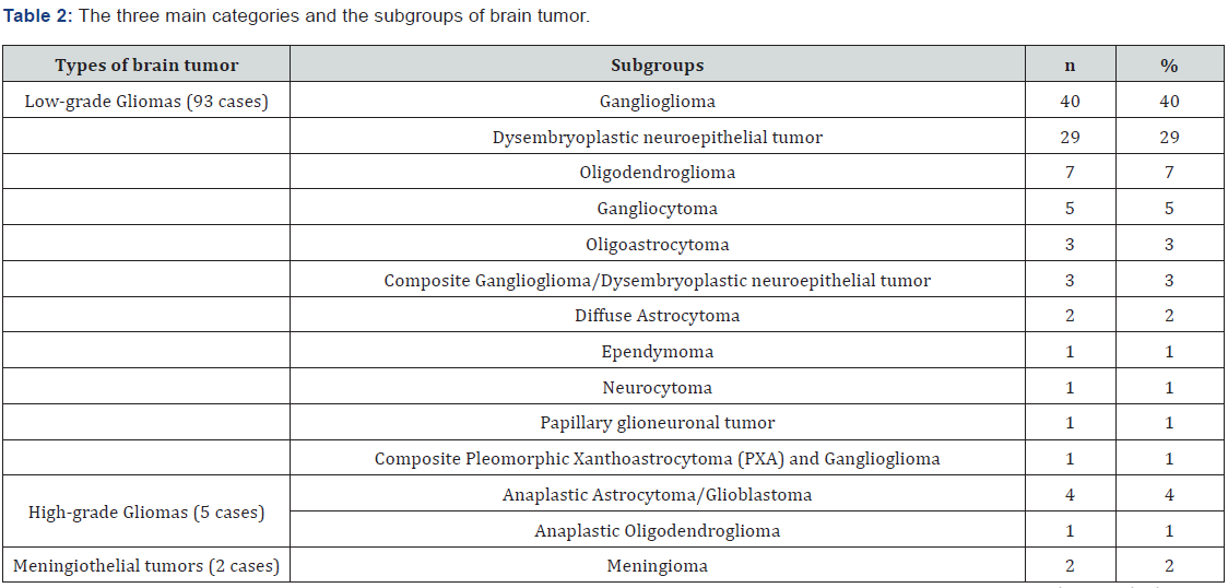
According to histopathological findings, the cases of brain tumors were grouped into three main categories (Table 2). Most of the tumors (93%) were low-grade gliomas or glioneuronal tumors (G/GNT) followed by high-grade gliomas (5%); only two meningioma cases (2%) were detected. Among the three main categories of brain tumors, low-grade G/GNT consisted of 11 entities. Under this category, 40 tumors were gangliogliomas, and 29 tumors were Dysembryoplastic neuroepithelial tumors (DNET). Among the five high-grade gliomas, three were astrocytic tumors. Interestingly, only two meningiomas were found. Table 2 details the pathological categories.

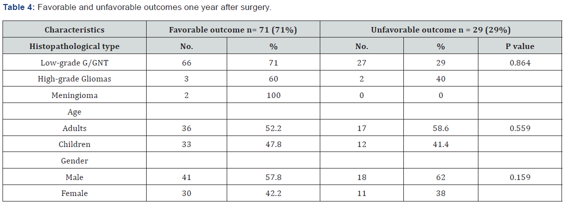
In this study, the primary postoperative outcome was evaluated using the modified ILAE classification described in the methodology section. During the first post-surgical year, most patients with low-grade G/GNT experienced favorable outcomes (classes 1 and 2, 61.3% and 9.7%, respectively) as shown in Table 3. In contrast, approximately 40% of the patients with high-grade gliomas showed favorable outcomes (ILAE class 1). Both meningioma cases (100%) showed outcomes of ILAE class 1. Among the 100 patients with brain tumors, 71 (71%) patients, including 66 patients with low-grade G/GNT, three patients with high-grade gliomas, and two patients with meningiomas, experienced favorable outcomes during the one-year post-surgical period (Table 4). Moreover, the incidence of favorable outcome was higher in adult patients than in children (52.2% versus 47.8%) and in male versus female patients (41% versus 30%). However, these differences in outcome dependent on histopathological type, age, and sex were not statistically significant (P = 0.864, 0.559, and 0.159, respectively).

Discussion

From the epilepsy registry at KFSH&RC, 100 patients who underwent surgery for tumor-related epilepsy were included in this study. Varying associations between different brain tumor types and epilepsy have been reported. Most studies show gangliogliomas to be the most common tumor type associated with epilepsy, followed by DNET, oligodendrogliomas, and astrocytomas [21-24]. These findings support our results, that is, most of our patients had low-grade G/GNT (93.0%), most of which were gangliogliomas (40%) followed by DNET (29%). Furthermore, Babini et al. reported that gangliogliomas (66.7% versus 40%) [25] were the most frequent tumors among their patients; however, their sample size was smaller (30 cases) than in our study. Contrary to our findings, Kahlenberg et al. reported that mixed oligo-astrocytomas were the most prevalent tumors followed by astrocytomas grade II and oligodendrogliomas of grade II [25]. The incidence of high-grade gliomas was lower (5%) in our study than in a study by Michelucci et al. in Italy (77.0%), a finding attributed to a significantly high prevalence of high-grade gliomas in that area [15].

Seizures have a great impact on patient and caregiver quality of life. Seizures affect all aspects of a patient’s life, such as employment, social life, driving, and entertainment. Epilepsy surgery as described in many recent studies, is considered to relieve tumor-related epilepsy and achieve favorable outcomes [15,26,27]. However, in some cases, epilepsy persist even after resecting the primary focus [25]. All of our patients underwent well-planned epilepsy surgery, which is defined as the resection of the tumor and peritumoral tissues [23]. Consequently, our patients showed varying degrees of improvement during the first post-surgical year depending on the type of brain tumor (low-grade G/GNT versus high-grade gliomas); however, the differences were not statistically significant (P = 0.864). For example, patients with low-grade tumors, which were the most common type of tumor in our study (93 cases), showed a favorable outcome during the first post-epilepsy surgery year with approximately 71% freedom from seizure (classes 1 and 2). Michelucci et al. reported findings that were similar to our study, that is, better outcomes was observed in patients with low-grade gliomas (76%) [15]. In contrast, Kahlenberg et al. showed that about half of their patients (30 out of 54; 55.6%) with brain tumor-related epilepsy showed good post-surgical outcomes (seizure-free periods > 12 months) [25]. These proportions were lower than those observed in our study (55.6% versus 71%).

In our opinion, the ILAE classification of epilepsy surgical outcomes should be simplified using only four categories rather than six to facilitate ILAE application. Hence, whenever patients have four or more seizures (outcomes 4, 5, and 6), they should be classified into one category (category 4). The currently used ILAE Commission on Neurosurgery in 2001[28] still has some elements that make the use of categories 4, 5, and 6 difficult to measure and implement in daily practice, particularly when including parameters related to quality of life. This new modified classification can help the researchers in their ongoing studies.

Furthermore, few patients in our study had high-grade gliomas (five) or meningiomas (two). These patients showed a favorable outcome during the first post-surgical year (60% and 100%, respectively). Michelucci et al. reported similar results in which 58% of their patients with high-grade glioma became seizure free after tumor removal [15].

Pediatric and adult groups showed no significant differences regarding seizure outcomes during the first post-surgical year (P = 0.559); thus, we cannot claim that surgery is more beneficial in pediatric patients. These findings were similar to those of other studies [23].

Our study has three main limitations:

1. the sample size was small and included only five patients with high-grade gliomas and two patients with meningothelial tumors (two cases); this made comparison with low-grade G/GNT insufficient although our sample size (100 cases) is comparable to that of other studies;

2. this series of tumor-related epilepsy does not represent the population with epilepsy in Saudi Arabia because not all patients with tumor-related epilepsy are eligible to be admitted to our institution, and thus, they are treated; and

3. the possibility of bias occurring during data collection in a retrospective study. Despite the above-mentioned limitations, we hope that our study provides valuable information on one of the most debatable topics in epilepsy surgery in the country and throughout the Middle East.

Conclusion

In this study, the most common tumor-related epilepsy was low-grade G/GNT. Outcomes of post epilepsy surgery of different brain tumors have been achieved with a favorable outcome in both children and adults. Thus, thorough pre-surgical evaluation of patients with brain tumor-related epilepsy in EMU is highly recommended to enhance better post-epilepsy surgical outcomes. Further prospective, multicenter studies are needed with a larger number of patients to allow the findings to be more generalizable in Saudi Arabia.

Click here: https://juniperpublishers.com/index.php 

Tuesday, November 29, 2022

Takotsubo Caused by Pulmonary Embolism - Juniper Publishers

 Juniper Online Journal of Case Studies - Juniper Publishers

Abstract

Takotsubo is a transient acute coronary myocardial infarction due to a catecholaminergic discharge accounting for 1 in 36,000 adults after intense physical or psychological stress. Most often found in women over 50 years of age. Its association with pulmonary embolism is very rare.

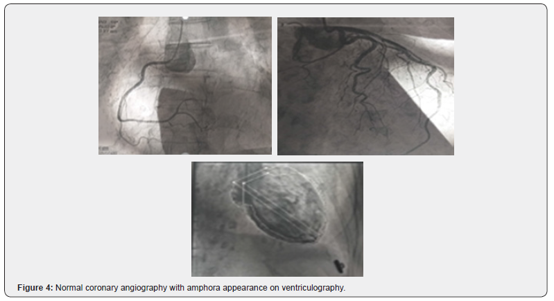
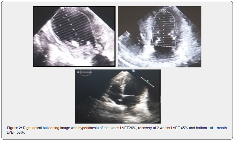
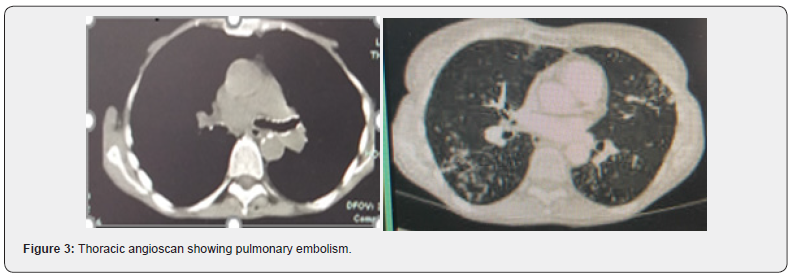
With this in mind, we report the case of a 76-year-old female patient with poorly followed chronic obstructive pulmonary disease (COPD). She presented to the emergency department with acute respiratory distress and lipothymia. Clinical examination revealed hypoxia with SaPO2 at 86% in free air, blood pressure at 120/80mmHg, tachycardia at 112 beats/min. The electrocardiogram showed S1Q3, hyper-right axial deviation, complete right bundle branch block with fragmented QRS, positive AVR with a tachycardia of 125 beats/min. A thoracic angioscan was performed, showing a bilateral pulmonary embolism of segmental and sub-segmental level. Ultrasensitive troponins were highly elevated at 1530ng/l with transthoracic echocardiography showing signs of acute pulmonary heart disease associated with apical ballooning, very akinetic with hyperkinesia of the bases, LVEF 26% suggestive of takotsubo confirmed by coronary angiography coupled with ventriculography giving an amphora-like appearance with a healthy coronary. The patient was initially admitted to the intensive care unit and then to the hospital for an intermediate-high risk pulmonary embolism complicated by takotsubo. The etiological work-up of the pulmonary embolism was normal. She received apixaban, Ramipril and bisoprolol. The evolution was marked by a recovery of the bi ventricular function with an LVEF of 58% in 1 month.

Takotsubo was secondary to respiratory failure caused by pulmonary embolism through catecholaminergic discharge resulting in a redistribution of beta receptors in the myocardium.

Keywords: Pulmonary embolism; takotsubo cardiomyopathy; Myocardial infarction

Abbreviations: COPD: Followed Chronic Obstructive Pulmonary Disease; APH: Acute Pulmonary Heart Disease; CICU: Cardiovascular Intensive Care Unit; LVEF: Left Ventricle; HPA: Hypothalamic-Pituitary-Adrenal; MI: Myocardial Infarction; CPA: Acute Pulmonary Heart; ARBs: Angiotensin 2 Receptor Blockers

Introduction

First described in a Japanese medical journal in 1990 about 5 cases, by the team of Hikaru Sato et al. [1-3] Takotsubo cardiomyopathy usually presents as transient left ventricular dysfunction with apical wall motion abnormalities associated with electrocardiographic changes similar to those of acute coronary syndrome in the absence of significant coronary disease [4,5]. It usually lasts about 15 days, without mortality or severity in the acute phase, and usually occurs in postmenopausal women, with 90% of cases in women aged 67-70 years [5], accounting for about 80% of cases in women over 50 years [4]. Takotsubo syndrome accounts for approximately 1-3% of all patients worldwide, or 1 case per 36,000 adults. In the USA, it accounts for 0.02% of hospital admissions and 1-2% of coronary syndromes in FRANCE [4,6]. The pathophysiological mechanism of takotsubo cardiomyopathy remains unclear, and several possible theories have been put forward, such as excess catecholamines, coronary artery spasm, microvascular dysfunction and metabolic disorders [3]. However, many of these theories focus on the central role of the sympathetic nervous system which, in response to an emotional, physical or combined trigger, releases an excess of catecholamines that cause the disturbance in myocardial kinetics. The mechanism by which catecholamines cause these contraction abnormalities is currently unclear [1], let alone its relationship to pulmonary embolism or being triggered by it. It is with this in mind that we report a case of pulmonary embolism causing takotsubo.

Case Report

We report the case of a 76-year-old female patient with poorly monitored COPD. She presented to the emergency department with acute respiratory distress and lipothymia. The clinical examination revealed hypoxia with SaPO2 at 86% in the open air and 97% under oxygen at 6litre/minute, blood pressure at 120/80mmHg, tachycardia at 112 beats/min. The electrocardiogram showed S1Q3, hyper-right axial deviation, complete right bundle branch block with fragmented QRS, positive AVR with a tachycardia of 125 beats/min. A thoracic angioscan was performed, showing bilateral segmental and sub-segmental pulmonary embolism. Ultrasensitive troponins were highly elevated at 1530ng/l with transthoracic echocardiography showing signs of acute pulmonary heart disease (APH) associated with apical ballooning, very akinetic with hyperkinesia of the bases, LVEF 26% suggestive of takotsubo confirmed by coronary angiography coupled with ventriculography giving an amphora-like appearance with a healthy coronary. The patient was initially admitted to the Cardiovascular Intensive Care Unit (CICU) and then to the hospital for an intermediate-high risk pulmonary embolism complicated by takotsubo. The etiological work-up of the pulmonary embolism was normal. She initially received oxygen therapy for 72 days, apixaban (Eliquis) 10mg x2/dr for 7 days then 5mg x2/dr for 6 months. Ramipril 5mg/dr, bisoprolol 2.5mg/dr. The evolution was marked by a recovery of the biventricular function at 1 month of the treatment with a LVEF at 45% in 2 weeks then at 58% in 1 month.

Discussion

Takotsubo cardiomyopathy is a transient stress cardiomyopathy, the symptomatology of which is highly suggestive of acute myocardial infarction [2]. It usually occurs in postmenopausal women and accounts for about 90% of women with a mean age of 67-70 years. A woman over 55 years of age is 5 times more likely to develop takotsubo than a younger woman, and 10 times more likely than a man [1,2]. Several factors are incriminated in the occurrence of takotsubo. Among them we have :

a) Contributing factors:

i. Falling blood levels of estradiol at the menopause (estradiol seems to protect the microcirculation from the vasoconstrictive effect of adrenaline);

ii. Genetic predisposition, supported by the existence of family cases;

iii. A history of psychiatric illness observed in 42% of cases (e.g. depression in 20% of cases, anxiety) or neurological illness.

b) Triggering factors:

i. Physical stress (stroke or TIA, subarachnoid haemorrhage, acute respiratory failure, accident, strenuous sports activity, cancer chemotherapy, even coronary disorders).

ii. Negative psychological stress (bereavement, divorce, anger, anxiety, financial or professional problems, floods, earthquakes, etc.), but also positive (happy surprises) [2].

Studies that have investigated the pathophysiology of Takotsubo syndrome highlight the central role of strong sympathetic stimulation and parasympathetic depression [2]. Indeed, there are two initial elements of physiology to consider. The first is the cognitive centres of the brain and the hypothalamic-pituitary-adrenal (HPA) axis, and the amount of epinephrine and norepinephrine released in response to a given stress (i.e. the "gain" of the HPA axis). The second is the response of the cardiovascular system (including the myocardium, coronary arteries and peripheral vasculature) and the sympathetic nervous system to sudden sympathetic activation and the surge in circulating catecholamines. Serum catecholamine levels at presentation are significantly higher than resting levels in the same patient or in comparable patients with acute heart failure due to acute myocardial infarction (MI), suggesting a potential for excessive HPA gain and epinephrine release. However, there is currently no proven pathophysiological mechanism to clearly explain Takotsubo syndrome. There may be a synergistic combination of more than one factor, and mechanistic studies have produced conflicting results [7]. The main manifestation of takotsubo is an acute coronary syndrome characterised by angina, repolarisation disorders on the electrocardiogram with, in particular, the pathognomonic sign of an AVR lead with positive T waves, combined with the absence of negative T waves in the V1 lead [3,4]. Elevated cardiac biomarkers and kinetic disturbances are associated with severe left ventricular dysfunction such as transient akinesia or dyskinesia of the apical segments, resulting in ballooning and base preservation [1,2,8,9]. Coronary angiography usually finds healthy coronary arteries in 70-90% of cases, with ventriculography usually showing a characteristic amphora pattern and left ventricular wall motion abnormalities [2]. Early cardiac MRI shows global kinetic disturbances in the apical and medial segments with edematous T2 hypersignal of the apex and middle part of the left ventricle without late enhancement or perfusion abnormalities suggestive of myocarditis or infarction [10].

Pulmonary embolism is a serious and fatal condition, representing the third leading cause of death worldwide after cardiovascular disease and cancer, according to the French Federation of Cardiology in 2021 [11]. Its association with takotsubo is unclear, but increased catecholamine levels during severe pain or respiratory distress associated with pulmonary perfusion defects related to pulmonary embolism appear to lead to the development of left ventricular wall motion abnormalities [12].

Our patient is a 76 year old woman, menopausal, presenting with physical stress such as respiratory distress which constitute three factors favouring takotsubo. The diagnosis in our case was oriented by an electrocardiogram which showed a positive AVR lead although we noted an S1Q3 aspect, a hyper-right axial deviation with a complete right bundle branch block associated with repolarization disorders with fragmented QRS in favour of a pulmonary embolism. Biological markers were strongly positive and rarely encountered in pulmonary embolism. On transthoracic echocardiography, apart from the signs of CPA, we noted severe dysfunction of the left ventricle involving the apical and medial segments with apical ballooning and conservation of the bases, with diagnostic confirmation on coronary angiography coupled with ventriculography as reported in the literature, which objectified healthy coronaries with an amphoric aspect of the left ventricle. Pulmonary embolism in us being at high risk, seems to be at the origin of takotsubo, given the extent of the pulmonary artery involvement and the severity, causing respiratory failure that may be at the origin of a catecholaminergic storm. This causes a redistribution of myocardial beta receptors with a predominance of Gs forms (negative inotropes) at the apex, while G1 type beta receptors (positive inotropes) remain dense at the base. This mechanism is responsible for a dysfunction of the left ventricle with an aspect of apical ballooning in systole causing a decrease in coronary perfusion by a phenomenon of microvascular spasm, responsible for direct lesions of the myocytes as well as a metabolic disorder in the myocardium [13].

The treatment of takotsubo is mainly based on the use of ACE inhibitors and angiotensin 2 receptor blockers (ARBs) as an improvement in one-year survival has been observed with a decrease in recurrence. Whereas bêta-blockers, proposed in the therapeutic strategy, do not seem to be effective in the long term with a recurrence rate of 30%. Antiplatelet agents and anticoagulants are used on a case-by-case basis in combination with treatment of the cause [1,5]. In our case, patient was treated with Ramipril, bisoprolol for takotsubo and apixaban for pulmonary embolism.

Conclusion

Pulmonary embolism associated with takotsubo cardiomyopathy is rarely described to date given its mechanism of occurrence and the severity of the two pathologies that can cause sudden death. This second entity is rarely encountered and sometimes unrecognised, and may have a poor immediate vital prognosis with rapid recovery.

Click here: https://juniperpublishers.com/index.php 

Monday, November 28, 2022

Behind A White-Collar Crime - Juniper Publishers

 Forensic Sciences & Criminal Investigation - Juniper Publishers

Abstract

Money, a masterpiece of paper which is ruling the world economy. This masterpiece also plays a central and fundamental role in “White Collar Crime” [1]. white collar crimes are committed by the people of high social standing [2]. Money is the main reason which makes people who are considered as flawless in many other ways to commit a crime (or) an illegal act against law [1]. Actually, there are many other reasons rather than a coin. Many sociologists worked on this White-Collar Crimes and defined this in their very own ways. White Collar Crimes are corporate crimes which are referred as “CRIME IN THE SUITS” [1]. These are the crimes committed by a person of respectability and high social status. South Africa stands highest in the white-collar crimes according to the reports of 2018 [1]. After going through brief research by reading various articles published about White-Collar crimes, their statistics, various psychological and objective reasons behind them, this is a review article about the reasons “Behind A White-Collar Crime” [3].

Keywords: White Collar Crime; Money; Other Reasons; Crime in The Suits; High Social Standing People; Corporate Crimes; Criminological Vernacular; Bankruptcy Fraud; Anxiety; Hunger of Wealth

Introduction

White Collar Crime is a crime done by people who are well educated and known. Yes, you have read that right. It’s a crime which is committed by people of high social status. If we start from the history of white-collar crime, there will be a lot to be discussed [4]. Edwin H. Sutherland in 1949, published his well renowned classic book named “WHITE COLLAR CRIME” More than fifty-five years after the introduction of the expression “WHITE COLLAR CRIME” into criminological vernacular, it became so difficult for many government officials to define this particular term [1]. This term was defined by many sociologists and government officials in their very own way. Some examples of the types of crimes generally categorized as “WHITE COLLAR CRIMES” are consumer fraud, illegal competition, price fixing, deceptive practices, embezzlement, check and credit card fraud, tax evasion, bankruptcy fraud, corporate bribery, kickbacks, payoffs, bait, and switch frauds, computer crime, pilferage, insurance fraud, securities fraud, political corruption, and fraud against the government [2]. A WHITE-COLLAR CRIME costs both financial and social costs [2]. Many people described it has non- violent results, but this crime has indirect violent results in the life of a victim [2,1]. Many people who have been affected by these white-collar crimes have ended up losing all their life savings, which has led to ruining their families and ultimately pushing them to commit suicide [4].

Even though white-collar crimes are indeed less violent, they are still very dangerous and threatening the well-being of many people and companies within a society [1]. As we jump into the reasons BEHIND A WHITE-COLLAR CRIME there various. Money plays a vital role in this crime but there are many other reasons like control in society, fear and anxiety of decreasing from the social status, lack of social consciousness and integrity, weak and ineffective internal controls of organizations and departments, greed and hunger of wealth, monetary and financial gains and ineffective corporate culture in economy [5,1]. A considerable percentage of white-collar crimes offenders are gainfully employed middle Caucasian men who usually commit their first white collar offense sometime between their late thirties through their mid-forties and appear to have middle class backgrounds. Most have higher education, are married, and have moderate to strong ties community, family, and religious organizations [6,3]. “WHITE COLLAR CRIME” is a word that we read daily in various sections of our newspapers, but we don’t even notice that these white-collar crimes are the reasons for the economic changes in the whole world. Some people don’t even know that a single white collar crime in large scale can impact the life of every individual citizen in various ways. These are the crimes mostly done by the people who rule us and who work for us in the name of the government. But we don’t even care about future, all of us want our short-term goals to be done. The increase in white collar crimes can totally collapse our whole economic scale which we have been building from years and years.

Material and Methods

This research paper is based on various articles from internet and newspapers. Many lines were extracted from the effective research papers written by the authors like Susan P. Shapiro, Edwin Sutherland, Robin Singh, Rgatz, Laurie, Cleff etc. There are numerous articles based on the reasons behind a white-collar crime and the psychological reasons behind a white-collar crime. There is an article where one can extract or acknowledge a lot of information about white collar crimes, the name of the article is “the conceptualization and research of white-collar crime” which is written by Susan P. Shapiro. this article consists mostly about the concept and history of white-collar crimes. This is the basic stage i had followed because here one can learn the roots of this white-collar crime and then eventually get into deeper. Mainly i preferred old research journal or article so that i can present the information in a most realistic way and interesting too. The old journals or articles are quite different from the latest ones the main difference is knowledge. We get to learn more information from the old journals or books written by people like Edwin Sutherland. By reading various articles, journals and books related to the preferred subject we will be able to know some basic subject vernaculars. Then we can dig deeper into the subject by reading tons and tons information provided by the technology now a days. This research article is written by following the same method.

Firstly, all you have to do is to study as many articles related to the selected topic, so that we can get a great idea to start our article. And the most important thing is to note the important things while reading these articles. So that we can frame sentences easily with those important lines and knowledge collected. Collection of knowledge is very important to write something, whatever it is. Articles are the blend of several important and great innovations of things that are collected from other sources like journals, other articles, and books. At the starting of this [7] this article, i questioned myself about these white-collar crimes, i.e., what drives people who are often seen as exemplary in many other ways to commit criminal acts? this lead me to the reasons behind a white collar crime. Then i started writing this article. When it comes to my case i collected as much as matter i can, from all the sources. I first noted all the important lines from the very article i read about white collar crime, and then written the introduction with those important points which i have noted earlier while reading articles and journals related to white collar crimes, but i also added my own lines to the article and made it a bit more interesting and different from others [1].

Results

Through the whole research we will get to know about the reasons behind a white-collar crime. These reasons include psychological, social and all other reasons behind this crime [4]. The reasons are such as lack of accountability, opportunity to commit, peer support, greed and loopholes, Money plays a vital role in this crime but there are many other reasons like control in society, fear and anxiety of decreasing from the social status [5], lack of social consciousness and integrity, weak and ineffective internal controls of organizations and departments, greed and hunger of wealth, monetary and financial gains and ineffective corporate culture in economy [2,3]. Money plays a vital role in this crime but there are many other reasons like control in society, fear and anxiety of decreasing from the social status, lack of social consciousness and integrity, weak and ineffective internal controls of organizations and departments, greed and hunger of wealth, monetary and financial gains and ineffective corporate culture in economy [5,1].

Discussion

The purpose of the study is to know the reasons behind the white-collar crime, most of the people know that the reason behind the white-collar crimes is money. But this article describes about many other reasons which lead to a white-collar crime. It has found that lack of social consciousness can also impact a person to commit crimes. And there are many reasons behind a white-collar crime, as most people quote that it’s just money but not there are many things which influence a person to commit a crime. This study has a significant reason that is it focuses mainly on the reasons behind a white-collar crime. As there are many crimes but white-collar crimes are something most of the people are not aware of white-collar crime. But this study makes you understand about the white-collar crime and its reasons and impacts too in some lines. Results included in this article reveal as many reasons as possible behind this crime but there can be any other reasons or situations that lead to the actual crime. Any situation which cannot be handled by a man leads him to the edge of breaking the law, whatever the crime is he will not be able to control it. There can be many other reasons which can be a reason to a crime but when it comes to white collar crime, even though it is done by high professionals, the greed stands first in the reasons. And the money comes the next when it comes to my opinion. When you read some of the white-collar crimes, you will realize that even though a man reaches highest position in his life or career, he is not satisfied. He always aims for another level after that. And sometimes when that thoughts over crosses it leads him to greed of becoming the only highest. But he will not be able to decide that it leads to his destruction. A greedy man who seeks more than worth, he dies disvalue. And the crimes committed by all those greedy men mostly are called white collar crime [8-14].

Conclusion

There can be no doubt that there are many other reasons behind a white-collar crime, than money even though it plays an important role in white collar crime. there are many other reasons like control in society, fear and anxiety of decreasing from the social status, lack of social consciousness and integrity, weak and ineffective internal controls of organizations and departments, greed and hunger of wealth, monetary and financial gains and ineffective corporate culture in economy [5,1].

Click here: https://juniperpublishers.com/index.php 

Thursday, November 24, 2022

Happy Thanksgiving Day


It’s time to be thankful to all who helped us for the better progress. Juniper Publishers wishes everyone Happy Thanksgiving Day

Wednesday, November 23, 2022

Maxillary Sinus Augmentation in the Presence of Retention Cyst: A New Clinical Approach - Juniper Publishers

 Open Access Journal of Surgery - Juniper Publishers

Abstract

In case of acute atrophy of superior maxillary, the intra-sinus bone regeneration represents a necessary procedure in the execution of a rehabilitation prosthetic - implantation of lateral posterior sectors [1,2]. The presence of big cystic neoformation within maxillary sinus does not permit, except if it is preceded by a surgical review of sinus cavity, the realization of regeneration technique [3,4]. The presence of large cystic neoformations within the maxillary sinuses does not allow, if not preceded by a surgical revision of the sinus cavities, the implementation of the regenerative technique [5,6]. This article describes an innovative approach in the execution of the maxillary sinus lift technique in the presence of endo-sinus retention cysts, with simultaneous therapy of the neoformation mucosa and crestal atrophy.

Keywords: Intra-sinus bone; Surgical revision; Maxillary sinus; Crestal atrophy

Materials and methods

A 52-year-old female non-smoker patient came to our observation for the resolution of an intercalated edentulous of the first quadrant. After a careful medical history compatible with implant surgery, a physical examination was performed aimed at evaluating a dental condition and in particular gnathological and occlusal conditions favorable to an implant-prosthetic therapeutic approach. However, the subsequent first level radiological examination showed an atrophy of the alveolar ridge vertically at the level of implant sites 1.5 and 1.7. A partial stenosis of the corresponding maxillary sinus was also found which necessitated a diagnostic investigation by means of a CT scan of the maxillary arch. The analysis of the cross sections allowed the measurement of the vertical bone volume, quantified in 4 mm at the 1.5 site level and 3 mm at the 1.7 site level.

A dome-shaped neoformation was also observed at the level of the right maxillary sinus, with a large implant base with homogeneous parenchymal-like density and a convex surface with regular and smooth margins. In the absence of obvious symptoms, potential odontogenic etiological factors and an aggravation of the lesion revealed by the analysis of a previous radiographic examination, a diagnosis of intra sinusal retention cyst was made.

Given the need to resort to a large maxillary sinus lift technique, we then proceeded to measure the volume to be increased for the insertion of standard-sized implants and the consequent cranial displacement that the pseudocyst would have undergone following the procedure. Having identified the risk of a potential obstruction of the ostium ad antrum with possible sinus superinfection, the need for a reduction in the neoformation was agreed so that the regenerative technique could be carried out. Although the operational steps described in the literature involved the intervention of an otolaryngology specialist for the revision of the sinus cavity and subsequently, following a membrane regeneration of at least six months, the implementation of the techniques to increase the intrasinusal bone crest [7], it was decided in agreement with the patient to perform a simultaneous surgical approach in order to reduce the duration of treatment and therapeutic morbidity. Therefore, after antibiotic prophylaxis with amoxicillin 875mg plus clavulanic acid 125mg (augmentin) two tablets taken in 12 before surgery, a first quadrant plexus anesthesia was performed using articaine with adrenaline 1: 80000 and a full-thickness mucoperiosteal flap was elevated. with crestal incision and two release incisions mesial to element 1.4 and distal to element 1.7.

Skeletonized the lateral wall of the maxillary sinus up to a height corresponding to the central portion of the cystic lesion, about 22 mm from the alveolar bone crest, it was made using a diamond ball drill with a diameter of 1.5 mm at a speed of 10,000 rpm. min a perforation of the cortex and subsequently a break in the underlying Schneider membrane; at this point the needle of a 5 ml disposable syringe was inserted through the bone and mucous gap through which the entire cystic liquid content was aspirated. We therefore continued, 2 mm caudally with respect to the bone perforation performed, with the design of the hatch for access to the sinus cavity according to the technique of large sinus lift described by Caldwell and Luc. The Schneider membrane was thus detached with instruments at an angle and incremental diameter, paying the utmost attention to coronal dislocation and closure by elastic contraction of the tissue of the access perforation to the cystic cavity. After membrane mobilization they were collected distally to the antrostomic window of the autologous bone chips which, mixed with heterologous bone chips (Bio-oss) in a ratio of about 1: 2, were positioned at the level of the sinus cavity and thickened by means of bone compactors. At the end of the procedure, no resorbable membrane was applied to close the access hatch. Finally, the primary flap was closed by first intention using single detached stitches with 4.0 silk.

Antibiotic therapy with Amoxicillin 875 mg ed. was prescribed. B.C. clavulanic 125mg two tablets a day for 5 days combined with anti-inflammatory and pain-relieving therapy for 48h and rinses with 2% chlorhexidine three times a day for two weeks (Figures 1-4).

Results

The biochemical analysis of the cystic fluid, taken in an amount of about 5 ml during the reduction of the lesion, he confirmed the radiological and clinical diagnostic suspicion, presenting almost all cholesterin crystals, serum and mucous residues [8]. The postoperative course had an optimal course, with limited morbidity and absence of nasal fluid loss. The sutures were removed 15 days after surgery. Six months after the first surgical phase, the absence of complications made it possible to proceed with the insertion of two implants with a diameter of 4mm by 10 mm in length in position 1.5 and a diameter of 4mm by 10 mm in length in position 1.7.

The implant insertion and the maintenance of the reduction of the lesion were evaluated on the panoramic examination following surgery. The intrasinus graft appeared sufficiently integrated so as to allow a valid primary stability of the implant fixtures with an insertion torque greater than 30 Nm in both sites. A careful radiographic analysis of the right sinus cavity was therefore carried out, which underlined a clear radiolucency of the slaughtered sinus as evidence of the absence of recurrence of the lesion allowing a clear identification of the lower limits of the cavity. From a clinical point of view, the patient did not report any symptoms of sinus stasis, therefore the absence of pain corresponding to the maxillary sinus in question, the absence of nasal mucus and correct functionality of the corresponding ostium ad antrum. The persistence of an otolaryngological state of health meant that we proceeded four months later with the third surgical phase consisting in the uncovering of the implant fixtures and subsequently with the realization of the prosthetic products first provisional and after about 30 final days (Figure 5).

Discussion

A success factor in modern implant therapies is certainly the operative speed combined with the reduction of morbidity. From the introduction of modern bioactive surfaces to flapless techniques up to immediate implant-prosthetic rehabilitations, the scientific community has directed much of the research to resolve the discomfort caused by the absence of a dental element in the shortest possible time and with minimal invasiveness and encouraging the patient to solve this type of problem. Cases of bone atrophy are still conditions that require procedures with prolonged healing times, especially when the technique involves implant placement at a different time than the regenerative technique [9].

If, in addition to the regenerative procedure, in the case of a large maxillary sinus lift, surgical otolaryngological therapies are to be performed that prepare the patient to accept the insertion of an intrasinus graft [10], the implant-prosthetic rehabilitation could be lengthened temporally up to an overall period about 18 months [11].

We therefore wanted to thoroughly analyze the therapeutic approach in the case of maxillary sinus pseudocysts during therapy with large sinus lift. The indications for sinus lift in patients with retention cysts are not clearly defined in the literature. Although some authors have stated that the presence of an antral cyst could be a contraindication for the predictability of the “sinus lift” procedure, the results obtained from other studies [12,13], including that of Ofer Madinger et al., Argue instead, that the the presence of a cystic lesion in the maxillary sinus does not affect the possibility of carrying out the procedure.

From a clinical point of view, intrasinus cysts have an absolutely benign course, characterized by phases of ectasia and reduction of the lesion volume, up to sometimes the complete disappearance of the lesion itself. Sometimes the spontaneous opening of the cystic wall and the drainage of its contents from the nose determine both clinical and radiographic regression. Therefore, periodic monitoring of lesions is preferred, unless the patient complains of symptoms of a certain severity. In the case described, the dimensions of the lesion make it necessary to treat the neoformation which may consist in the removal or drainage of the lesion, in fact, as stated by Ziccardi and Betts, since a sinus cyst reduces the size of the sinus antrum, performing a floor elevation could further reduce the breast size resulting in obstruction of the ostium and accumulation of fluid, creating the conditions for a potential iatrogenic sinusitis [14]. Consequently, maintaining the patency of the ostium ad antrum is essential to ensure that the sinus can drain the physiological mucous reservoirs of the mucociliary system and thus maintain its physiological function as an air filter [15].

Conclusion

An intrasinus retention cyst is not an absolute contraindication for the creation of a large sinus lift. The low frequency of sinus membrane perforation and post surgical sinusitis makes the treatment safe. However, in patients with large lesions and where the diagnosis is unclear, further and thorough evaluation should be done prior to any intervention, with careful evaluation of the potential obstruction of the ostium ad antrum due to displacement of the mass in the cranial sense [16]. If necessary, the contextual treatment of the cystic lesion and the carefully planned regenerative therapy is an achievable therapy. The planned operation allows to reduce the implementation times of the implant-prosthetic rehabilitation and also allows the operator to interface in the sinus lift phase with an intact membrane and not in the post-ENT surgery regeneration phase.

Click here: https://juniperpublishers.com/index.php 
Tuesday, November 22, 2022

Diversity and Taxonomical Identification of Rays in Pondicherry (Puducherry) Coastal Waters - Juniper Publishers

 Oceanography & Fisheries - Juniper Publishers

Introduction

Elasmobranchs indicate (shark, sawfishes, rays, and skates) some of the most endangered marine species across the world [1]. They are slow growing and yield relatively few young, which makes them very vulnerable to human activities such as fishing; masses of sharks and rays are caught by fishing vessels every year. India is among the top three elasmobranch fishing countries, and these species contribute to source of revenue and food security of thousands in the country. However, catches in the country have been declining over the past few decades, indicating that elasmobranch populations are under serious threat [2]. The Elasmobranchs contributed approximately 4% of the India and 3% of the Tamil Nadu catches [3]. Among the total elasmobranchs catches, 1.6% (823.6 t) of the catches was from Kasimedu, Chennai, and Tamil Nadu. Catch using trawl nets was highly dominated by sting rays (74.1%), whereas Carcharhinid sharks (51.1%) were dominant in the catch by mechanized gillnet.

The elasmobranchs fishery in Chennai constituted 13 species of sharks, 13 species of rays, and 4 species of guitar fishes [4]. Documenting chondrichthyans (Elasmobranchs) regions and understanding their taxonomy and diversity in specific ecosystems are very important for safeguarding and management of these decreasing resources. Elasmobranchs research is limited in India even though it is rich diversity, long history, and huge fishery. Especially in Pondicherry coastal waters there is no proper documentation of elasmobranchs. Due to very less work and fulfilling the lack of study on diversity of elasmobranch special reference with diversity and taxonomical characters of ray fishery were carried out.

Material and Methods

Puducherry region is situated on the coromandel cast between 11° 45’ and 12°03’N latitudes and 79°37’ and 79°53’E longitudes with an area of 293 km2 and the coastal shoreline area assessed 24 km. The present study was carried out for a period of 18 months from November 2019 to April 2021 in four landing centres (Periyakalapet (12° 2' 13'': 79° 51' 58'' NW9194), Pillaichavadi (12° 0' 31'': 79° 51' 31''NW 4892), Veerampattinam (11° 53' 31'': 79° 49' 37'' NW4778) and Nallavadu (11° 51' 32'': 79° 48' 56''NW 3543)) to study the ray fish diversity in Puducherry coastal waters. Samples were collected and observed every three days once all landing centres. Rays were identified using FAO sheets and CMFRI special publication.

Results

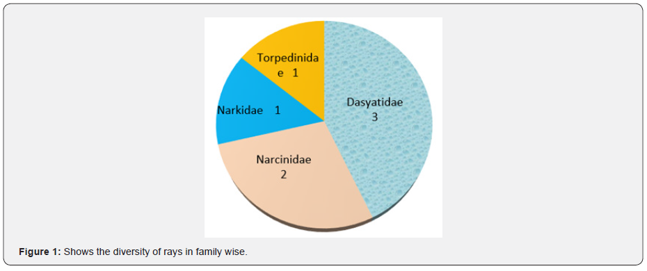
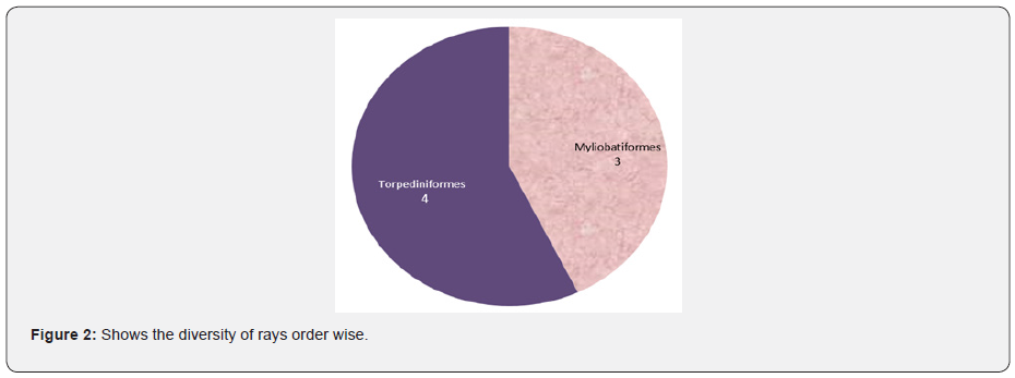
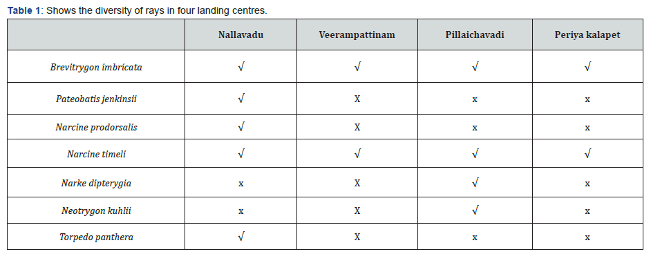
In the present study totally 7 species were recorded from four landing centre of Pondicherry coastal waters. Three species belongs to Dasyatidae family, two species belong to Narcinidae family, one species belongs to Narkidae family, and one species belongs to Torpedinidae family. Species recorded in order Torpediniformes are Narcine prodorsalis, Narcine timeli, Narke dipterygia and Torpedo panther. Brevitrygon imbricate, Pateobatis jenkinsii, Neotrygon kuhlii were belongs to order Myliobatiformes. Brevitrygon imbricate and Narcine timeli recorded in all landing centre. Narke dipterygia and Neotrygon kuhlii recorded in Pillaichavadi landing centre. Pateobatis jenkinsii, Torpedo panthera and Narcine prodorsalis recorded in Nallavadu landing centre.

Taxonomical identification characters

Brevitrygon imbricate

The scaly whipray (Brevitrygon imbricata) is a species of stingray in the order Myliobatiformes and family Dasyatidae. Disc width equal to disc length; tail shorter than body; ventral surface of disc entirely white. Small whipray with sub-oval disc, pointed snout, disc slightly longer than width. Cross-shaped band of denticles on disc, no large sized dorsal denticles. Moderately elongate tail, with a row of spear shaped thorns (up to 6); tail base depressed with 1 or 2 spines. Apex of pectoral broadly rounded. Dorsal surface pale brown to yellowish brown, margins pale, ventral side white.

Pateobatis jenkinsii

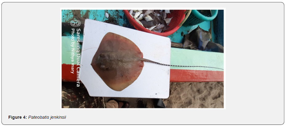
Large sized whipray with broad rhomboid disc, with a short, broad snout, band of flat denticles on central disc (poorly developed in young); granular patch on upper surface of disc and row of heart shaped dermal denticles from below the spiracle extending on to tail. Pectoral fin with rounded outer margin, pelvic fin small and narrow. Cylindrical and depressed tail with no fin folds; tail with row of upright thorns; tail shorter than length of disc. Dorsal surface uniformly yellowish brown (rarely with small dark spots on disc near tail base), black beyond tail sting, ventral white. IUCN Red List status : Vulnerable.

Narke dipterygia

Small ray with a sub circular disc with a very short snout; body entirely naked above and below, without dermal denticles or thorns. Mouth very small and not strongly arched. Eyes very small, partly/sometimes embedded in skin, spiracles larger than eye. Nostrils slit-like. Single dorsal fin originating over pelvic free rear tips. Pelvic fins broad. Dorsal surface plain to reddish brown with white bars/blotches on sides of tail extending anteriorly to above rear pelvic fin bases; and on rear of pectoral disc; ventral surface white.

Neotrygon kuhlii

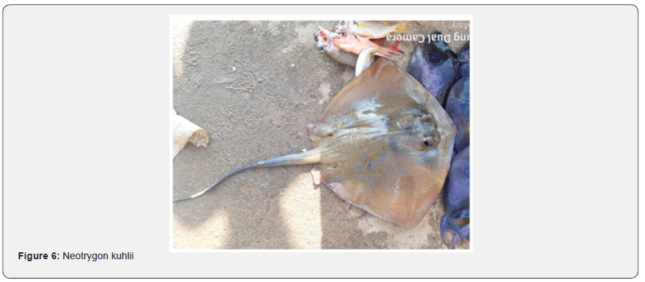
Small stingray with a smooth rhomboidal disc with somewhat angular apices; anterior margin almost straight; snout broadly rounded; tip seldom pointed. Eyes large and protruding. Denticles confined to single row of short, thorn-like structures along disc midline; cutaneous tail folds prominent, fold located beyond sting. Tail relatively broad-based, slightly depressed, slender, and compressed beyond sting; banded beyond sting. Dorsal surface greyish/greenish, or brownish with prominent bluish spots or bluish-white dark-edged ocelli; spots and ocelli variable in size and number; ventral surface mostly pale.

Narcine timlei

Disc soft, oval or Sub trapezoidal, widest near mid-length; eyes usually smaller than spiracles; eyes and spiracles joined together; spiracles rounded, with elevated smooth rims; 1st dorsal fin originating slightly posterior to pelvic fin insertion; mouth width slightly wider than internarial space; tooth bands sub equal about half of mouth width. Colour: Dorsally uniform yellowish, brownish, or purplish brown; posterior margins of dorsal fins, lateral tail region and posterior pelvic borders whitish; ventrally creamy white.

Torpedo panthera

Medium sized torpedo with small clusters of isolated, and sometimes blurry whitish spots over the disc, pelvic fins, and tail. Disc fleshy and broadly circular. Margin of spiracles with 7 short tentacles or papillae. Inter dorsal distance roughly equal to the distance between the 2nd dorsal and caudal fin. The body colour pale to reddish brown with white markings.

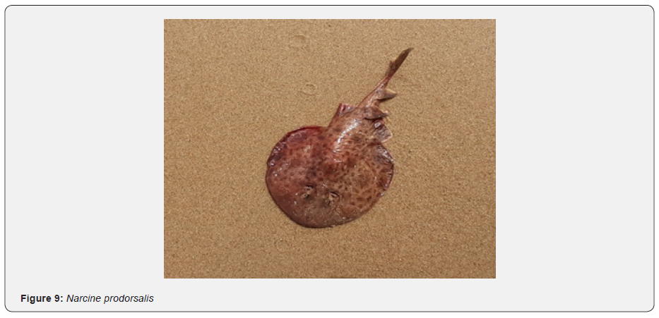
Narcine prodorsalis

The Narcine prodorsalis is medium size animals characterized by having numerous small regular dark black and brown spots and the background is light brown and an oval to heart shaped disc. The species may resemble with Chinese numbfish and small spot numbfish but relatively distinguishable by the colour pattern and smaller spots [5] (Table 1) (Figures 1-9).

Discussion

Diversity of rays on the south coast of India is denoted by 65 valid ray species [6,7]. India is one of the leading elasmobranch-fishing countries [5], with catch data showing a continuous decline in landing over the last two decades [8]. When trawl fishing is well-known for its lack of selectivity [9], a significant portion of rays was captured from trawl nets, gill nets and long lines [10]. Bengal whipray (Brevitrygon imbricate) is a common commercial ray fish in the south coast, especially in Tamil Nadu. They are rarely documented as discards in Muttom and Colachel harbours. The species is scattered in the Indo-west Pacific. They are demersal fishes occupying at depth up to 55 m. feeding of the ray fish is not well known [5]. It predominantly feeds on benthic invertebrates [11]. In the present study also Brevitrygon imbricate recorded in all landing centre.

The Brown numb fish, Narcine timlei was recorded from the southwest coast of Kerala and Tamil Nadu. The species showed less abundance in the trawl bycatch. This species is dispersed in the Indo-west Pacific region. They are benthic and more numbers in shallow waters. Little is known about the biology and fecundity ranges from 2–3 pups in each gestation [5]. Feeds mainly on bottom- dwelling organisms [12]. In the present study Narcine timeli recorded in all landing centre. Narcine prodorsalis is not very common to site and due to few reports and species-specific studies not much information on biology, full dispersal range and threats are available. Hence, N. prodorsalis has been entitled as data deficient the International Union of Conservation of Nature (IUCN) in 2009 [13]. Alifa Bintha Haque and Nazia Hossain report the 1st record of a female N. prodorsalis in Bay of Bengal, off the coasts of Bangladesh and prolonging the range of the geographical distribution of the species further west to Andaman Sea, within the Bay of Bengal.

In the present study also Narcine Prodorsalis recorded first time in Pondicherry coastal waters. Karuppasamy et al. [14] reported Neotrygon kuhlii from southwest and southeast coast of India. In the present study Neotrygon kuhlii recorded in Coromandel Coast. Eschmeyer et al. [15] reported the Pateobatis jenkinsii inGanjam coast of Orissa State, India. Similarly, Pateobatis jenkinsii recorded in the present study Nallavadu landing centre. Sujatha et al [16], studied and discussed the taxonomy and length-weight relationship of torpedo electric rays of the genus Torpedo (Pisces: Torpedinidae) off Visakhapatnam coast of India. In the present study Torpedo panthera recorded in Nallavadu landing centre. Ali Momeninejad et al. [17], recorded Narke dipterygia as a first report in Iran water. In the present study Narke dipterygia recorded in Pillaichavadi landing center. So, the present study confirming the diversity and taxonomical identification of rays in Pondicherry coastal waters.

Monday, November 21, 2022

The Inactivation of Severe Acute Respiratory Syndrome Coronavirus 2 (SARS Co-V2) by microSURE™ Hand & Skin Sanitizer; A Proprietary Engineered Antimicrobial Silica Solution with Benzalkonium Chloride - Juniper Publishers

 Biotechnology & Microbiology - Juniper Publishers

Abstract

As coronavirus disease 2019 (COVID-19) infections and related deaths continue to negatively impact the world, methods for adequate personal protection and hygiene remain a crucial topic of discussion. In the United States of America, current recommendations related to hand hygiene include washing hands with soap and water or to use a hand sanitizer with approved FDA ingredients when soap and water are not readily accessible. Today, a novel technology that utilizes a proprietary formula consisting of reengineered amorphous colloidal hydrated silica in combination with minimal amounts of agency approved inert and active ingredients has proven to not only be effective on contact but continues to protect long after it has dried. This technology was developed over a decade ago and over that period has proven to be both safe and effective. As the world is faced with yet another global pandemic the need for proven biocidal solutions are vital. The purpose of this short literature is to lay out the facts associated with both alcohol-based and non-alcohol-based hand sanitizers and present the findings demonstrated by microSURE™ Hand and Skin Sanitizer in a bio safety level-3 (BSL-3) laboratory, when tested directly against Severe Acute Respiratory Syndrome coronavirus 2 (SARS Co-V2), the virus responsible for causing (COVID-19). The details presented throughout this paper will provide insight as to why Benzalkonium chloride (BZK) must not be overlooked when deciding on the best method for protecting your body from harmful microbes and protecting your skin from potential damage.

Keywords: Coronavirus; Benzalkonium chloride; Hydrated silica; Colloidal silica; Amorphous silica; Antimicrobial; Hand sanitizer; Hand hygiene

Abbreviation: BZK: Benzalkonium Chloride; CDC: Center for Disease Control and Prevention; COVID-19: Coronavirus disease 2019, FDA: United States Food and Drug Administration; SARS-Co-V2: Severe Acute Respiratory Syndrome Coronavirus 2

Background

Over the past two decades, society has had to combat three extremely pathogenic coronaviruses that have all carried detrimental effects on the human population. [1,2] Coronaviruses are a family of viruses that are understood to cause illnesses which can vary from the common cold to severe diseases such as Middle Eastern Respiratory Syndrome (MERS) and Severe Acute Respiratory Syndrome (SARS). [1-4] Current literature suggests that each of the mentioned coronavirus outbreaks originated from an animal source, the first (SARS-CoV) is said to have transmitted from civet cats in 2002, (MERS-CoV) is said to have transmitted from camels in 2012 and the novel (SARS-CoV2), the virus responsible for coronavirus 2019 (COVID-19) is said to have been transmitted from bats in 2019[4-6]. Both of the SARS associated coronaviruses have been linked to originating in China and MERS in Saudi Arabia, however all three members of the coronavirus family began to rapidly infect individuals throughout the globe and have collectively been responsible for hundreds of thousands of deaths [7]. Currently, there are no approved vaccines or antiviral drugs for the (SARS Co-V2) virus and because of this, having the ability to control and or contain the virus in order to limit dissemination is vital. It is for this very reason why proven solutions for personal protective equipment, sanitization, cleaning, and disinfecting are eminent means of safety measures.

Although members of the coronavirus family share several commonalities, this literature will focus primarily on (SARSCoV2). Transmission of the virus is predominantly spread from human to human via respiratory droplets, which are naturally expelled as an infected individual sneezes or coughs [1-3]. Recent studies have proven that the virus has the ability to persist on inanimate surfaces such as plastic, stainless steel and cardboard and can remain on these surfaces for days at a time [3]. Other studies have concluded that the novel virus has the ability to linger on human skin far longer than the commonly known flu virus can, stating that the virus has the potential to remain on human skin for up to 28 days [8]. Benzalkonium chloride (BZK) is an active ingredient present in many of today’s consumer products. The use of BZK was first reported as a promising skin disinfectant in 1935 [9]. Since then its use has evolved to being much more than just a skin disinfectant. BZK is currently being utilized not only in personal care products such as hand sanitizers, soaps, lotions, and shampoos, but it is also found in spray disinfectants, pharmaceutical products such as eye drops, skin and wound antiseptics, mouthwashes, surgical disinfection products, burn treatments and more. One of the major actions of BZK relates to its antimicrobial capabilities, the biocidal actions of BZK create a dissociation of the unwanted microbe’s membrane lipid bilayers and stimulates the leakage of cellular contents, therefore compromising its permeability and eliminating its presence [10].

Research Based Evidence

BZK has been proven effective against bacteria, viruses, fungi and protozoa [11]. The reason for its widespread use of application aside from its antimicrobial efficacy is based on the fact that it is easier on the skin than the majority of other skin disinfectants, especially when being compared to alcohol. Unlike alcohol, BZK can be used directly on open skin or an open wound without causing damage to the wound bed or creating a painful sting and or burning sensation. When it comes to hand sanitizers, there has been a lot of debate as to whether an alcohol-based sanitizer or non-alcohol-based sanitizer is the better option.

In 1998, a study using non-alcoholic based sanitizer with benzalkonium chloride as the active ingredient was completed via FDA performance standards and determined that the benzalkonium chloride-based sanitizer performed better than alcohol-based hand sanitizer after repeated use [12]. It wasn’t until recently that BZK has begun to once again become a common topic of discussion as it relates to the current ‘coronavirus disease 19’ (COVID-19) pandemic caused by ‘Severe acute respiratory syndrome coronavirus 2 (SARS Co-V2) and the measures society is taking in order to eliminate the risk of spreading or contracting microbes responsible for causing illness and disease. Of late, there has been a need for more BZK studies and research regarding hand sanitizers, as the amount of data available is still limited when compared to the more commonly used alcohol-based products.

As history is often known to repeat itself, newer studies continue to prove what previous research pointed out, and that is that benzalkonium chloride hand sanitizers demonstrate greater effectiveness than alcohol-based hand sanitizers. For example, in an infection control study performed by Bondurant et. al. to evaluate the effectiveness of BZK as the active ingredient in reducing transient skin contamination with staphylococcus aureus in health care workers, as compared to the effectiveness of an ethanol-based hand sanitizer, research found that the benzalkonium hand sanitizer significantly reduced staphylococcus aureus versus an ethanol sanitizer and also concluded that BZK had greater skin presence and persistence [13]. This same research study was able to reference multiple experiments which have confirmed that BZK- manufactured products demonstrated ‘persistent antibacterial efficacy’ even up to four hours after bacterial contact with skin, as opposed to alcohols efficacy duration, which has only been documented to reach around 10 minutes maximum [13]. Although efficacy is extremely vital when it comes to antimicrobial properties associated with hand sanitizer, the other crucial aspect is the affect it has on people’s skin and overall health. It is important to note the differences in the amount of active ingredient present between each set of hand sanitizers, BZK is usually only around .13% of the final end product, while alcohol normally exists in excess or 60% or greater of the final end product. Today, society seems to be much more focused about the ingredients present in consumer products, as well as living a health-conscious and safer lifestyle. There has been an increase in the number of vegetarians, vegans and plantbased diets, there has been an increase in the number of health club memberships, and social media has been helped educate people on the different changes they can make to live a more health-conscious lifestyle.

With that said, appearance and personal care has also become very important and because individuals have become so concerned with what products may harm their bodies, the use of alcohol-based hand sanitizers is no longer the only go to option. Many big-name hand sanitizers use alcohol as an active ingredient and the reasons for this are quite clear. Alcohol is ridiculously cheap, it is effective at eliminating many of the common germs responsible for causing the spread of infection directly on contact, and it is widely available. In addition to these factors, alcohol has been around for a much longer time period and because of this it would be safe to assume that it has become widely accepted by society as the ‘only’ reliable option. Unfortunately, alcohol is not as safe as many people believe it is and there have been several studies which prove this.

Research has proven that the continued use of alcoholbased hand sanitizers is responsible for several underlying skin reactions. These reactions include dryness, itching, irritation, cracking and even bleeding. [14] To most individuals, the notion of dry or cracked skin may not seem to be that serious of an issue, but this perception drastically changes when realizing the detrimental health effects that can arise from these ‘little’ issues.

The cracking and dryness of the skin leads to changes in the skin flora, resulting in more frequent bacterial colonization and bacterial susceptibility, particularly by staphylococcus aureus and gram-negative bacilli. [15] This means that as consumers continue to use alcohol-based hand sanitizers, over time, as the skin dries and the more alcohol-based sanitizer you use increases, so does the risk for infection. Therefore, the product that was originally being used to help eliminate the risk of infection, instead becomes the culprit responsible for inviting infection.

Current Guidelines

Current Center for Disease Control and Prevention (CDC) guidelines suggest that alcohol-based hand sanitizer with a high concentration and or antiseptic hand soap for hand washing are the best options for hand hygiene. [16] Alcohol has been proven to be effective at killing bacteria when concentrations are between 60% and 90% and works mainly because of its ability to denature proteins. [5,9] However, studies have actually shown these higher concentrations of alcohol essentially lose their effectiveness because water is needed as an adjunct in order for maximum potency to be achieved [17]. In 2016, the United States Food and Drug Administration (FDA) removed 19 antimicrobial ingredients from the list of allowed consumer products, BZK was not one of them [18]. Although BZK was not removed from the FDA’s allowed antimicrobial list, The CDC notes that the reasons for its statements regarding high concentration alcohol-based hand sanitizers instead of BZK or other various quaternary ammonium products is because such products necessitate further studies [17,18].

Materials and Methods

In September of 2020 test results of microSURE™ Hand & Skin Sanitizer directly against the SARS Co-V2 virus were completed and released. This testing was accomplished to demonstrate residual efficacy of the solution and was conducted in a bio-safetylevel 3 (BSL3) laboratory. A very concise protocol was followed, as indicated below.

Petri dishes were coated with the following treatments:

a. diH20- deionized water (Negative treatment control)

b. MicroSURE, formulation of IP material and Benzalkonium chloride (BZK)

The coating procedure was as follows:

a. 100mm and 60mm petri dishes were used to create a sandwich to expose virus to treated surfaces

b. The inner bottom surface of 100mm dishes were filled with 1.0 ml material, to cover the surface. The bottom of a 60mm dish was placed in the material, exposing the bottom to the material.

c. The petri dishes sat for 10 minutes and then the coating materials were removed, and the dishes allowed to dry.

d. Petri dishes were packaged in pairs with the 60mm dish inside the 100mm dish with the lid on the 100mm dish and stored at room temperature.

e. Coated petri dishes were then subjected to viral inactivation testing at several different times points after coating: 3 hours, 1 day, 2 days and 8 days.

Materials were transferred to the BSL3, and inactivation tested as follows at 4 different time periods after coating–3 hours, 1 day, 2 days and 8 days:

a. Quintuplate dish sets were transferred into the BSC.

b. 20 μl SARS CoV2 virus (5e6 FFU/ml, so total of 1e5 FFU) placed on treated 100mm petri dish.

c. The 60mm petri dish was placed on top, sandwiching the inoculum between the dishes.

d. The dishes were incubated at RT in humidified box for 30 minutes.

e. The dishes were separated and rinsed 5x with 0.5 ml of infection medium (DMEM with 2%FBS and antibiotics).

f. The 0.5 ml of infection media was transferred to sterile tubes and tested in a FFU assay.

FFU assay (immunostain for focus-forming units)

a. To each well of a 96-well plate seeded with Vero cells, add 50 μl neat media (from coverslip rinsing) and dilute 10-fold to 10-5

b. Incubate plates for 1 hr and then overlay with 50 μl Methycellulose medium

c. Incubate approximately 24 hr at 37°C, 5% CO2

d. Remove media, washing with PBS and fix plates with 80:20 MeOH: Acetone

e. Remove plates from BSL3 following approved protocol

f. Immunostain plates with anti-SARS-CoV-2 Spike mAb, 1C02, using anti-human IgG-HRP to visualize FFU. Count FFU comparing diH2O-treated to test material-treated coverslips.

Results

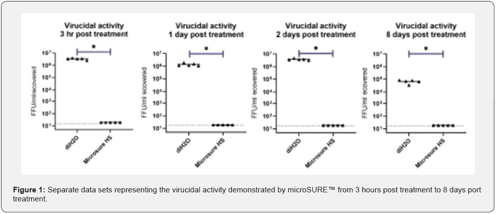
Treatment of the surfaces by the test material microSURE™ significantly reduced the amount of infectious material, SARSCoV- 2, at all-time points after coating compared to the control, diH2O-treated surfaces, with a 30minute virus contact time. These results, which show a >3-4 log reduction at all time periods, is both statistically significant (p<0.05 t-test, post hoc Mann- Whitney test), as well as biologically relevant (Figure 1).

Conclusion

The microSURE Hand and Skin Sanitizer proved to be efficacious against the novel SARS CoV-2 virus throughout the entire 8-day testing period. Based on this testing, this solution that consists of a proprietary formulation in combination with BZK is a superior alternative to alcohol-based solutions as it relates to efficacy, specifically virucidal residual efficacy. Overall, when deciding on the best hand sanitizer to use, it is important that the product is both efficacious and as safe as possible for the skin. Although both BZK and alcohol have positive effects when being used as an active ingredient in hand sanitizer, there are several factors which differentiate the two. Alcohol has been around for a much longer time period and has been studied in much greater detail because of this. Alcohol is also readily available, inexpensive, and widely accepted by the general public because there was never enough literature to suggest a superior alternative, until now. As mentioned previously, BZK has been around for over 90 years, yet the amount of research and experiments were never at a level like that of alcohol.

Yet, even with limited data, the FDA still did not remove the ingredient from its approved list of antimicrobial active ingredients for accepted use in hand sanitizers, and the reasons for this are obvious. BZK has been proven to be more effective than alcohol as an active ingredient throughout numerous studies. In comparison to alcohol, BZK has specifically been proven to combat some of the more problematic bacterial infections, such as those associated with staphylococci and gram-negative bacteria. Furthermore, when looking the results seen with the microSURE Hand and Skin Sanitizer with BZK, this solution not only demonstrated superior efficacy against the novel coronavirus, but it also demonstrated a residual efficacy that is unmatched by any other alcohol-based sanitizer and other sanitizers with BZK as an active ingredient. In terms of health effects, the skin reactions documented with alcohol-based sanitizers tend to be much more common and serious when compared to BZK. Although current CDC guidelines suggest using a high concentration alcohol-based hand sanitizer in an attempt to prevent bacterial infection, countless studies have been conducted and prove that high concentration alcohols are not potent by themselves and that alcohol-based hand sanitizers come with the increased risk for skin reactions to occur. The continued use of alcohol-based hand sanitizer has been well documented, and the negative affects it has on skin can be debilitating when it comes to providing a reliable defense against infection. This is because as consumers continue to use this type of hand sanitizer, the skin dries and cracks, altering the normal skin flora and therefore becomes susceptible to unwanted and harmful microorganisms. The important concept to remember is that there are other available options besides alcohol-based hand sanitizers. Time and time again, BZK-based hand sanitizers have been proven to be both efficacious and safer on skin than alcohol-based sanitizers. New literature is beginning to focus more on BZK and the hope is that as studies continue to become available and findings are more publicized, the general public will learn that there is another alternative to using alcohol-based hand sanitizers and this alternative is one that has continues to show preeminence over the commonly used alcohol based hand sanitizers currently available.

Click here: https://juniperpublishers.com/index.php   
Artificial Intelligence System for Value Added Tax Collection via Self Organizing Map (SOM)- Juniper Publishers

  Forensic Sciences & Criminal Investigation - Juniper Publishers Abstract Findings:  Based on our experiments, our approach is an effec...