Friday, May 31, 2024

Characterization of Headache in Patients with Temporomandibular Joint Disorders - Juniper Publishers

 Advances in Dentistry & Oral Health - Juniper Publishers

Abstract

Objective: The incidence and characteristics of headache in patients with temporomandibular joint disorder (TMJD) had not been described previously. The aim of the current study was to discuss this issue.

Materials and methods: The study involved 200 patients with TMJD and 200 control subjects. The incidence and characteristics of headache, as well as associated symptoms were compared between the two groups.

Results: The incidence of chronic headache was significantly higher in the TMJD patient group (88%) than in the control group (37%); (P=0.001). The headache patterns included unilateral throbbing migraine headache, as well as bilateral regional headache.

Conclusion: The results lend support to the theory of overactivity of pericranial muscles in TMJD, as well as extracranial –intracranial cross-talk in sensory nociceptive nerve fibers.

Keywords: Headache; Temporomandibular Joint Disorder; Chronic pain

Abbreviations: TMJD: Temporomandibular joint disorder; CNS: Central nervous system; TTH: Tension-type headache; TMD: Temporomandibular disorder; TMJs: Temporomandibular joints; DC: Diagnostic Criteria; MRI: Magnetic resonance imaging; ICHD: International Classifications of Headache Disorders; CT: Computed tomography

Introduction

The pathogenesis of headaches, especially primary headaches, is probably multifactorial. Various structures in the cranium have been implicated in the causation of headache; with central nervous system (CNS) plastic changes contributing to the chronicity of the condition [1,2]. The cranial calvarial bones are covered from outside by skin and pericranial myofascial structures. Myofascial trigger points are prevalent in both tension-type headache (TTH) and migraine [3,4]. Underneath the calvarial bones is the dura mater. The dura mater has a rich supply of nociceptors and mechanoreceptors; both of which are implicated in the pathogenesis of headaches [5,6]. Animal studies, as well as human imaging studies, have stressed the importance of sterile dural inflammation as a universal finding in primary headaches; thereby sensitizing the receptors [7,8]. The dural inflammation is associated with an increased barrage of action potentials, along nociceptive fibers to the trigeminal ganglion [9,10]. Furthermore, non-trigeminal nociceptive afferents from the occipital dura mater and cervical structures contribute to headaches; being mediated through the upper three cervical nerves [11,12]. Nociceptive afferents from head and neck structures converge onto the subnucleuscaudalis in the brain stem and upper cervical spinal cord [13].

Temporomandibular disorder (TMD) is an umbrella term for pain and dysfunction involving the masticatory muscles and the temporomandibular joints (TMJs) [14]. Most studies on TMD patients report that headache is a frequently associated symptom [15-17]. The Diagnostic Criteria for Temporomandibular Disorders (DC / TMD) considers headache as one of the diagnostic categories of TMD; in addition to TMJ and masticatory muscle disorders [18]. Temporomandibular joint disorder (TMJD) represents a major category of TMD; although, occasionally, it is not associated with painful symptoms [19]. Radiological studies, especially magnetic resonance imaging (MRI) have confirmed the various clinical signs elicited in patients with TMJD [20]. Pathophysiologically, a dysfunctional, abnormal TMJ cannot be considered as an isolated disorder. Many factors, including bruxism, malocclusion, lateral pterygoid muscle dysfunction (which is centrally-mediated) contribute to the joint dysfunction [21,22]. These factors cause abnormal stresses on the TMJ, which are associated with ligament laxity, a low-grade joint inflammation, and deterioration in joint lubrication; all of which predispose to a dysfunctional joint [23,24]. Pain in TMJD is an intringuing issue, and there is often a poor correlation between the degree of pain over the joint and the degree of joint pathology [25,26]. A recent systematic review on structural and functional brain MRI studies revealed aberrant CNS changes in TMJD with pain. The CNS aberrations involved sensory, motor, and affective regions in the brain. Interestingly, the imaged brain aberrations were frequently resolved after intra-oral splint therapy [27].

From a clinical perspective, headache disorders tend to be under-recognized and under-treated, especially in developing populations [28]. The reason for this is that the diagnosis of primary headaches, which is clinically based, frequently does not fulfill the strict criteria provided by the International Classifications of Headache Disorders (ICHD). In these situations, the diagnoses of (probable TTH) and (probable migraine) are often made [29,30]. Furthermore, TTH and migraine frequently coexist [31]. In contrast to the rather diffuse head pain in primary headaches, TMJD pain is localized infront of the ear, being frequently associated with joint noises and functional limitations [32,33]. Additionally, TMJD is commonly associated with otoneurological and alleged sinus symptoms; known as Costen’s syndrome [34-36]. Although the headache associated with masticatory myofascial pain had been previously characterized, headache associated with TMJD had not been previously specifically addressed. Notably, headache associated with masticatory myofascial pain most commonly resembles TTH with a bilateral temporal location [37-39]. On the other hand, the TMJ contains; in addition to nociceptors, mechanoreceptors (proprioceptors), which guide mandibular movements through their inputs to the central pattern generator in the brain stem [40]. The distribution of the proprioceptors in the joint is altered in cases with joint pathology [41]. According to the (gate control theory), altered proprioceptive input to the CNS can alter pain sensation experienced in patients with TMJDs [42]. In addition to exaggerated nociceptive input from the TMJ in TMJD, altered proprioceptive input to the CNS may also contribute to CNS plastic changes causing a peculiar type of pain, in addition to abnormal jaw mechanical behavior [43]. From these perspectives, the aim of the current study was to characterize the incidence and pattern of headache in patients with documented TMJD. As far as the author is aware, and after a Pub Med literature search from 1970 till 2023, this issue had not been previously addressed.

Materials and Methods

The current study was a clinical study conducted at the craniofacial pain clinic at El-Sahel Teaching Hospital in Cairo, Egypt. The study period was 6 months, starting on the 1st of January 2023. Ethical approval was obtained from the Ethics Committee of the General Organization for Teaching Hospitals and Institutes in Cairo (Approval number: 83- 2022). Informed consent was obtained from the patients and control subjects to participate in the study. The patient group consisted of consecutive adult subjects with chronic jaw pain, who reported the experience of joint sounds during mandibular movements. The presence of joint sounds was later confirmed on examination. This criterion allowed objective evidence of TMJD for inclusion in the study. Patients who had jaw pain (arthralgia and/or myalgia) without joint sounds were not included in the patient group. The control group consisted of adult relatives of the patients presenting to the clinic, who experienced no sounds elicited on movement of the mandible. The absence of joint sounds in the control group was later confirmed on examination, revealing clinical evidence of normal TMJs. Exclusion criteria in both groups consisted of any acute disorder in the head and neck, as well as previous macrotrauma to the jaw.

A questionnaire and examination findings sheet were filled in by the author. The main criterion investigated in the current study was the reporting of chronic headache for at least 6 months in both cohorts of participants. In the participants suffering from headache, the characteristics of the head pain were noted according to the description of the subject (pressing or throbbing). Furthermore, the laterality of the head pain was noted (unilateral versus bilateral). The most commonly invloved region in the head was addressed (diffuse, frontoorbital, temporal, occipital, or at the vertex). The presence of otalgia, facial pain, and/or neck pain in association with the headache was reported. Questioning also included the presence or absence of autonomic symptoms accompanying the headache, including nasal congestion, rhinorrhea, and/or lacrimation. Additionally, conditions possibly predisposing to TMJD were asked about (bruxism and dental status). Relevant to TMD, a diagnosis of depression was noted if present. For the reporting of depressive symptoms, the patient had to have been diagnosed by a psychiatrist or reported daily weeping. A previous history of open-lock was addressed. Otological symptoms pertaining to Costen’s syndrome (subjective hearing loss, subjective tinnitus, and/or vertigo) were explored.

Examination of the subjects followed a standard protocol. The TMJ was palpated, noting for any exquisite tenderness (by observing a palpebral reflex). Tenderness over the TMJ was assigned as arthralgia. Any TMJ noises elicited during opening and closing the mouth were classified as click (snapping noise), or crepitus (grating noise). An audible noise was confirmed by palpation of the joint. The presence of joint noises was used to categorize the subject as a TMJD patient. Next, the temporalis and masseter muscles were palpated, noting for any exquisite tenderness (eliciting a palpebral reflex), and any referral pattern for the pain (denoted as myalgia). The oral cavity was observed for the dental status; and accordingly, the subjects were classified as having normal dentition, being partly edentulous, or totally edentulous. While the mouth was fully opened, any trismus was noted (inter-incisal distance less than 40 mm). While the mouth was closed, any open-bite deformity was observed. Any gross deviation of the mandible during opening and closing the mouth was noted. Following examination of the stomatognathic system, examination of the ears, nose, pharynx, and eyes was performed.

Statistical analysis

Results were expressed as mean ± standard deviation or number percent (n %). Comparison between categorical data (n %) was performed using Chi square test. Comparison between mean values of age in the two groups was performed using unpaired t test. Statistical analysis was performed using SPSS computer program (version 19 windows). P value ≤ 0.05 was considered significant.

Results

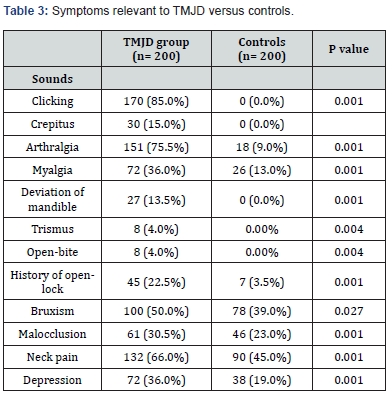
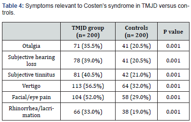
The current study comprised 200 TMJD patients and 200 control subjects. The mean age of the patient group was 40.34 ± 12.36 years, while the mean age of the control group was 39.1 ± 12.38 years (P= 0.319). There was no significant difference in the male-to-female ratio between the two groups (Table 1). The incidence of chronic headache in the patient group (88%) was significantly higher than in the control group (37%); (P= 0.001). Migraine headache was unilateral and throbbing. Its incidence was significantly higher in the patient group (13%) than in the control group (6%); (P= 0.017). The incidence of bilateral regional headache was significantly higher in the patient group than in the control group for temporal, fronto-orbital, and occipital locations (Table 2). Table 3 reveals clinical features pertinent to TMJD in patients versus controls. Notably, clicking was noted in 85 % of TMJD patients, while crepitus was elicited in 15 % of TMJD patients. The control subjects showed clinically normal TMJs. All other symptoms and signs related to the stomatognathic system had higher incidence in TMJD patients versus control subjects (Table 3). Table 4 reveals the incidence of symptoms related to Costen’s syndrome in patients and control subjects. All otological and alleged sinus symptoms had a higher incidence in TMJD patients versus controls.
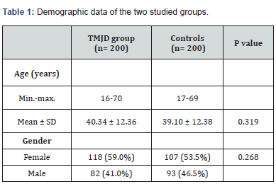
Data are expressed as mean ± SD or number (%).

TMJD= temporomandibular joint disorder.

p> 0.05= not significant.

Discussion

Temporomandibular joint disorder (TMJD) involves a set of functional and pathological changes affecting the TMJ [44]. As the two TMJs cannot function independently during mandibular movements, TMJD is very frequently a bilateral disorder [45]. The current concept is that the pathogenesis of TMJD involves excessive or abnormal stresses on the TMJ, that exceed the adaptive capacity of the joint tissues [46]. These abnormal stresses acting on the TMJ lead to laxity of the joint ligaments, thereby predisposing to disc displacement [47]. Furthermore, an associated low-grade inflammation in the TMJ has been reported to initiate a cascade of events, including impaired lubrication and increased friction coefficient in the joint [48,49]. The result of increased friction in the joint is a disturbance in disc-condyle complex harmony and predisposition to disc displacement and degenerative joint disease [50,51]. Moreover, increased tension in the lateral pterygoid muscle had been postulated to account for the most common form of disc displacement (anteromedially) [52]. Clinically, reducing disc displacement manifests as clicking, whereas degenerative joint disease manifests as crepitus [53]. In the current study, the TMJD patient group demonstrated clicking in 85% of the patients, whereas crepitus was present in 15% of the patients. In the control group, there was absence of joint noises during mandibular movement. A painful, tender TMJ is designated as TMJ arthralgia. The current view is that the degree of TMJ arthralgia is correlated with the levels of inflammatory mediators and neuropeptides, which sensitize and activate afferent nociceptive nerve fibers in the joint [54,55]. The current study revealed TMJ arthralgia in 75.5% of patients, versus 9% of control subjects (P= 0.001).

A continuous barrage of action potentials from the sensitized and activated TMJ nociceptors contribute to the chronicity of TMJ pain [56]. In pain literature, chronic pain is defined as a multidimensional experience involving negative sensory, affective, and cognitive aspects [57]. These aspects are associated with corresponding abnormal structural and functional brain regions, seen on neuroimaging [58]. Currently, there are two proposed theories for these brain changes in chronic painful TMJD. The first theory involves sensitization and activation of CNS pathways relevant to chronic pain in its three dimensions, consequent on a continuous barrage of action potentials from the peripheral TMJ nociceptors [59,60]. The alternative hypothesis is the activation of a pre-set brain pain neuromatrixby stress and peripheral pain [61-64]. In both theories, the result of chronic painful TMJD is the unwanted experience of pain, alteration in homeostatic mechanisms; and importantly, an altered motor output to the masticatory and cranial muscles [65-67].

The altered motor output to these muscles contributes to trigger points, commonly observed in patients with TMJD associated with headache [68,69]. Temple headaches, frontoorbital headaches, and occipital headaches represent common presentations of headache disorders. Various studies propose that these regional headache disorders primarily arise due to entrapment of respective sensory nerve fibers by the overcontracting pericranial muscles. These nerve fibers include various trigeminal and upper cervical nerve fibers which course through the muscles [70-72]. In the current study, 26% of patients with TMJD had headache at the temples; 21% had headache with a fronto-orbital location; and 12% had headache at the occipital region (Table 2). In the control subjects, 12.5% had headache at the temples, 10.5% had headache at the fronto-orbital region; and 2% had headache at the occipital region. These regional headaches had a pressing, tightening quality and were of bilateral location. The difference in incidence of these respective headaches between the TMJD patient group and the control group was statistically significant (Table 2).
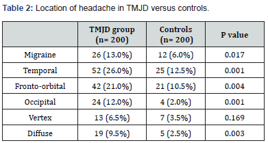
Data are expressed as number (%).

TMJD= temporomandibular joint disorder.

p> 0.05= not significant; p≤ 0.05= significant.
Data are expressed as number (%).

TMJD= temporomandibular joint disorder.

p≤ 0.05= significant

TMJD may act as a migraine trigger in genetically predisposed individuals [73]. A recent large longitudinal population study had suggested that the relation between TMJD and migraine is bidirectional [74]. A biological explanation for the association between TMJD and migraine is that extracranial and intracranial trigeminal innervation forms a functional unit [75]. This concept is supported by numerous animal studies, which reveal that dural afferents project through the calvarial bones to innervate extracranial structures [76]. It is hypothesized that there would be a physiological and pathological cross-talk between trigeminal nociceptive nerve fibers in both the intracranial and extracranial compartments [77,78]. Activation of the meningeal and meningovascular nociceptive afferents, with the release of various neuropeptides, results in a sterile dural inflammation [79]. Activation of dural mechanoreceptors, in the context of dural inflammation, explains the throbbing pain, experienced in some forms of headache, especially migraine [5]. Migraine headache is typically a unilateral throbbing headache, associated with photophobia, phonophobia and nausea/vomiting [80]. In the current study, headache typical of migraine was experienced in 13% of TMJD patients versus 6% of controls (P=0.017).

Totally, 88% of TMJD patients in the current study reported chronic headache, in contrast to only 37% of control subjects (P=0.001). Importantly, 66% of TMJD patients also reported chronic neck pain versus 45% of control subjects (P=0.001). Neck pain is mediated via the upper cervical nerves, and it is commonly associated with headache and TMD [12]. Indeed, the cranio-mandibular-cervical complex has been considered as an anatomical and functional unit, with neuromuscular restoring forces stabilizing this unit against the force of gravity [81,82]. Epidemiological, clinical, and imaging studies have revealed a significant association between TMJD and neck pain [83-86]. It is postulated that this association might be due to the convergent nociceptive input from the TMJ and neck structures onto the subnucleuscaudalis in the brain stem [87]. An alternative theory that was also postulated involves abnormal proprioceptive afferents from the TMJ that activate the trigemino-cervical reflex, resulting in abnormal neck muscle contraction and neck pain [88]. The trigemino-cervical reflex could also be implicated in migraine and TTH [89,90], thereby providing a putative explanation for the association of TMJD, headache, and neck pain.

Further adding to the morbidity in patients with TMJD, is the frequent association of symptoms pertaining to Costen’s syndrome. In 1934, James Costen reported (a syndrome of ear and sinus symptoms dependent upon disturbed function of the temporomandibular joint) [91]. These symptoms have recently been validated by audiometric tests and computed tomography (CT) scans of the paranasal sinuses [34-36]. In the current study, hearing loss, subjective tinnitus, and vertigo were reported in 39%, 40.5%, and 56.5% of TMJD patients, respectively. The incidence of these symptoms in control subjects was 20.5%, 21%, 32%, respectively (Table 4). The difference in incidence of these symptoms between the two groups is statistically significant. The origin of these symptoms in TMJD patients was postulated to be due to an increased tension on the malleus of the middle ear from a displaced TMJ disc, leading to abnormal position of the stapes at the oval window [92-94]. This, in turn, would lead to altered firing patterns from inner-ear hair cells [95]. Additionally, inputs from higher brain centers onto relevant brain stem nuclei may exaggerate these symptoms in conditions of pain, stress, anxiety, and lack of sleep, via top-down mechanisms [96,97]. Alleged sinus symptoms, especially facial pain, are also common in patients with TMJD [36]. In the current study, facial/eye pain was present in 52% of TMJD patients, versus 29% of control subjects (P=0.001). Facial/ eye pain had been attributed to the convergence of nociceptive branches of the trigeminal nerve onto the subnucleuscaudalis in the brain stem [98]. Jones described (midfacial segment pain), as a condition analogous to tension-type headache, with a similar pathophysiology, in patients with normal nasal endoscopy and clear sinus CT scans [99-101]. He postulated that this condition was attributed to abnormal nociceptive afferents from a regional source. Interestingly, Costen, in his original reports, mentioned that the otologic and alleged sinus symptoms were alleviated by correction of dental bite, in his patients with malocclusion [91]. Correction of the dental bite probably also ameliorates TMJD pain and headache [102,103]. The incidence of malocclusion in the current study was 30.5% in TMJD patients, versus 23% in control subjects (P=0.001).
Data are expressed as number (%).

TMJD= temporomandibular joint disorder.

p≤ 0.05= significant

Physical and emotional stress appear to be a common denominator in patients with headache, neck pain, and TMJD [104,105]. The stress response involves a pronounced sympathetic outflow and the release of stress hormones, and it is associated with a documented overactivity of head and neck muscles [106,107]. Moreover, the stress response is associated with peripheral and central sensitization in pain pathways, thereby contributing to the chronicity of headache, neck pain, and jaw pain [108,109]. Biomechanically, overactivity of masticatory muscles, as in bruxism, causes abnormal stresses on the TMJ, leading to its deterioration [110]. Overactivity of the lateral pterygoid muscle leads to the most common form of disc displacement (anteromedially) [111]. Clinically, bruxism, as a model of stress, is associated with TMJD [112,113]. In the current study, a report of bruxism was found in 50% of TMJD patients, versus 39% of control subjects (P=0.027).

The main strength of the current study was that it shedded light on the patterns of headache and associated symptoms in patients with TMJD, a previously undiscussed topic. The presence or absence of TMJ noises allowed an objective clinical distinction between TMJD patients and control subjects, respectively. Furthermore, the cohorts of TMJD patients and control subjects in the current study were of comparable socio-economic conditions, thereby allowing reliable distinctions to be made between both groups. The major limitation of the current study was that the reporting of symptoms depended entirely on the recalling of symptoms by the participants, which was totally subjective. From this view, the diagnosis of headache depends mainly on the verbal description of the patient. Apart from migraine, which has a classic presentation, no attempt was made to classify the headache from a diagnostic point of view. Rather, a description of headache sites was used in the current study, which was generally of a pressing, tightening quality and of bilateral location. The variable locations of headache in the study lends support to the theory of sensory nerve entrapment by respective over-contracting muscles. This notion emphasizes the recently-documented fact that extracranial and intracranial nociceptive nerve fibers act as a functional unit. A further limitation of the current study was that the author was not blinded, as regards the participant’s clinical features. However, the author has several publications regarding TMJD, which may validate the clinical signs elicited in the current study, as well as the recording of the relevant symptoms.

Conclusion

The current study revealed a higher incidence of headache in patients with TMJD, compared to control subjects. In addition to the typical migraine unilateral, throbbing headache, the patterns of headache frequently revealed a bilateral regional distribution with a pressing, tightening quality. This lends support to the theory of overactivity of pericranial muscles in TMJD, causing entrapment of isolated sensory trigeminal or cervical nerve branches, thereby explaining the regional headache patterns. Moreover, the connections between the extracranial and intracranial sensory nerve fibers account for the frequency of migraine in several participants.

To Know More About Advances in Dentistry & Oral Health Please click on:
https://juniperpublishers.com/adoh/index.php


For more Open Access Journals in Juniper Publishers please click on: 

Thursday, May 30, 2024

An Overview of the Latest Insights and Innovations in Pylephlebitis: A Comprehensive Review - Juniper Publishers

 Gastroenterology & Hepatology - Juniper Publishers


Abstract

Pylephlebitis, a rare condition marked by infective thrombosis in the portal vein, presents diagnostic and therapeutic challenges. This review explores its epidemiology, etiology, clinical manifestations, diagnosis, and treatment. Primarily a complication of intra-abdominal infections, diverticulitis and appendicitis are prevalent causes. Clinical presentation mirrors intra-abdominal infections, demanding a high index of suspicion. Diagnosis involves clinical examination, laboratory investigations, and imaging, with CT and ultrasound commonly employed. Treatment integrates broad-spectrum antibiotics and surgical interventions targeting the infection source. Prognosis, influenced by prompt diagnosis and complications, reports mortality rates of 7-30%. Early recognition and aggressive management are pivotal for favorable outcomes, emphasizing a comprehensive approach to Pylephlebitis prevention, diagnosis, and treatment.

Keywords: Pylephlebitis; Portal vein thrombosis; Intra-abdominal infections; Leukocytosis; Gastrointestinal distress

Abbreviations: CT: Computed Tomography; MRI: Magnetic Resonance Imaging; CRP: C-Reactive Protein; ESR: Erythrocyte Sedimentation Rate; ALP: Alkaline Phosphatase; ALT: Alanine Aminotransferase; AST: Aspartate Aminotransferase; GGT: Gamma-Glutamyl Transpeptidase; US: Ultrasonography; PET: Positron Emission Tomography

Introduction

Pylephlebitis, a rare condition, refers to infective suppurative thrombosis within the portal vein, typically arising as a complication of intra-abdominal suppurative processes. Its incidence is low, estimated at approximately 0.37-2.7 cases per 100,000 person-years. Virtually any intra-abdominal or pelvic infection with drainage through the portal venous system can lead to pylephlebitis. Notably, diverticulitis (26.5%) and acute appendicitis (22%) are the most frequent underlying causes attributed to the anatomical proximity of the inflamed structures to the portal system. Additionally, contiguous infections such as cholangitis/cholecystitis (3.5%) or pancreatitis (5.5%) can precipitate pylephlebitis. In 8.5% of cases, hepatic abscesses are implicated. Pathogens commonly associated with pylephlebitis include Escherichia coli, Bacteroides spp., and Streptococcus spp., while fungal and parasitic infections are occasional etiologies [1,2]. Symptomatically, patients typically present with fever, abdominal pain, nausea/vomiting, and diarrhea. Laboratory investigations often reveal leukocytosis, elevated C-reactive protein (CRP) and erythrocyte sedimentation rate (ESR), hyperbilirubinemia, anemia, hypoalbuminemia, and thrombocytopenia. Diagnosis hinges on confirming portal vein thrombosis in febrile patients with bacteremia, achieved through pathogen identification via culture and imaging studies such as CT scan, MRI, angiography, endoscopic ultrasound, or positron emission tomography [1,2]. Treatment entails broad-spectrum antibiotics tailored to the suspected source of infection and likely pathogens, with adjustments based on culture and antimicrobial susceptibility testing results. Therapy typically spans four to six weeks post-symptom onset, initially administered intravenously until significant clinical improvement is observed, followed by oral administration, often a combination of metronidazole and fluoroquinolones, to complete the course [1,2].

Etiology & Pathogenesis

Pylephlebitis is a rare condition associated with intra-abdominal infections in areas neighboring or drained by the portal vein and its tributaries [3,4]. A retrospective study with one hundred relevant cases identified diverticulitis as the leading cause of pylephlebitis, followed by appendicitis [5]. Another study with ninety-five patients highlighted pancreatitis as the main predisposing factor [6]. Bacteremia was present in 88% of cases, with Bacteroides fragilis and Escherichia coli being the most common agents [7,8]. Pylephlebitis results from thrombosis of a vein adjacent to an infected area, extending to main branches and damaging additional tissue and organs. In cases involving cholangitis/cholecystitis and hepatic abscess, it is challenging to determine if these conditions caused or resulted from pylephlebitis. Some cases have no established source of infection [8]. This infectious pathology can lead to portal vein thrombosis. Its multifaceted etiology includes bacteria as a primary cause [3]. Bacteria colonizing smaller thrombophlebitis veins can lead to septic emboli and pylephlebitis [6]. A recent systematic review showed that out of 220 individuals, a single pathogen was found in 155 cases (70.5%), with a median age in the 50s and the youngest at 20 years old [3-5]. Studies revealed diverticulitis and appendicitis as leading causes of pylephlebitis, with the former prevalent in younger individuals and the latter in seniors [3]. Other highly contagious infectious diseases, such as Gastroenteritis, Cholangitis, pancreatitis, invasive liver procedures, endometriosis, Upper urinary tract infections, and umbilical catheterization, also elevate the risk [3,5].

Epidemiology & Risk Factors

Pylephlebitis, a rare medical condition, exhibits a multifaceted epidemiology with various associated risk factors. A retrospective study involving one hundred cases identified diverticulitis as the predominant cause, followed closely by appendicitis [7]. Furthermore, pancreatitis emerged as a significant predisposing factor in a study involving ninety-five patients [9]. Bacteremia was notably present in 88% of cases, with Bacteroides fragilis and Escherichia coli being the most frequently isolated agents [10]. Pylephlebitis, characterized by thrombosis of veins adjacent to infected areas, leads to extension into main branches, damaging surrounding tissues and organs [1]. The causal relationship with pylephlebitis remains challenging to ascertain in cases involving cholangitis/cholecystitis and hepatic abscess [10]. Additionally, recent systematic reviews emphasize the prevalence of diverticulitis and appendicitis, particularly in younger and senior populations respectively [6]. The etiology of Pylephlebitis is intricate and primarily rooted in bacterial infections. Notably, bacteria within smaller thrombophlebitis veins contribute to septic emboli formation, further exacerbating the development of Pylephlebitis [9]. A systematic review of 220 individuals demonstrated that a single pathogen was identified in 70.5% of cases, with males and individuals in their 50s mainly affected [1]. Beyond diverticulitis and appendicitis, other infectious diseases, including Gastroenteritis, Cholangitis, pancreatitis, invasive liver procedures, endometriosis, Upper urinary tract infections, and umbilical catheterization have been identified as additional risk factors [1,7]. This intricate interplay of factors underscores the complexity of Pylephlebitis epidemiology and necessitates a comprehensive understanding of effective clinical management [1].

Clinical Manifestations

In the clinical presentation of Pylephlebitis, upon admission, fever and abdominal pain stand out as the most reported symptoms. Additionally, patients often report symptoms such as nausea, vomiting, and diarrhea. A comprehensive clinical examination frequently reveals tenderness in the right upper quadrant or the abdomen, accompanied by potential findings of hepatomegaly, splenomegaly, and, occasionally, unusual jaundice [1]. Jaundice, specifically, tends to manifest when hepatic involvement becomes disseminated [7]. It's crucial to note that these symptoms can contribute to clinical confusion, as they closely resemble the presentation of intra-abdominal infections, which play a pivotal role in the pathogenesis of pylephlebitis [1]. As the condition progresses, patients may develop signs indicative of sepsis and multi-organ failure [3]. Given the nonspecific nature of the initial symptoms, maintaining a high suspicion index is essential to facilitate early diagnosis and prevent complications [11]. The similarity in presentation to intra-abdominal infections underscores the need for a thorough clinical evaluation and diagnostic workup to differentiate Pylephlebitis from other abdominal conditions and initiate timely and appropriate interventions.

Diagnosis and Laboratory Evaluation

The diagnosis of pylephlebitis continues to be a challenge. At present, there are no established criteria for diagnosing pylephlebitis. During the history and physical exam, patients usually exhibit nonspecific symptoms, including fever, fatigue, nausea, vomiting, diarrhea, abdominal pain, abdominal tenderness, weight loss, enlargement of the liver and spleen, ascites, and jaundice if there is a widespread liver involvement. Pylephlebitis is diagnosed based on imaging findings of a thrombus in the portal vein in conjunction with sepsis and corresponding clinical manifestations of the disease [1,2,12]. However, thrombosis in the portal vein alone is insufficient to validate the diagnosis of pylephlebitis [2]. The patients initially exhibit leukocytosis during the early stages of the disease [1,2,12]. However, it is essential to note that the absence of leukocytosis, or the presence of average white blood cell count, leukopenia, or neutropenia, does not definitively exclude the possibility of the condition.

Elevated levels of liver enzymes, including alkaline phosphatase (ALP), alanine aminotransferase (ALT), aspartate aminotransferase (AST), and gamma-glutamyl transpeptidase (GGT), are frequently observed in these patients. Bilirubin levels often remain within the normal range in these patients unless there is extensive liver involvement [2]. Most patients with pylephlebitis have positive cultures, although there have been instances where blood cultures were negative [1]. Therefore, the absence of positive blood cultures does not preclude the presence of pylephlebitis [1]. Before commencing antimicrobial medication, it is crucial to acquire blood cultures. Polymerase chain reactions have demonstrated enhanced sensitivity in identifying the etiological microorganisms in individuals with sepsis-associated pylephlebitis [1]. Commutated tomography (CT), color flow Doppler ultrasonography (US), magnetic resonance imaging (MRI), and positron emission tomography (PET) scans are all viable methods for diagnosing pylephlebitis and detecting portal vein thrombosis [1,2,12,13]. Studies indicate that CT and US are the most commonly utilized imaging techniques [1,2,12,13]. CT has demonstrated its superiority over the US due to its capacity to identify other related problems, such as hepatic abscesses, mesenteric ischemia, and intestinal ischemia [1,2].

Prevention & Treatment Strategies

Preventing and treating Pylephlebitis involves a multifaceted approach to address the underlying intra-abdominal infections. Early recognition and aggressive management of infections, particularly diverticulitis and appendicitis, are paramount in preventing the progression to Pylephlebitis [14]. Antibiotic therapy plays a central role in preventing and treating Pylephlebitis, targeting the bacterial agents involved in the infection. Timely administration of appropriate antibiotics is crucial to minimize the risk of septic complications and reduce the extension of thrombosis in the portal vein [10]. In cases where a specific pathogen is identified, tailored antibiotic therapy based on susceptibility testing is recommended [15]. Furthermore, interventions addressing the source of infection, such as surgical removal of an inflamed appendix or diverticula, can contribute significantly to preventing recurrence and reducing the likelihood of Pylephlebitis [14]. In the treatment phase, managing sepsis and multi-organ failure becomes imperative. Aggressive supportive care improves patient outcomes, including hemodynamic stabilization, fluid resuscitation, and organ support [9]. Regular monitoring of clinical and laboratory parameters is essential to assess the response to treatment and identify potential complications early on [15]. A comprehensive approach to preventing and treating Pylephlebitis involves promptly recognizing and managing underlying intra-abdominal infections and targeted antibiotic therapy. Surgical interventions addressing the source of infection play a crucial role in preventing recurrences. Timely and aggressive supportive care is essential in the treatment phase, emphasizing the importance of close monitoring for a favorable clinical outcome.

Prognosis

The prognosis of Pylephlebitis is contingent upon various factors, including the promptness of diagnosis, the effectiveness of the initiated treatment, and the presence of underlying comorbidities. While advancements in diagnostic techniques and therapeutic modalities have improved outcomes, the condition's severity often dictates the prognosis. Mortality rates have historically been reported to range from 7% to 30%, underscoring the potential seriousness of Pylephlebitis [16,17]. Early recognition and aggressive management of intra-abdominal infections, the primary culprits of Pylephlebitis, are pivotal in influencing the clinical trajectory and mitigating complications [18]. Cases where sepsis and multi-organ failure ensue tend to carry a more guarded prognosis, emphasizing the importance of timely interventions and vigilant monitoring [19]. Despite the challenges, successful outcomes have been reported with antibiotic therapy, surgical interventions addressing the source of infection, and supportive care [14].

Conclusion

Pylephlebitis, a rare condition resulting from infective suppurative thrombosis in the portal vein, presents a diagnostic and therapeutic challenge. The condition primarily emerges as a complication of intra-abdominal infections, with diverticulitis and appendicitis being the most prevalent underlying causes. The clinical manifestations include nonspecific symptoms such as fever, abdominal pain, and gastrointestinal distress, mimicking intra-abdominal infections, necessitating a high index of suspicion. The diagnostic process involves a combination of clinical examination, laboratory investigations, and imaging studies, with commutated tomography (CT) and ultrasound being commonly utilized. Treatment comprises broad-spectrum antibiotics tailored to the suspected source of infection, along with surgical interventions addressing the underlying cause. Prognosis varies, influenced by factors like promptness of diagnosis and the presence of complications, with mortality rates historically ranging from 7% to 30%. Early recognition and aggressive management are pivotal in improving outcomes, highlighting the need for a comprehensive and timely approach to Pylephlebitis prevention, diagnosis, and treatment.

To Know more about Advanced Research in Gastroenterology & Hepatology
Click here: https://juniperpublishers.com/argh/index.php

To Know more about our Juniper Publishers
Click here: https://juniperpublishers.com/index.php


Wednesday, May 29, 2024

The Harmonious Intersection: Exploring the Relationship Between Music and Studying - Juniper Publishers

 Psychology and Behavioral Science - Juniper Publishers

Abstract

This review explores the complex relationship between studying and music, looking at how it affects academic achievement, emotional moods, and cognitive processes. It starts out by talking about the “Mozart Effect,” a phenomenon that first suggested that listening to Mozart’s music could improve one’s capacity for spatial-temporal understanding. On the other hand, more investigation has produced contradictory findings about its generalizability and reproducibility. Research has repeatedly demonstrated the positive cognitive effects of background music on study sessions, including enhanced motivation, attention, and memory recall. Important roles are played by variables including temp, genre, and personal preferences in adjusting these effects. Music has a broad spectrum of emotional effects, from calm to exhilaration, and it can help manage stress and foster a positive learning atmosphere. Research has indicated that specific genres of music might trigger feelings of relaxation, hence reducing the detrimental impact of stress on academic performance. Background music affects academic performance; some research suggests that it improves focus and productivity, while other studies raise concerns about possible distractions. A critical analysis of these results emphasizes how crucial it is to take task demands and individual differences into account. Investigating the underlying systems provides opportunity for more focused interventions by illuminating arousal levels and mood control. Relevant practicalities include selecting music based on personal tastes and task requirements while being aware of possible distractions. Teachers and students can both gain from using background music to increase motivation and focus during study sessions. To completely comprehend music’s educational value, more research is necessary, as is the necessity to create individualized plans for incorporating music into productive learning environments.

Keywords: Music; Studies; Emotions

Introduction

For students, music has been an essential component of human society, providing amusement, a means of expression, and a source of inspiration. It has an impact on many aspects of life, including education. Researchers have long been interested in the connection between music and studying, and they have studied the effects of music and academic performance, emotional states, and cognitive processes. In the 1190s, the “Mozart Effect” attracted a lot of attention when research revealed that listening to Mozart’s music could momentarily improve one’s capacity for spatial-temporal reasoning [1]. Although this discovery was exciting at first, more investigation into its generalizability and reproducibility has produced conflicting results [2]. Notwithstanding this debate, there is still a lot of interest in the possible cognitive advantages of music, which is driving research into how it affects studying. An investigation into the function of background music as a learning behavior mediator was conducted [3]. According to their research, listening to music while studying may have a positive impact on cognitive functions including motivation, focus, and memory recall. This emphasizes how music can influence study spaces and enhance educational opportunities. Furthermore, studies show that the impacts of music on studying can be modulated by a number of variables, including tempo, genre, and personal preferences [4,5]. Studying the effects of music on mood and emotional states has also drawn attention. According to a study, listening to music can cause a variety of emotions, including exhilaration and relaxation [6]. Research has shown that music can be used as a stress-reduction strategy, with some musical genres evoking feelings of relaxation [7]. According to these results, music may be able to lessen the detrimental effects of stress on learning and foster a more favorable learning environment. Numerous research has looked into the connection between academic performance and musical performance.

Background music may have an impact on productivity and focus, according to research on how it influences elementary school students’ task performance [8]. Lesiuk [9] investigated the impact of music on adults’ work performance and found that it may have positive impacts on mood and reduce stress. These results suggest that music may be useful in enhancing study habits and academic performance. But the research on how music affects learning is extensive and diverse, including a range of approaches, target audiences, and conclusions. While some research indicates that music improves academic performance and cognitive tasks, other studies point to possible drawbacks or negative effects, especially when taking individual differences and task demands into account (Furnham & Trew, 1999) [10]. A thorough grasp of the connection between music and studying necessitates a critical evaluation and synthesis of these data. The purpose of this review is to offer a thorough analysis of the literature on the benefits of music for studying. We want to clarify the mechanisms behind music’s impact on cognitive functions, affective states, and academic achievement by analyzing data from various studies. Effective ways for incorporating music into study environments will be informed by the discussion of practical consequences for educators, researchers, and students. Our goal in doing this investigation is to add to the current conversation about how music might improve academic performance and learning environments.

Literature Review

Researchers in a wide range of fields are very interested in how music affects learning. The goal of this extensive review of the literature is to offer a thorough overview of the studies that have already been done on how music affects academic performance, emotional states, and cognitive functions. We analyze how music affects learning and investigate the possible advantages and drawbacks of incorporating music into study environments, drawing from a wide range of studies. According to the theory known as the “Mozart Effect,” listening to Mozart’s music could momentarily improve one’s capacity for spatial-temporal reasoning [1]. This discovery generated a lot of interest, but further investigation into its generalizability and reproducibility has yielded conflicting results [8]. Researchers are still looking into the cognitive advantages of music, even in light of the controversy surrounding the Mozart Effect. The impact of background music on learning behavior was investigated in a study [3]. They discovered that listening to music during study sessions may have an impact on cognitive processes, possibly improving motivation, focus, and memory recall. Furthermore, Kotsopoulou and Hallam [4] stressed the significance of taking into account a variety of aspects, including pace, genre, and personal preferences, which might modify the impact of music on studying. It was noted that a variety of emotional reactions, from excitation to relaxation, can be evoked by music [6]. Research has shown that music can be used as a stress-reduction strategy, with some musical genres evoking feelings of relaxation [7]. According to these results, music may be able to improve the learning environment by reducing the detrimental impacts of stress on studying. Research on the connection between academic achievement and music has also been done in great detail. An investigation on the effects of background music on primary school students’ task performance revealed that it may have a good effect on productivity and concentration [8]. Lesiuk [9] investigated the impact of music on adults’ work performance and found that it may have positive impacts on mood and reduce stress.

The research on music’s study-enhancing benefits is not without debate, either. According to a study, music may cause distractions or have detrimental effects, especially when taking individual differences and job demands into account (Furnham and Trew, 1999). Because of the variation in results and approaches, a study stressed the significance of critically assessing the conclusions of such investigations [10]. In conclusion, music has a wide range of complicated and multidimensional effects on studying, affecting academic achievement, emotional states, and cognitive processes. While some research indicates that listening to music while studying might be beneficial, other studies point to possible drawbacks or harmful consequences. To fully comprehend music’s impact on studying, future research must take into account individual characteristics, task demands, and the context in which music is used. We can continue to investigate the possible advantages and restrictions of including music into learning environments by doing additional research and critical analysis.

Results

A comprehensive analysis of the research on music’s pedagogical benefits has uncovered a complex interplay spanning academic, affective, and cognitive realms. The main conclusions drawn from the studies under examination are outlined in this section, along with the possible advantages and drawbacks of incorporating music into learning environments. First, the idea of the “Mozart Effect,” which proposed that listening to Mozart’s compositions could momentarily improve one’s capacity for spatial-temporal reasoning, was discussed. Subsequent research on the impact of music on cognition has highlighted the significance of background music in educational practices. According to a study, background music may improve focus, motivation, and memory recall during study sessions by influencing cognitive processes [3]. Furthermore, the significance of a number of variables, including pace, genre, and personal preferences, which might alter the impact of music on learning, was emphasized [4]. It has been discovered that music has a significant emotional impact, evoking a variety of reactions from enthusiasm to relaxation. These varied emotional reactions to music were noted in a study [6]. Research has shown that music can function as a stress-reduction strategy, with specific genres evoking feelings of calm. According to these results, music may be able to improve the learning environment by reducing the detrimental effects of stress on studying [7].

Regarding academic achievement, studies have demonstrated that music can have both beneficial and detrimental effects on task performance. According to a study that looked into how background music affected elementary school students’ task performance, it may have a good effect on productivity and concentration [8]. Lesiuk [9] investigated the impact of music on adults’ work performance and found that it may have positive impacts on mood and reduce stress. Nonetheless, given individual variations and work demands, worries regarding possible distractions or detrimental effects of music have been voiced (Furnham and Trew, 1999). Because of the variation in results and approaches, a study emphasized the significance of critically assessing such findings [10]. In conclusion, a thorough analysis of the research on music’s educational benefits demonstrates a nuanced and intricate link. Certain studies show potential distractions or bad effects, while other studies reveal good effects on academic performance, emotional moods, and cognitive processes. To create techniques for successfully incorporating music into study environments and to comprehend the subtle effects of music on learning, more research is required.

Findings

A survey of the literature reveals a complex interaction that includes intellectual, emotional, and cognitive aspects between music and studying. When studies on the “Mozart Effect” were first conducted, they revealed that listening to Mozart’s music may improve one’s capacity for spatial-temporal reasoning [1]. Subsequent research, however, produced contradictory results in terms of its generalizability and reproducibility [2]. However, research on how music affects cognition has illuminated the possible advantages of having background music playing while studying. Background music has been shown in a study to improve motivation, focus, and memory, hence modifying study conditions to maximize learning outcomes [3]. Furthermore, it has been demonstrated that the effects of music on studying might vary depending on tempo, genre, and personal preferences [4]. Apart from its cognitive impacts, music evokes a wide array of emotional reactions that can impact academic performance. Research has shown that music can elicit a wide range of feelings, from enthusiasm to relaxation [6]. This was further bolstered by research showing that some musical genres might cause relaxation reactions, hence serving as a stress-reduction tool [7]. According to these results, music may be able to lessen the detrimental effects of stress on learning and provide a more favorable atmosphere for learning. Investigating the connection between academic achievement and music yields fascinating discoveries.

Research on the effects of background music on elementary school students’ task performance revealed beneficial benefits on focus and output [8]. Similarly, a study examined music’s effects on work performance among adults, indicating potential benefits in enhancing mood and reducing stress [9]. However, given individual variations and job demands, worries over possible distractions or detrimental effects of music were voiced (Furnham and Trew, 1999). Because of the variation in results and approaches, a study emphasized the significance of critically assessing such findings [10]. All things considered; the conclusions drawn from the examined research offer a sophisticated picture of how music affects learning. Certain studies show potential distractions or bad effects, while other studies reveal good effects on academic performance, emotional moods, and cognitive processes. Thus, the goals of future research should be to clarify the mechanisms underlying these effects and provide methods for incorporating music into study spaces in an efficient manner. The results also have practical ramifications. To improve focus, motivation, and memory recall during study sessions, educators and students should think about playing background music. To optimize the advantages of music, it is crucial to take into account personal tastes, task requirements, and the situation in which it is utilized. Teachers should also adjust their usage of music to account for possible distractions and harmful effects. The results of the analyzed studies, in summary, demonstrate the nuanced connection between studying and music. A greater comprehension of music’s involvement in studying can be attained by additional research and critical analysis, which will pave the way for the creation of learning environments and tactics that are more productive.

Discussion

A thorough analysis of the research on music’s studyenhancing benefits reveals a nuanced link with consequences for academic, emotional, and cognitive domains. This part covers the main conclusions drawn from the papers that were examined, looks into possible mechanisms that could be causing these effects, and suggests future paths for study as well as practical applications. The results of research on the cognitive effects of music on learning underscore the potential advantages of background music in terms of improving motivation, focus, and memory recall [3]. The “Mozart Effect” has produced conflicting results, nevertheless, indicating that task demands, and individual differences could have a variable impact on how music affects cognition [2]. To maximize the benefits of music in study situations, certain variables must be taken into account. Furthermore, the requirement for individualized approaches to music selection during studying is highlighted by the impact of pace, genre, and personal preferences on the cognitive impacts of music [4]. It has been demonstrated that music can evoke a range of emotions in listeners, from enthusiasm to relaxation [6]. Research has shown that listening to music can help people cope with stress, which is especially important when studying because stress can have a negative effect on performance [7]. According to these results, adding music to study spaces may help reduce the negative impacts of stress and foster a more favorable environment for learning.

There is a complicated relationship between academic performance and music, with research pointing to both beneficial and detrimental effects. Although background music has been linked to increased productivity and work performance, possible distractions have drawn criticism, especially in situations where task demands and individual characteristics vary (Furnham & Trew 1999) [8]. The necessity for a critical review of these findings was emphasized, given the variation in the results and techniques, and the possibility that different contextual circumstances could influence the impact of music on academic performance [10]. Further understanding of the benefits of music for studying may come from investigating possible mechanisms. The arousal hypothesis, for instance, postulates that music may improve cognitive function by raising arousal levels, which in turn promotes better focus and attention [6]. Furthermore, according to Labbé et al. [7], the mood regulation theory suggests that emotional states are influenced by music, and these moods then impact cognitive processes.

Comprehending these pathways may facilitate the creation of more focused therapies aimed at maximizing the benefits of music for studying. The studied findings have practical consequences for instructors and students. Considering elements like speed, genre, and personal preferences, teachers could think about adding background music to study sessions to improve motivation and concentration [4]. But it’s important to be aware of the possible drawbacks and detrimental effects of music, especially in situations where people differ from one another and the demands of their tasks change (Furnham & Trew, 1999). Personalized approaches to music selection during study sessions, tailored to individual student preferences and the specific task at hand, may also prove advantageous. The results of the analyzed studies, in summary, demonstrate the complexity of the relationship between studying and music. Although music can improve academic achievement, emotional states, and cognitive processes, its effects might vary based on a number of factors. Subsequent investigations ought to clarify the processes that underlie these impacts and devise tailored approaches for successfully incorporating music into learning environments. A greater comprehension of music’s involvement in studying can be attained by additional research and critical analysis, which will pave the way for the creation of learning environments and tactics that are more productive.

Conclusion

The investigation of music’s impact on studying indicates a nuanced and intricate relationship that takes academic, emotional, and cognitive aspects into account. Although the “Mozart Effect” first generated enthusiasm due to its possible cognitive benefits, further research on the subject of its generalizability and reproducibility has shown conflicting findings. On the other hand, studies on the cognitive impacts of background music during study sessions have consistently shown improvements in motivation, attention, and memory. In adjusting these effects, it is important to take into account a number of variables, including tempo, genre, and personal tastes. It has been discovered that music elicits a wide spectrum of emotional reactions, from exhilaration to relaxation. It can be used as a coping strategy for stress, reducing its detrimental effects on studying and fostering a more favorable learning environment. While some research on the effects of background music on academic performance indicates that it can improve focus and productivity, others raise concerns about potential distractions, especially in different situations.

The significance of taking individual differences and job demands into account is highlighted by a critical review of these findings. Investigating the mechanisms behind music’s enlightening impacts on studying offers promising new perspectives on mood management and arousal levels, as well as ways to improve these benefits through more focused interventions. Teachers and students alike can practically profit from the understanding gained from this review. Study sessions may benefit from background music, but it’s important to choose music that fits the needs of the activity at hand and your personal tastes. Effectively integrating music into learning spaces requires awareness of potential diversions and bad effects. The analyzed studies conclude by emphasizing the necessity for more investigation to completely comprehend music’s educational significance. Teachers and students can design more productive learning environments by clarifying the mechanics behind its impacts and creating customized integration techniques. We can use music to improve academic results and learning experiences by conducting more research and critical analysis.

To Know more about Psychology and Behavioral Science International Journal
Click here: https://juniperpublishers.com/pbsij/index.php

To Know more about our Juniper Publishers
Click here: https://juniperpublishers.com/index.php


Tuesday, May 28, 2024

Innovative Ethanol Sensors Utilizing Organic Material Patterned Surface Acoustic Wave Detection - Juniper Publishers

 Current Trends in Biomedical Engineering & Biosciences - Juniper Publishers

Abstract

In this paper, sensor based on surface acoustic waves for ethanol vapors detection, was fabricated and studied. For room temperature detection of the volatile compound, a novel carbyne nanofilm was deposited on the surface of a SAW transducer. The conventional electrodes were replaced by conductive polymeric coting of poly(3,4-ethylenedioxythiophene):poly (styrenesulfonate) (PEDOT:PSS) to minimize the microstructural and elemental difference between the electrode and gas sensitive material. The experimental results showed that the sensors had a good response to ethanol at room temperature. The maximum response of the sensors was at 600 ppm, where the time delay between the output and the input signals of the transducer was ~ 160 μs. The parasitic contact resistance and interface capacitance were found insufficient to affect the accuracy of the measurement, although their values are higher as compared to the sensors with metal electrodes.

Keywords: Ethanol sensor; Organic materials; PEDOT:PSS; Surface acoustic waves; Synchronous changes

Abbreviations: SAW: Surface Acoustic Wave; QCM: Quartz Crystal Micro Ballance; IDT: Interdigitated; EIS: Electrical Impedance Spectroscopy

Introduction

The constant drive for advancements in sensing technology has led to the development of ethanol sensors that leverage surface acoustic wave (SAW) detection principles. Ethanol sensing is a critical area of research with applications ranging from breath analyzers for alcohol detection to critical medical conditions monitoring. Tracking ethanol levels in the environment helps in assessing air quality, tracking pollution sources, and implementing measures to mitigate the impact of ethanol on air quality and public health [1].

Electrochemical sensors utilize the changes in electrical properties (such as current or voltage) of an electrode surface upon interaction with ethanol molecules. These sensors offer high sensitivity and selectivity, making them suitable for real-time ethanol monitoring applications [2]. Infrared spectroscopy relies on the absorption of infrared radiation by molecules, including ethanol, to determine their presence and concentration [3]. Ethanol molecules exhibit characteristic absorption patterns in the infrared spectrum, allowing for accurate identification and quantification of ethanol in a sample. Quartz crystal microballance (QCM) sensors utilize the piezoelectric properties of quartz crystals to detect changes in mass or viscosity that occur upon adsorption of ethanol molecules on the crystal surface [4]. The frequency shifts in the crystal resonator at the presence and concentration of ethanol in the sample. By utilizing SAW technology, which relies on the conversion of electrical energy into mechanical waves along the surface of a piezoelectric substrate, it is aimed to achieve improved sensitivity, selectivity, and response times in ethanol detection.

While some of the early mentioned sensor architectures are complex and expensive, requiring skilled operators and sophisticated instrumentation. SAW sensors offer a simpler design, cost-effective fabrication, and ease of integration, making them more accessible and practical for widespread deployment [5]. Techniques like IR spectroscopy and mass spectrometry may have longer response times due to sample preparation steps and analysis requirements. SAW sensors typically offer rapid response times, enabling real-time monitoring of ethanol concentrations with minimal delay. Instruments based on certain detection principles, may lack portability and require laboratory settings for operation. SAW sensors are often compact, lightweight, and suitable for on-site or field applications where portability is essential. Certain sensor architectures may be prone to drift, signal degradation, or calibration issues over time, impacting the long-term reliability of the sensor system. SAW sensors can exhibit high durability, stability, and reproducibility in detecting ethanol concentrations, ensuring consistent performance and minimal maintenance requirements.

The use of organic materials offers advantages such as biocompatibility, low-cost fabrication, and potentially higher sensor performance compared to traditional inorganic counterparts. Organic materials typically have tunable properties, such as conductivity, permittivity, and mechanical characteristics. The manufacturing processes for organic materials can be simpler, more scalable, and potentially more cost-effective, making the production of sensors more accessible and economical [6]. Organic materials may exhibit lower levels of interference from background signals or cross-reactive compounds, leading to improved selectivity in ethanol sensing. This reduced interference can enhance the sensor’s ability to distinguish ethanol from other compounds, contributing to more reliable and accurate detection results. Carbyne, a one-dimensional allotrope of carbon with remarkable mechanical and electronic properties, has attracted significant interest in the field of sensor technology as a sensing layer. Due to its ultra-thin structure composed of sp-hybridized carbon atoms arranged in a linear chain, carbyne exhibits exceptional strength, stiffness, and tunable electrical conductivity, making it an intriguing candidate for enhancing sensing capabilities in various applications [7]. Bottom-up approaches, such as molecular self-assembly or surface-assisted synthesis, can be employed to build carbyne structures layer by layer on a substrate. By carefully manipulating precursor molecules or atoms, researchers can guide the formation of carbyne thin films with controlled properties. It has been demonstrated its sensitivity toward organic volatile compounds, which makes the carbyne preferable candidate for biomedical engineering and health-care applications [8].

Metal oxide semiconductors such as tin oxide (SnO2), zinc oxide (ZnO), and tungsten oxide (WO3) are widely used as sensing layers in gas sensors. These materials exhibit changes in electrical conductivity in the presence of target gases, enabling the detection and quantification of gases like nitrogen dioxide (NO2), carbon monoxide (CO), and volatile organic compounds (VOCs) [9]. Conductive polymers like polyaniline, polypyrrole, and polythiophene are employed as sensing layers in chemical and biological sensors. These polymers undergo changes in conductivity or optical properties upon interaction with specific analytes, making them suitable for detecting gases, humidity, pH levels, and biological molecules. Poly(3,4-ethylenedioxythiophene): poly(styrenesulfonate) (PEDOT:PSS) is a conductive polymer composite commonly used as an electrode material in sensors due to its excellent electrical conductivity, transparency, flexibility, and stability. PEDOT:PSS is biocompatible and non-toxic, making it ideal for interfacing with biological systems in biosensors and medical devices [10]. Its biocompatibility reduces the risk of adverse reactions or tissue damage when used in implantable sensors or diagnostic tools. The surface properties of PEDOT:PSS can be tuned by modifying the composition, doping level, or surface treatment of the polymer, allowing for tailored interactions with specific analytes or target molecules in sensors. This tunability enhances the sensitivity and selectivity of PEDOT:PSS-based sensors.

In this paper, a novel approach that incorporates organic materials such as PEDOT:PSS patterned through the lift-off technique for improving the sensor’ environmental and cost effectiveness, was developed and studied. Through this work, it was explored the feasibility and effectiveness of organic materialpatterned SAW devices for ethanol sensing applications. The novelty lies in the specific combination of organic material electrode patterning through lift-off, because normally the resolution of patterning cannot exceed few hundred micrometers, because the organic solutions are often ink-jet printed or spray coated with no need of patterned. However, this is not the case of SAW, where the electrode topology must be interdigitated (IDT) with defined lower size and shape features to avoid measurement errors and information losses. Being of the same classes – organic materials – carbyne and PEDOT:PSS have similar thermal and mechanical characteristics. Thus, during heating with a microheater for initial measurement conditions restoring, due to their close thermal coefficient of expansion, the interface stress is expected to be minimal, which will contribute to the long-term reliability of the sensor devices. In addition, if the technology is translated to wearable and the films grow on a flexible substrate, their mechanical stability will enhance the device durability.

Materials and Methods

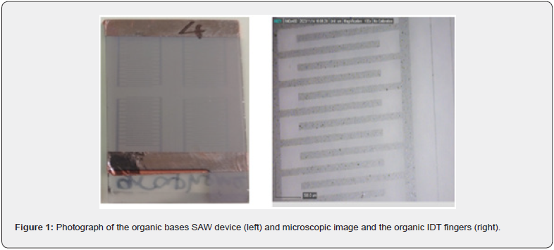
Quartz substrates were immersed in acetone cleaning solution in supersonic bath and dried with ethanol vapors. The substrates were ozone treated for adhesion promoting of the next layer. Ultrathin gold film (12 nm) was vacuum DC sputtered on the quartz substrate and patterned by conventional photolithography with inverse image of regarding the SAW IDT electrode fingers. This film will serve as a sacrificial for transferring the pattern into the organic electrode. The deposition sequence and conditions for carbyne film fabrication are presented elsewhere [11]. PEDOT:PSS thin film was sprayed on its top, afterwards, the gold underlayer was lifted off in potassium iodide etching solution, which is not aggressive to the organic film. The optimized atomizing conditions was set, like spraying pressure of 4 bars, spraying angle of 45o and nozzle - substrate distance of 8 cm at temperature of pulverization of 105 oC. Photograph of the organic bases SAW device and microscopic image and the organic IDT fingers, are shown in Figure 1 left and right, respectively. Electrical impedance spectroscopy (EIS) was conducted by Hioki Impedance analyser IM3590 in the frequency range from 1Hz to 200 kHz. Testing of the signal transducing at different ethanol vapor concentrations was realized by signal generator up to 20 MHz and the output waveform voltage was monitored with a digital storage oscilloscope DQ2042CN with a frequency bandwidth up to 50MHz. A lab-made chamber with controlled environment was used, which accommodates the measured device and commercial hygrometer reading the amount of water vapor present in the air in percentage.


Results and discussion


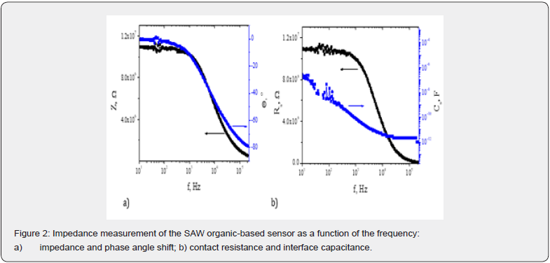
A decrease in impedance by one order of magnitude around 1 kHz in the surface acoustic wave-based sensing device can result from resonance effect (Figure 2a). The observed frequency of 1 kHz may coincide with a resonant frequency or a critical point in the device’s impedance-frequency response curve. At resonance, the impedance can decrease sharply, indicating that the device is most responsive or efficient at that specific frequency. Due to the lower conductivity of the electrode material as compared to the metals, the impedance of the device is in MΩ range. The quality of electrode deposition, adhesion to the substrate, and uniformity across the device’s surface can influence impedance levels. Poor electrode adhesion or non-uniformity can introduce noisy curves with multiple ripples. The stability of the impedance curve in each frequency range is an indication for the presence of a uniform electrode and sensing layers, as well as stable contact in between. The synchronous changes in phase angle shift and impedance at a specific frequency could indicate a resonant behavior of the device at that frequency. Electrodes may introduce parasitic effects, such as stray capacitance, which may interfere the impedance matching process.

As is seen in Figure 2b, there is only serial interface capacitance, which is in the expected range and decrease from nF to pF with the frequency increase. The presence of interface states or contaminants at the contact interface can impact resistance properties and introduce frequency-dependent behavior. At higher frequencies, the effects of interface states, or surface impurities may become more pronounced, contributing to contact resistance variations. It may due to the incomplete lift-off the sacrificial layer during the organic electrode patterning. Overall, the SAW line is in working condition with negligible distortions of the signals that can be ascribed to the non-conventional choice of electrode material and the adapted technology for its patterning.

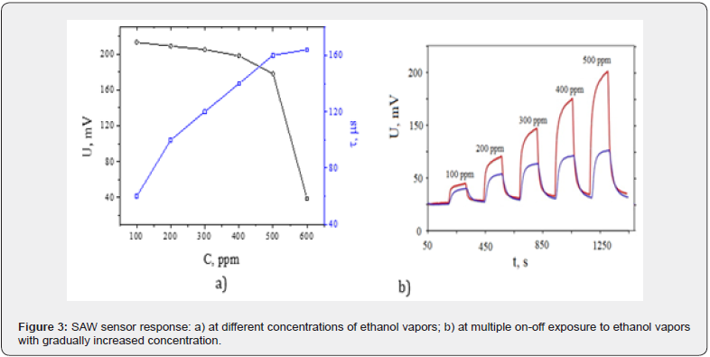
Voltage differences between the output and input signals of the SAW and time delay of the signal vs. ethanol concentration is shown in Figure 3a. According to the mass-loading conditions (concentration changes), the output voltage of the SAW line attenuates and the signal delays proportionally to the increased density on the path of the elastic wave’s propagation. The sensor is relatively linear in the range 100-500 pm and exhibited a sensitivity of 40μV/ppm. In the range 500-600 ppm the sensitivity increased to 1.4mV/ppm. This increased mass loading leads to a greater change in the sensor’s resonant frequency, resulting in a more pronounced shift that enhances sensitivity. Higher ethanol concentrations lead to a more complete and dense coverage of the sensor surface by ethanol molecules. This higher surface coverage increases the sensor’s response to ethanol and enhances sensitivity by producing a more substantial change in the detected signal. In SAW devices, acoustic waves propagate at a constant velocity along the surface of the substrate. This fixed propagation speed ensures that the time delay between signals remains consistent and linear, making time-based measurements more reliable and predictable compared to voltage-based measurements, which can be influenced by signal amplification or attenuation.


In addition, time-based measurements in SAW sensors may exhibit greater noise robustness compared to voltagebased measurements. The distinct time delay responses are less susceptible to noise interference and variations, allowing for more accurate and sensitive detection of signal differences. Therefore, on the same plot, the time delay between the signals in microseconds is also presented as a measure for concentration detection. Figure 3b shows the dynamic responses of all sample sensors to 100-600 ppm ethanol at an operating temperature equal to the room temperature. By increasing vapors concentration, the sensors are still able to recover to the initial state. As compared to previously studied SAW ethanol sensors with metal electrodes, the fabricated full organic-based sensor device is able to detect in the same concentration range, at similar time delay sensitivity of average 0.26μs/ppm vs. 0.23μs/ppm for the metal electrodes. This is a proof for successful realization of workable ethanol vapor sensor that can be used for close to realtime measurements determining the health condition of patients. The sensors demonstrate negligible hysteresis and restores its initial conditions after shading their surface from ethanol vapors. The devices are relatively inert and their response and recovery times varied from 40s and 50s at low concentration to 50 s and 70 s at higher concentrations beyond 300 ppm. As a modified electrode has lower conductivity, which cannot accelerate the transfer of charge carriers, the response time and recovery time are increased, as compared to the typical values of 10-20 s for SAW mechanism.

Conclusion

In summary, PEDOT:PSS nano coatings were successfully prepared by a spray deposition method and the prepared SAW sensor was applied to the vapor sensing test of ethanol. The samples achieved a response of 100-600 ppm at room temperature, with relatively linear concentration response in the range of 10–500 ppm ethanol vapors. In addition, the sensor had satisfactory time parameters (the response time and recovery time to 100 ppm at room temperature were 40 and 50 s, respectively).

To Know more about Current Trends in Biomedical Engineering & Biosciences
Click here: https://juniperpublishers.com/ctbeb/index.php

To Know more about our Juniper Publishers
Click here: https://juniperpublishers.com/index.php

Monday, May 27, 2024

Clinical and Biochemical Changes Among Nonspecific Low Back Pain Patients Treated with Wet Cupping Therapy: A Prospective Interventional Study - Juniper Publishers

 Orthopedics and Rheumatology Open Access Journal - Juniper Publishers

Clinical and Biochemical Changes Among Nonspecific Low Back Pain Patients Treated with Wet Cupping Therapy: A Prospective Interventional Study

Kaki Abdullah1*, Rohaiem Sawsan2, Mourad Samiha3, Al Jaouni Soad4, Mohsen Abdul Elalah5 and Sabrin Kholey6

1Abdullah M Kaki, Consultant and Professor, Department of Anesthesia & Critical care, Faculty of Medicine, King Abdulaziz University; Consultant of Pain Medicine, ALsalama Hospital, Jeddah, Saudi Arabia

2Sawsan Rohaiem, Professor, Department of Physiology, Faculty of Medicine, King Abdulaziz University, Jeddah, Saudi Arabia

3Samiha Mourad, Cupping Clinic, King Abdulaziz University Hospital, Jeddah, Saudi Arabia

4Soad Al Jaouni, Professor of Paediatric Haematology, Department of Haematology, Faculty of Medicine, King Abdulaziz University, Jeddah, Saudi Arabia

5Mohsen Abdul Elalah, Cupping Clinic, King Abdulaziz University Hospital, Jeddah, Saudi Arabia

6Sabrin Kholey RN, Cupping Clinic, King Abdulaziz University Hospital, Jeddah, Saudi Arabia

Submission: March 30, 2024; Published: April 10, 2024

*Corresponding author: Abdullah Kaki, Department of Anaesthesia & Critical Care, Faculty of Medicine, King Abdulaziz University, Saudi Arabia

How to cite this article: Kaki Abdullah*, Rohaiem Sawsan, Mourad Samiha, Al Jaouni Soad, Mohsen Abdul Elalah and Sabrin Kholey. Clinical and Biochemical Changes Among Nonspecific Low Back Pain Patients Treated with Wet Cupping Therapy: A Prospective Interventional Study. Ortho & Rheum Open Access J. 2024; 22(5): 556105. DOI: 10.19080/OROAJ.2024.22.556105

Abstract

Background: Wet cupping is an alternative treatment option, used to treat several medical conditions. The present study aimed to assess the effect of wet cupping therapy on nonspecific low back pain with respect to both clinical and laboratory profiles.

Methods: A total of 444 patients with low back pain were included in this study. Five wet cupping sessions were performed on the bladder meridian of each patient on a monthly basis. Pre-and post-cupping assessments of low back pain were performed using a visual analogue scale, and blood samples were collected to evaluate biochemical changes.

Results: The mean age of the included patients was 47.9±13.8 years, and 76.3% were female. The baseline pain score at rest was 5.6±3.0, which reduced significantly to 2.7±2.8 after the third cupping session (p <0.05). Similarly, the average baseline pain score with activity was 4.8±3.3, which reduced significantly after the third session to 1.9±2.3. Blood chemistry analysis revealed a significant decrease in blood urea, creatinine, total protein, and albumin levels, while there was a significant increase in total cholesterol and HbA1C. The correlation between the laboratory values and the magnitude of reduction in pain score in the first session revealed that only the calcium levels exhibited an inverse significant correlation with the magnitude of reduction in pain score.

Conclusions: The results of the present study support the use of wet cupping as a therapy to relieve nonspecific low back pain as well as improve body physiology. Saudi Clinical Trial Registry no 21092302.

Keywords: Nonspecific; Low Back Pain; Wet Cupping; Clinical; Biochemical

Abbreviations: NLBP: Nonspecific Low Back Pain; Hb: Hemoglobin; Hct: Hematocrit; BUN: Blood Urea Nitrogen; HbA1C: hemoglobin A1C; ALP: Alkaline Phosphatase; AST: Aspartate Aminotransferase; ALT: Alanine Transaminase

Introduction

Chronic nonspecific low back pain (NLBP) is one of the most common health problems worldwide, with approximately 80% of the population suffering from NLBP at least once during their lifetime [1,2]. In most patients, the symptoms are not attributed to etiological or neurological causes [3]. Conventional treatments, including medications [4] and surgical interventions, [5] have demonstrated efficacy in certain groups of patients. Unfortunately, these treatment modalities are not always effective and may cause serious side effects [5,6]. For these reasons, some patients have moved towards complementary and alternative medicine [7]. Wet cupping (Hijama) is one of the traditional Chinese medicinal techniques, known for thousands of years and used to treat several medical conditions, such as pain, hypertension, and stroke [8- 10]. Despite many published articles describing the mechanisms behind the action of wet cupping and correlating it with modern medicine, we still lack a clear pathway explaining the mechanism of action of wet cupping. In this single arm prospective study, we aimed to evaluate the effect of wet cupping on low back pain and blood laboratory values among a group of patients with NLBP.

Materials and Methods

Following ethics and research committee approval (Ethics and research committee, King Abdulaziz University hospital, reference no 851-12), a prospective interventional study was conducted at King Abdulaziz University Hospital, Jeddah, Saudi Arabia, from 2014 to 2019. The study was carried out in accordance with the principles stated in the Declaration of Helsinki. Informed written consent was obtained from all patients aged 18 years and above, suffering from low back pain for 3 months or longer, diagnosed by an expert pain specialist, and referred to the cupping clinic upon their request to try alternative and complementary therapy.

Pregnant, lactating women; patients with mental or psychological illnesses; patients using opioids or psychotropic medications; patients with local infection at the site of cupping; cancer patients; patients with chronic renal, hepatic, or cardiac diseases; patients with hematological diseases or using anticoagulants; and patients who complained of other acute or chronic pain conditions were excluded from the study. Baseline measurements were performed three days prior to the performance of wet cupping, and further samples were obtained one week after each cupping procedure for three consecutive sessions. Blood samples were collected after 12 hours of fasting from every patient to assess biochemical changes following wet cupping therapy. Data were collected by a research assistant interviewing the patient at each visit using a well-structured questionnaire. The questionnaire contained two parts: the first part dealt with demographic data, anthropometric data, and comorbidities, while the second part of the questionnaire covered the assessment of pain using the visual analogue scale. Data was collected prior to cupping session and one week after the intervention. This process was repeated for each session. Any complications resulting from the cupping procedure were documented.

The visual analogue scale (VAS) for pain assessment ranges from 0 to 10 cm, with 0 indicating no pain and 10 indicating the worst pain imaginable. This scale was explained to the patient, and baseline values were obtained before the performance of the procedure, both at rest and during activity, and repeated after each session monthly for 4 consecutive months. Cupping was performed by an expert therapist using sterile disposable plastic cups, placed bilaterally at the bladder meridians (BL 23, BL 24, and BL 25) according to the World Health Organization guidelines for acupuncture point locations [11,12]. Skin sterilization was performed, and slight skin laceration was performed using a sterile 22-gauge surgical lancet followed by application of plastic cups to the bloodletting points and suction was activated. Bleeding from the dermal microcirculation was initiated by reapplying the plastic cups on the skin scarification points to encourage bloodletting. After 5-10 minutes when the blood in the cup became solid, the cup with the blood was removed, the skin was cleaned with alcohol, and covered with gauze for 24 hours. Blood samples were collected as a baseline prior to cupping and repeated monthly following the second and third sessions. The obtained blood samples were evaluated for hemoglobin (Hb) concentration, hematocrit (Hct) values, electrolyte levels, calcium, blood urea nitrogen (BUN) and creatinine levels. In addition to the above, liver function test, thyroid function test, hemoglobin A1C (HbA1C) assessment, and lipid profile analysis were also performed.

Statistical Analysis

Given that research on wet cupping is limited, a pilot study was conducted to estimate the sample size. Considering 80% as an acceptable power to detect the difference in VAS pain score with 95% confidence (significance set at p value < 0.05), a sample size of 47 patients was found to be appropriate. All data analyses were performed using the Statistical Package for the Social Sciences (version 22.0; IBM Corporation, Armonk, NY, USA). Participant demographic data were analyzed using descriptive statistics. The Wilcoxon signed-rank test was used to analyze pain scores, as pain score is an ordinal variable and the readings obtained were paired, that is, each patient had two readings, one before and one after the cupping procedure.

Results

A total of 444 patients with NLBP were assessed for severity of pain using VAS before being subjected to wet cupping and repeated after each session of the procedure. Pain scores were recorded at rest and during activity. Most of the patients were women (76.3%) and the mean age of participants was 47.9±13.8 years, ranging between 18-88 years with no statistically significant differences between females (48.3±13.0 years) and males (46.7±16.0 years). The mean BMI was 30.0±7.8. At baseline, before starting wet cupping, the average pain score at rest was 5.6±3.0, which was reduced significantly after the first session to an average of 4.1±3.2. The same was observed after the second and third sessions, where the average pain score decreased from 4.7±4.2 to 3.2±3.0 in the second session, and from 4.6±3.5 to 2.7±2.8 in the third session (p <0.05).

Meanwhile, it was found that although the average pain scores were reduced in the fourth and fifth sessions, these reductions were not statistically significant (p >0.05) (Table 1). Similarly, it was found that the average pain score with activity at the baseline before starting wet cupping was 4.8±3.3, which was reduced significantly after the first session to an average of 2.9±2.5. Further reduction was noted in the second session from 3.6±3.0 to 2.2±2.4, and in the third session, there was a reduction from an average of 3.4±3.1 down to 1.9±2.3 (p <0.05). Meanwhile, the reduction in the average pain scores in the fourth and fifth sessions was not statistically significant (p >0.05) (Table 2). Although the average reduction in pain score was found to be higher among females (1.6±3.5) than males (1.1±3.1), this difference was not statistically significant (p >0.05).


Meanwhile, as shown in Figure 1, the magnitude of reduction in pain score was higher among young patients and decreased with age; nevertheless, this inverse correlation was not statistically significant (p >0.05). Similarly, there was no correlation between BMI and the magnitude of pain reduction, as shown in Figure 2. Blood chemistry analysis was carried out for all patients during the first three sessions, and Table 3 shows the changes in the laboratory readings following wet cupping. There was significant decrease in BUN, creatinine, total protein, and albumin, while there was a significant increase in cholesterol level and HbA1C (p <0.05). The correlation between the laboratory parameters and the magnitude of reduction in pain score after the first session showed that only calcium level exhibited an inverse significant correlation with the magnitude of reduction in pain score (Table 4). No complications were reported in any of the patients.


Discussion

The main finding of our study is the significant reduction in NLBP as well as the reduction in the readings of blood laboratory values following the performance of multiple sessions of wet cupping. The relief in lower back pain after wet cupping therapy has been documented in many of the previously published studies [13-18]. Although the number of wet cupping sessions carried out were different among these studies, the positive effect of wet cupping on low back pain was the same.

In our study, reduction in VAS score was noticed after the 4th and 5th sessions of wet cupping, both at rest and with activity, but it was not significant and further investigation is needed to clarify the underlying mechanism and exclude any contributing factors. We observed a significant reduction in the levels of BUN, creatinine, total protein, and albumin, while there was a significant increase in cholesterol and HbA1C levels following wet cupping. When the laboratory changes were correlated with the changes in pain after the first session of wet cupping, calcium levels exhibited an inverse relationship with the reduction in pain score. Husain et al. reported similar changes in biochemical profile in 31 healthy adults and reported a significant reduction in serum urea and creatinine levels following wet cupping therapy, while the fasting blood sugar was found to be lower at one, three, and four months after therapy when compared to baseline [19].

The changes in blood sugar are opposite to our finding, where HbA1C increased with repetitive sessions of wet cupping. In a study by Al showafi, venous blood samples of 30 healthy subjects were collected 10 days after wet cupping and a significant decrease in serum creatinine, cholesterol, triglyceride, and lowdensity lipoprotein with an increase in the serum high-density lipoprotein were documented; meanwhile, the effect on urea and blood glucose was not significant [20]. Ranaei-siadat et al. used a similar study design with a smaller sample of healthy subjects and showed a regulatory effect of wet cupping on fasting blood sugar and cholesterol, while no positive effect was reported on liver function test, renal profile, thyroid profile, calcium, Hb, or Hct [21]. Mashlool and Aowada showed a significant decrease in blood sugar levels post cupping compared to the pre-cupping in 100 volunteers. A significant increase in albumin level was reported when compared to the pre-cupping level [22].

In another study, Abbas et al. observed a significant increase in red blood cell number after cupping and a significant decrease in Hct, Hb, platelets, neutrophils, and lymphocytes. Cholesterol levels, as well as low-density lipoprotein and triglycerides were found to be lower. Liver enzyme alkaline phosphatase (ALP), creatinine, BUN, and blood glucose levels decreased as well, while total protein, aspartate aminotransferase (AST), and alanine transaminase (ALT) increased after wet cupping. Serum electrolyte levels showed a significant decline in serum calcium ion levels [23]. A similar finding regarding calcium levels was found in our study, but it was statistically insignificant. Looking at the designs of these studies, the sample sizes, the assessment tools, and the cultural background of the involved subjects, we might find an explanation for the reported differences in the results. Some studies have reported findings that are consistent with our findings, while others have reported contrary results; however, the general consensus is that there is a significant decrease in creatinine, urea, and albumin levels. While fasting and random blood sugar levels were decreased in previous studies, HbA1C levels increased in our study. Unfortunately, none of the studies excluded the effects of exercise, diet, nutritional status, physical and emotional status of the subjects. Thus, further studies are needed to estimate the effect of wet cupping on biochemical profile after considering all these factors.

Although the changes in calcium level were not statistically significant, there was an inverse relationship between the level of calcium and pain relief after the first session of wet cupping. The exact mechanisms behind the development of chronic pain remains unclear and may depend on the type of pain and the initiating causes but there is considerable evidence pointing towards the role of intracellular calcium in the development of persistent pain. Calcium-permeable ion channels and receptors have been implicated in the initiation of pain, as well as neuroplasticity [24-27]. The inverse relationship between the level of calcium and pain relief might be one of the mechanisms behind the positive effect of wet cupping on pain. More clinical trials are needed in the future to confirm this relationship.

Study Limitations

One of the limitations of the present study was the absence of a control group. The second limitation is the difficulty in differentiating the effect of wet cupping from the placebo effect, Lastly, the effect of other environmental factors that alter the body physiology and might contribute to the changes in the laboratory measurements is another limitation of the study.

Conclusion

The results of the present study support the use of wet cupping to relieve NLBP and regulate certain physiological parameters. Further studies with a larger sample size and better design are needed to confirm the effects of wet cupping on various body functions.


To Know more about our Juniper PublishersJuniper Publishers
Click here: https://juniperpublishers.com/index.php


Artificial Intelligence System for Value Added Tax Collection via Self Organizing Map (SOM)- Juniper Publishers

  Forensic Sciences & Criminal Investigation - Juniper Publishers Abstract Findings:  Based on our experiments, our approach is an effec...