Thursday, October 22, 2020

New Original Images of the Macro and Microscopic Retina by Pixelometry of the Digital Optical Biopsy - Juniper Publishers

 JOJ Ophthalmology - Juniper Publishers


Short Communication

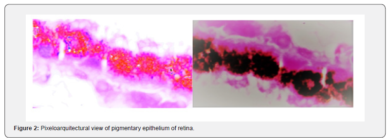
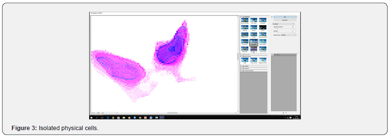
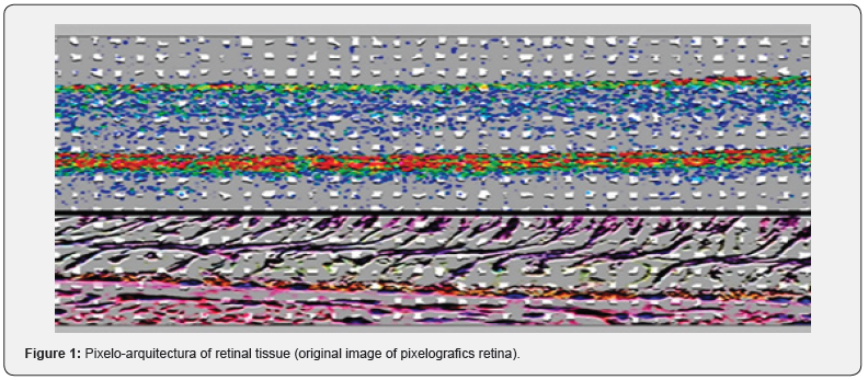
In this work, we present some of the physical morphological results obtained from the sequencing of images with digital optical biopsy [1], using pixelometric and pixelographic criteria [2-4]. The cellular and tissue images, although they have a known pattern, show the difference of a pure, active image, captured by a tomography (OCT), or optical coherence tomography. The geometry of the pixels denotes a certain combination of eucldiana and two-dimensional elliptical, especially with options 3D allowed in the construction scheme geometric Riemannian, and the stocks of subpixels (red, green and blue) dead pixels and stuck, expressing where color and resolution monitors have reached almost improbable geometric expressions, graphics cards such as the S3, NVIDIA, or ATI among others., giving the opportunity to overcome infinitely genome combination possibilities of identification, in this case with 16.8 million colors (32 bits). The QRS, the old fingerprint, facial detectors FBT face detection, bar codes, different forms of interferometry and spectrometric, have led to the possibility of using non-invasive methods of protein identity uncalculated limits or the DNA (Figures 1-3).
JOJ Ophthalmology
JOJ Ophthalmology
JOJ Ophthalmology
We used the pixelometric new non-Euclidean geometry described, about the pixel, as measurement converter, density, metric, shapes, etc. Information generates “every being-every particle, every force field, even the time-space continuum. “The smallest scale in the universe-the one governed by the laws of quantum physics-seems a challenge to common sense”. Subatomic objects (pixel representation) can be in more than one place at a time, two particles at opposite ends of a galaxy can share information instantly, and the mere fact of observing a quantum phenomenon can radically change. It we present some of the physical morphological results obtained from the sequencing of images with digital optical biopsy [5-8], using pixelometric and pixelographic criteria of the retina, and specially pigmentary epithelium. The cellular and tissue images, although they have a known pattern, show the difference of a pure, active image, captured by a tomography (OCT), or optical coherence tomography. The smallest scale in the universe-the one governed by the laws of quantum physics-seems a challenge to common sense. Subatomic objects (pixel representation) can be in more than one place at a time, two particles at opposite ends of a galaxy can share information instantly, and the mere fact of observing a quantum phenomenon can radically change it.


To Know more about our Juniper Publishers

Click here: https://juniperpublishers.com/index.php   


No comments:

Post a Comment

Juniper Publishers FAQs: Your Guide to Common Publishing Questions and Solutions

  Juniper Publishers FAQ's Q: What are the Manuscript Guidelines in Open Access Journal Publishers? A: Manuscript guidelines in ope...