Wednesday, December 18, 2019

Periodontitis in a Group of Patients with Cardiovascular Disease-Juniper Publishers

Anatomy Physiology & Biochemistry International Journal

The association between periodontal disease and cardiovascular disease is based on the systemic inflammation which also involves the periodontopathogenic bacteria. Our research study investigated the prevalence of periodontal disease in a group of 20 patients with cardiovascular disease, divided into 2 groups according to the presence of periodontal disease, demographic and environmental factors. Periodontal and cardiovascular disease were established according to the current international classifications. The systemic inflammation was assessed by the following markers: leukocytes number, C-Reactive Protein, ESR and fibrinogen. The periodontal disease was assessed based on the bacterial plaque index, gingival bleeding index and clinical attachment loss.
Our results indicated that the prevalence of periodontal disease in patients with cardiovascular disease was 55%, and higher in females than males. Among patients with cardiovascular disease, those in the rural area had a poor oral hygiene. Patients with hypertension showed a higher number of teeth with periodontal pockets compared with patients with other cardiovascular diseases. There were no significant changes in systemic inflammatory markers related to the presence of periodontal disease.
Keywords: Periodontitis; Cardiovascular disease; Prevalence; Periodontal pockets; Systemic inflammatory markers
Abbrevations: AC: Anticoagulants; AHA: American Heart Organization; CAL: Clinical Attachment Loss; CHF: Chronic Heart Failure; CI: Calculus Index; CIHD: Chronic Ischemic Heart Disease; CRP - C Reactive Protein; CVD: Cardiovascular Disease; CVE: Cerebrovascular Event; DI: Debris Index; DSR: Digital Subtraction Radiography; ESR: Erythrocyte Sedimentation Rate; GBI: Gingival Bleeding Index; GDPR: General Data Protection Regulation; HBP: High Blood Pressure; IL-1β: Interleukin -1 Beta; IHD: Ischemic Heart Disease; MI: Myocardial Infarction; OHI-S: Oral Hygiene Index-Simplified; PA: Platelet Antiaggregants; PD: Periodontal Disease; TNF-α: Tumor Necrosis Factor Alpha; US: United States; WBC: Leukocyte; WHO: World Health Organization

Introduction

Periodontal disease (PD) is a chronic inflammation that occurs in response to the periodontopathogenic bacteria and causes the progressive and irreversible destruction of the periodontal tissues and eventually, leads to tooth loss [1]. Numerous studies have demonstrated the association between PD and CVDs [2] such as arteriosclerosis [3], dyslipidemia [4] and hypertension [5]. According to prospective observational studies, this association is based on the systemic inflammation initiated by the microorganisms in the biofilm on the dental surfaces. However, a clear link has not been yet established [6]. PD and heart disease could have simultaneous onset, but singularly, one disease cannot induce the another [7]. This blurring persisted for several decades until several important studies established the influence of periodontal disease treatment on cardiovascular disease outcomes [8]. Since periodontal disease does not induce pain in the early stages and has a slow evolution, it is often left untreated [9,10].
According to the WHO, cardiovascular diseases are the number one cause of death globally [11]. In 2012, there were 17.5 million deaths globally, accounting for 31% of the total deaths [12]. Among cardiovascular diseases, IHD and cerebrovascular disease are the most common causes of death worldwide. According to the AHA, 795,000 people suffer a stroke annually [13].
The oral cavity is a reflection of a patient’s overall health, harmful habits and nutritional status. It is the entry as well as the site of microbial infections that also alter the general health [14]. Several theories explain the link between periodontal disease and heart disease. One theory is that oral bacteria entering the blood stream could affect the heart by attaching to the atherosclerotic plaques in the coronary arteries and leading to clot formation. Coronary artery disease is characterized by thickening and hardening of the coronary arteries walls due to the formation of plaques. Blood clots may obstruct the normal blood flow, restricting the amount of nutrients and oxygen required for the heart to function properly. This may lead to heart attacks. Another possibility is that the systemic inflammation caused by periodontal disease increases plaque formation, which may contribute to inflammation of the arteries walls. Researchers have found that people with periodontal disease are almost twice as likely to suffer from coronary artery disease as those without periodontal disease [15].
The inflammatory potential of periodontal disease is manifested at macromolecular level through systemic dissemination of local mediators such as CRP, IL-1β, IL-6 and TNF-α. According to Chambers et al., this goal together with the increased number of inflammatory molecules could be involve in cardiovascular disease [16]. There is strong relationship between the periodontal and cardiovascular diseases and two directions have been the focus of delineating this relationship: bacteria from the oral cavity directly exacerbate cardiovascular disease or alter the systemic risk factors of cardiovascular disease; the chronic periodontal inflammation at the focus of infection increase circulating levels of inflammatory mediators, and/or bacteria disseminated into the circulation elicit elevations in systemic host inflammatory mediators that exacerbate cardiovascular disease directly or alter other systemic risk factors for cardiovascular disease [17].
The purpose of this study was to assess the prevalence of periodontal disease in a group of patients with cardiovascular disease and to establish the correlation between serum levels of inflammatory markers and periodontal status in these patients. The specific objectives of the present study were:
a. Evaluation of the prevalence of periodontal disease in a group of patients with cardiovascular disease.
b. Assessment of clinical parameters and severity of periodontal disease (bacterial plaque index, gingival bleeding index, clinical attachment loss) and the form of cardiovascular disease according to current international classifications.
c. Evaluation of systemic inflammatory markers (leukocytes, CRP, ESR, fibrinogen).

Materials and Methods

The study was conducted on 20 patients diagnosed with cardiovascular disease, 12 women and 8 men aged between 42 and 82 years, hospitalized in the Cardiology 1st Clinic, County Emergency Clinical Hospital Cluj-Napoca. All the patients signed the informed consent and the protection of personal data GDPR for participating to this study.
The patients were enrolled based on exclusion and inclusion criteria. The inclusion in the study was based on two criteria:
a. Minimum 6 teeth present in the oral cavity.
b. The diagnosis of cardiovascular disease.
The exclusion criteria were:
a. Complete edentation or presence of less than 5 teeth.
About half of all-American adults-117 million individuals— have one or more preventable chronic diseases, many of which are related to poor quality eating patterns and physical inactivity with more than two-thirds of adults and nearly one-third of children and youth being overweight or obese [8]. With the dietary link of diet and disease in mind, one must consider the application of diet upon biochemical processing overall and in particular for this work, with that of ADZ progression. Dietary demographics may play a pivotal role in the radically reduced rates of progression between the United States and that of other countries such as Singapore. It may be a point to consider that, although certain fish and shellfish contain greater levels of mercury content, they also contain essential fatty acids and are a rich protein resource that is needed to promote neuro-physiological processing through normal biochemical pathway processing [20,21].Research is now more than ever asking the question whether diet could play a substantial role in ADZ progression as well as many other dementias when considering the population demographics between East and West.
b. Severe general conditions that didn’t allow patient’s examination.
c. Other general diseases that could increase the levels of systemic inflammatory markers.
Using the Excel program, all data obtained in this study were organized in frequency tables, sectorial graphs (pie charts) and contingency tables. Information on the type of edentation according to the Kennedy classification, the appearance of the dental units, the presence of the dental fillings, their aspect and the material used, the aspect of the periodontium, the presence of gingival inflammation, bacterial plaque and tartar were recorded and compared. The major drawback in this study was the lack of periodontometry because this examination was performed in the hospital environment, not in the dental office. The periodontal clinical examination assessed the gingival bleeding, periodontal pockets depth, gingival retractions and tooth mobility.
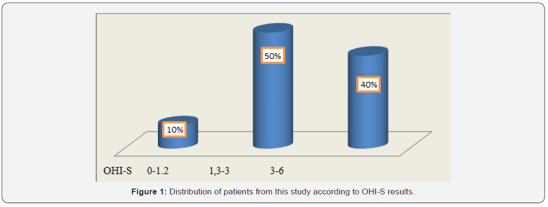
To evaluate oral hygiene, we calculated the OHI-S index (Greene and Vermillion, 1964) in patients with cardiovascular disease. Plaque examination was performed by inspection without staining solutions. The following dental surfaces were examined: the vestibular surface of teeth 1.6, 1.1, 2.6 and 3.1, and the lingual surfaces of teeth 3.6 and 4.6. The values of the debris index (DI) were registered as follows: 0 = absence of the bacterial plaque; 1 = supragingival plaque in the cervical 1/3 of the tooth, on the parcel; 2 = plaque in the middle 1/3 of the tooth; 3 = plaque in the occlusal or incisal 1/3 of the tooth. The values of the CI index were as follows: 0 = absence of the tartar; 1 = supragingival tartar in the cervical 1/3 of the crown; 2 = supragingival tartar in middle 1/3 of the crown and islands of subgingival tartar; 3 = tartar and in occlusal or incisal 1/3 of the tooth and a band of subgingival tartar. The values obtained in the assessment of the above areas were collected and divided by the total number of assessed areas for both DI and CI. Values obtained for DI and CI were collected and the OHI-S value was obtained. Interpretation of OHI-S index values was the following: between 0 and 1.2 = good oral hygiene; between 1.3 and 3 = satisfactory oral hygiene; between 3.1 and 6 = poor oral hygiene.
According to Maurizio S. Tonetti & al, understanding the periodontitis stages, as well as the correct treatment applied in each of these stages is crucial for an immediate treatment at these patients [18]. In order to assess the periodontal health status, we evaluated the GBI [19]. This is a reliable, easy-to-use indicator that reveals the presence or absence of gingival inflammation [20]. After about 10-15 seconds, the occurrence or absence of bleeding was recorded. The number of registered positive units was divided by the number of gingival margins examined and the result was multiplied by 100 for the expression of the percentage indicator. It was shown that scores obtained with this index were statistically correlated with those obtained from the application of the gingival index GI [21,22]. Several studies showed that the absence of gingival bleeding was a good indicator of the healthy marginal periodontium. Other studies demonstrated that the surfaces that bled upon probing did not show extensive tissue destruction in all cases. Moreover, this parameter is not correlated with the degree of gingival inflammation in smokers. The diagnosis of periodontal disease has been established based on the clinical criteria: multiple gingival attachment loss, grade 2 or 3 dental mobility with tooth malposition, bone resorption that could be clinically observed, gingival and periodontal pathological changes (presence of periodontal pockets, gingival bleeding, ulcers). Since the patients were hospitalized, the radiological examination could not be performed, in order to validate and complete the clinical diagnosis. The biochemical parameters assessed as markers of systemic inflammation included: WBC, CRP, Plasma fibrinogen and ESR.

Results

In this study, 20 patients were enrolled: 63% were women aged between 65 and 77 years and 37% men aged between 43 and 82 years. Among patients from the rural area, 73% were women and 27% were men. Among patients from the urban area, 55% were men. In this study group, 11 patients with CVD had PD (55% of which 35% were females and 20% were males). As represented in Figure 1, OHI-S indicated that 40% of all the patients achieved values ranging from 3 to 6, suggesting poor oral hygiene. Of these, 75% were females coming from rural areas. Among patients with both CVD and PD, 64% had a poor oral hygiene at the time of OHI-S assessment. These results suggested that in patients with CVD, the poor oral hygiene was correlated with the oral status and could be a risk factor for the occurrence and/or worsening of PD. The neglected oral hygiene observed in these patients might be the result of the associated general diseases.
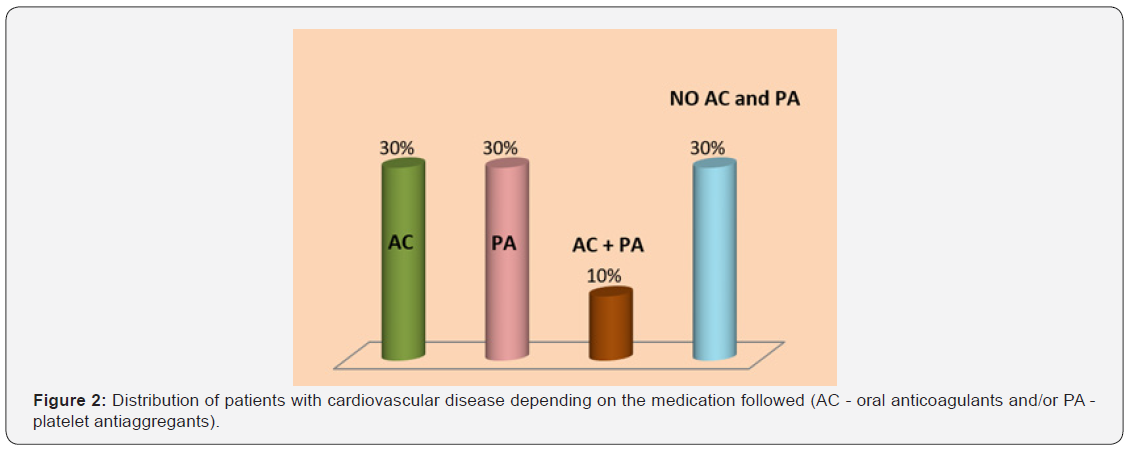
In Figure 2, 60% of patients with CVD were treated with AC (Acenocumarol or Apixaban) or PA (Acetylsalicylic acid, Clopidogrel or Ticagrelor) and 10% were administered drugs from both groups. This medication could be the explanation for the 85% of patients with CVD who experienced bleeding during periodontal examination.
Patients in this study presented different types of edentation according to the Kennedy classification. Most of the edentations were biterminal in both upper and lower jaws. Approximately 55% of the patients presented improper adaptation of prostheses on marginal profile, and 40% of them had no prosthetic crowns. Among patients with both PD and CVD, 45% had improper prostheses in terms of marginal closure, and the remaining 55% did not have prosthetic dentures. It is known that inappropriate prosthetic works are a local risk factor for PD, especially due to poor marginal adaptation that favors plaque retention on the prosthesis-tooth interface. Approximately 64% of patients with both CVD and PD had between 1 and 18 irrecoverable teeth that were either remnant roots or exhibited grade 2 or 3 dental mobility.
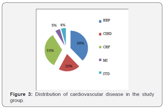
The CVDs diagnosed among the patients in this study included: HBP, CIHD, CHF, STD and MI (Figure 3). In patients with CVDs, the PD was found in 55% of patients with HBP, 40% in patients with CHF and 30% in patients with chronic CIHD.
Among patients with CVD and PD, 73% had between 1 and 12 teeth with inactive periodontal pockets, most of them with a depth of 2 to 4mm. As seen in Table 1, the largest number of teeth with periodontal pockets compared with the total number of teeth has been found in patients with HBP.
To evaluate the relationship between inflammatory markers and the presence of PD, the patients were divided into 2 groups. The first group included 11 patients with both CVD and PD, and the second group included 9 patients with CVD but without PD. There was no significant difference between the two groups regarding the inflammatory markers. However, as can be seen in Table 2, patients with both CVD and PD exhibited higher ESR and fibrinogen values compared with patients without PD.
There was no clear evidence if inflammation manifested by leukocytosis was the cause or the effect of atherosclerosis. Among patients with HBP, approximately 30% had leukocytosis. In the Table 3 can be seen the distribution of patients who had values of inflammatory markers evaluated above the normal range according to cardiovascular disease.
Although a clear link between PD and CVDs has not been yet established, several studies focused on the analysis of the relationship between PD and HBP [5,23,24]. Numerous clinical and epidemiological studies reported that the leukocyte count is an independent risk factor for coronary heart disease. In our study, of patients with CIHD, only 11% had leukocytosis (Table 3). According to a study investigating the relationship between white blood cells counts and CIHD, increased leukocyte counts were associated with an increased risk of cardiovascular morbidity and mortality in a group of undiagnosed patients. Leukocytosis influences the severity of coronary heart disease through multiple pathological mechanisms that mediate inflammation, causes the destruction of endothelial cells through oxidative stress and proteolysis, affects microcirculation by clogging the capillaries, arterioles and venules, induces hypercoagulability and contributes to the expansion of infarcted areas. Moreover, leukocytosis is a risk factor independent of atherosclerotic disease status [25]. Leukocytes play a pivotal role in normal host resistance at dental-plaque biofilm level and its dysfunctions are involved in periodontitis development [26].
Recently, numerous epidemiological studies confirmed that patients with elevated basal plasma levels of CRP have an increased risk of coronary artery disease and myocardial infarction. Prospective studies in the European and US countries have provided reliable data regarding the predictive value of CRP determinations on cardiovascular risk in both men and women. Thus, CRP is an indirect risk factor for coronary artery disease, and elevated levels may reflect some of the followings: inflammation of the coronary arteries in response to infectious agents; severity of inflammatory response in atherosclerotic vessels; extension of inflammation associated with myocardial ischemia; extension of inflammation associated with myocardial necrosis; the levels and activity of circulating proinflammatory cytokines.
There are two types of CRP tests: one of these has a measurable range that includes values obtained in patients with infectious or inflammatory processes (generally 0.3-20mg/dL) and the other can detect lower CRP levels (analytical sensitivity in around 0.01mg/dL) to estimate the risk of acute CVE. For this reason, the second test is termed CRP with high sensitivity (CRP ultrasensitive, “high-sensitive” CRP) [27]. At present, the value of the highly sensitive C-protein (hs-CRP) appears to be the most reliable predictor of acute CVE and is successfully used for the clinical assessment of patients with cardiovascular risk. Decision intervals for cardiovascular risk assessment are established according to the AHA: <0.1mg/dL - low risk; 0.1-0.3mg/dL - moderate risk; >0.3mg/dL - increased risk. For values higher than 1mg/dL, a non-cardiovascular cause should be considered.
Although there is clear evidence of the role of inflammation in coronary artery disease, the precise mechanism underlying the relationship between plasma CRP levels and the cardiovascular risk has not been yet established. It is still unclear whether increased CRP levels are the cause or the consequence of the disease or both. The inflammatory response associated with atheromatous lesions may trigger cytokine production in an amount sufficient to induce a measurable increase in plasma CRP. In turn, CRP, due to pro-inflammatory effects, may increase the formation of the dental plaque, or may have other effect that aggravate the PD.
In our study, 30% of the patients with CVD had CRP values ranging between 0.1 and 0.3mg/dL, which were associated with moderate cardiovascular risk. Among patients with chronic heart failure, 47% had CPR values higher than 1mg/dL. It has been reported that people with CRP serum levels that consistently exceed 1mg/dL had an increased risk of myocardial infarction. The following circumstances could influence the CRP values: pregnancy, obesity, the presence of an intrauterine contraceptive device, or the use of medications such as oral contraceptives, menopausal hormone replacement therapy, statins (drugs used to lower blood cholesterol), and anti-inflammatory drugs. The PD was present in 64% of rural patients and in 44% of urban patients. A recent transversal study based on the simple randomized method performed on 470 rural subjects, showed a 73.62% prevalence of PD [28]. These findings could be explained by the fact that the rural population neglects the oral health and is unaware of the seriousness of the oro-dental conditions, and also by the low accessibility to dental services.

Conclusion

Future studies should focus on a better understanding of the common pathogenic mechanisms and the interactions between cardiovascular disease and periodontal disease. In order to demonstrate the causal relationship between periodontal disease and cardiovascular disease, further studies should be performed on a large population to provide data enabling statistically significant correlations.
 
To Know More About Open Access Journals Please click on: ttps://juniperpublishers.com/index.php

Monday, December 16, 2019

Solvent Fractionation of Lignin from Sugarcane Bagasse-Juniper Publishers

Juniper Online Journal Material Science

The objective of this work was to examine lignin fractionation from sugarcane bagasse using three organic solvents. Lignin derived from lignocellulosic sources such as sugarcane bagasse can be used as sources of low molecular weight aromatic compounds. These compounds can be used as chemical building blocks for the production of high value materials. In this work, organic solvent fractionation using methanol, ethanol and acetone were used to fractionate lignin from sugarcane bagasse. It was demonstrated that among these solvents the acetone and ethanol fractionated higher concentrations of lignin from bagasse. This indicated that these two solvents can be used to obtain lignin components from which low molecular weight aromatic components can be derived that have various applications.
Keywords: Lignin; Organic Solvent Fractionation; Low Molecular Weight Aromatic Compounds

Introduction

Lignin is a complex aromatic heteropolymer, which forms a component of lignocellulose contributing to the structural integrity of plants. Bugg, Ahmad et al. [1], Xu, Arancon et al. [2] Due to its aromatic composition, it is seen as a “green” source from which various aromatic building block chemicals can be derived. Xu, Hanna et al. [3] These chemicals are described as molecules with multiple functional groups making them readily convertible to a variety of high-value materials. Farmer and Mascal [4], Isikgor and Becer [5] This is because the functional groups can form new families of useful molecules with a variety of applications. Farmer and Mascal [4] One example of a building block compound derived from lignin is vanillin. This compound possesses phenolic among other functionalities and is currently being investigated in polymer chemistry for production of polyesters among other types of polymers. Constant, Robitzer et al. [6], Sainsbury, Hardiman et al. [7], Fache, Boutevin et al. [8].
Currently the main source of lignin is from black liquor produced as a by-product from the pulp and paper industry. Park, Kim et al. [9] However, due to the heterogeneity of lignin and it low solubility the use of lignin as a source of aromatic compounds has been limited. Alternatively, methods using various organic solvents can fractionate components of lignin. This leads to more homogenous lignin fractions, from which consistent aromatic compounds can be derived. Zhao, Cheng et al. [10], Fiţigău, Peter et al. [11], Ragauskas, Beckham et al. [12], Sadeghifar, Wells et al. [13], Park, Kim et al. 2018 [9]. In this examination, organic solvent fractionation, which is a low energy process, was used to target the lignin component of bagasse. This process causes the disruption and removal of low molecular weight components of the lignin. Zhao, Cheng et al. [10], Fiţigău, Peter et al. [11] Organic solvents include alcohols such as methanol and ethanol, ketones such as acetone and glycols such as glycerol and ethylene glycol. Among these, commonly used organic solvents are methanol, ethanol and acetone. Fiţigău, Peter et al. [11], Sadeghifar, Wells et al. [13] These solvents work by cleaving ether and ester linkages in the lignin. Sugarcane bagasse is a lignocellulosic material which has high lignin content (11-25%, dry weight basis) and can be used as a renewable source of lignin. Chandel, da Silva et al. [14] Therefore the aim of this work was to examine the application of organic solvent fractionation using methanol, ethanol and acetone to derive low molecular weight lignin components.

Materials and Methods

All chemicals used in this work were obtained from Sigma Aldrich, St. Louis MO, USA unless otherwise stated.
The organic solvents consisted of two alcohols: methanol and ethanol and one ketone: acetone. Solvent water solutions were prepared at concentrations of: 10%, 30%, 50%, 70%, 90% and 100% (v/v). Zhao, Cheng et al. [10], Fiţigău, Peter et al. [11] Bagasse (1 g), was mixed with 50 mL of solvent solution in 125 mL flasks, which were set at room temperature for 24 hours without stirring. After this time, the bagasse residues were separated from the filtrate by gravity filtration. The concentration of lignin in this filtrate was estimated spectrophotometrically from a standard curve constructed using various concentrations (0 - 10 mg/ mL) of Kraft lignin in 1 M NaOH. Park, Kim et al. [9] The absorbance of lignin was determined by a wavelength scan from 190 - 800 nm, using single beam UV - Vis spectrophotometry (Agilent 8453 UV-Vis spectrophotometer, Shimadzu UV-1280). Fiţigău, Peter et al. [11], Sadeghifar, Wells et al. [13], Park, Kim et al. [9]. The liquor obtained from the fractionation process was analyzed spectrophotometrically to confirm the presence of lignin. This was done by evaporating the solvent under reduced pressure followed by lyophilization. The dried brownblack residue (5 mg) was dissolved in 1 M NaOH using a 10- mL volumetric flask. Fiţigău, Peter et al. [11], Wildschut, Smit et al. [15], Jääskeläinen, Liitiä et al. [16] Two ten-fold dilutions were made to produce a straw-colored solution for analysis. Additionally, Kraft lignin was utilized for comparison purposes. The absorbance of the solutions was found by wavelength scan from 190 to 1100 nm using UV- Vis spectrophotometry (Agilent 8453 UV-Vis spectrophotometer). Dominant peaks between 250 - 350 nm were related to lignin components extracted from the bagasse. Sun, Sun et al. [17], Tolbert, Akinosho et al. [18]. The program GraphPad Prism 7.01 was used for the statistical analysis and to construct the graphs displayed. In cases where the error bars are shorter than the height of the symbol, Prism does not draw those error bars.

Results and Discussion

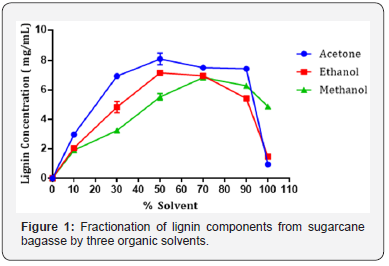
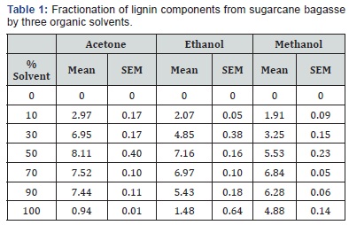
The presence of lignin components in the liquor derived from fractionation was confirmed by two dominant peaks at 291 and 331 nm. These results were compared to those collected for Kraft lignin alkali, which demonstrated dominant peaks at 219 and 288 nm. de la Torre, Moral et al. [19], Fiţigău, Peter et al. [11] This is the typical range at which lignin absorbs, demonstrating that the fractionation process removed components of lignin from the bagasse. Fiţigău, Peter et al. [11], Jääskeläinen, Liitiä et al. [16] The observed peak shift between solvent fractionated lignin and Kraft lignin is caused by the difference in lignin composition. Kraft lignin is derived from the Kraft pulping processes, which depolymerizes lignin into small aromatic derivatives using hot water, NaOH and Na2(SO3). Zhao, Cheng et al. [10], Espinoza- Acosta, Torres-Chávez et al. [20] These smaller fragments absorb UV light at a lower wavelength. This is relative to larger, more intact fragments of lignin, derived from the organic solvent fractionation process. These fragments tend to absorb UV light at a higher wavelength. Xu, Hanna et al. [3], Park, Kim et al. [9]. Further, the data collected from organic solvent fractionation of bagasse by acetone, ethanol and methanol, are displayed as mean ± SEM in Figure 1 and Table 1. Statistical analysis was performed to compare the means of lignin concentration fractionated from bagasse. For each of the three solvents, the highest mean lignin concentrations were compared using one-way ANOVA (alpha = 0.05).
The results presented in Figure 1 show that lignin is fractionated from bagasse in the order of acetone > ethanol > methanol between 10 - 70% solvent concentration. For acetone and ethanol, the highest concentrations of lignin were fractionated at solvent concentrations of 50% (Figure 1) and (Table 1). At concentrations above 90% (v/v), fractionating ability decreased. For methanol, the highest concentration of lignin was fractionated at 70% (v/v). It was also observed that after 50 - 70% solvent concentration, the lignin concentration generally decreased. This is related to the molecular weight of the lignin fragments. At solvent concentrations of 50 - 70%, maximal quantities of highly soluble lower molecular components are fractionated by the solvents examined. These components are high in free hydroxyl and carboxylic regions which significantly increase solubility. However, at higher solvent concentrations, higher molecular weight and less soluble lignin components are fractionated. Jääskeläinen, Liitiä et al. [16], Park, Kim et al. [9] This occurrence has been previously observed in fractionation of Kraft lignin with ethanol and also fractionation of several technical lignin’s with acetone Fiţigău, Peter et al. [11].
One-way ANOVA for the three solvents at 50% (v/v) showed that the effect of the solvent was significant (F (2, 6) = 21.28, p < 0.05). Post hoc analysis using Tukey HSD multiple comparison testing indicated that the concentrations of lignin fractionated by acetone at 50% v/v (Tukey, p < 0.05) was significantly higher than for methanol. Concentrations of lignin fractionated by ethanol at 50% v/v (Tukey, p < 0.05) were also significantly higher than for methanol. Conversely, the concentration of lignin fractionated by ethanol and acetone, both at 50% v/v were not significantly different (Tukey, p > 0.05). From this analysis, it was determined that either acetone or ethanol at 50% (v/v) could be selected a better solvent for the organic solvent fractionation of lignin components from bagasse Abdelaziz, Brink et al. [21], Fache, Boutevin et al. [8].

Conclusion

These results indicate that organic solvent fractionation can be applied directly to bagasse lignocellulosic material to obtain lignin components. Further, acetone and ethanol demonstrated similar ability to fractionate lignin from bagasse. The lignin components obtained can then be used as sources of low molecular weight aromatic compounds including vanillin, which can be used as chemical building blocks.
 
To Know More About Open Access Journals Please click on: ttps://juniperpublishers.com/index.php

Friday, December 13, 2019

Introduction to Therapeutic Antibodies-Juniper Publishers

Journal of Tumor Medicine & Prevention

The family of antibody biologicals has been growing and encompasses antibody-drug conjugates, radiolabeled antibodies for imaging, fusion proteins containing fragments of antibody, bispecific antibodies, and monoclonal antibodies. The rate of approval for these antibodies have exploded in the last century and are now utilized for treating cancer, cardiovascular disease, inflammatory disease, organ transplantation, infection, pulmonary respiratory disease as well as for diagnostic purposes. The present review aims to give a brief overview of the developmental history of these therapeutic antibodies along with a brief discussion of their pharmacodynamic and pharmacokinetic properties. Also, the review highlights the emerging limitations and the future strategies to overcome these limitations of antibody-based biologics.
Keywords: Monoclonal antibodies; Pharmacodynamics; Pharmacokinetics; Chimeric antibodies; Humanized antibodies; Murine antibodies; Human antibodies
Abbreviations: CDR: Complementary Determining Region; CDC: Complement Dependent Cytotoxicity; ADCC: Antibody Dependent Cellular Cytotoxicity; EGFR: Epithelial Growth Factor Receptor

Introduction

Antibodies are immunoglobulins that are part of the humoral immune response and are secreted by the B-cells (plasma cells). Antibodies act by binding to either soluble antigens or ligands that are expressed on the surface of organisms or cells. In terms of structure, antibodies are Y-shaped glycoproteins made up of two heavy chain polypeptides and two light chain polypeptides that are held together by disulfide bridges. The light and the heavy chain are made up of constant regions and variable regions with light chain having one variable and one constant region and the heavy chain having one variable and three to four constant regions (part of which forms the Fc, crystallizable portion). The variable region of the light chain and the heavy chain together forms the antigen-binding site (Fab, antigen binding portion). At the end of each variable region is the hypervariable region (CDR, complementary determining region) and it is this region that allows for numerous conformations for infinite antibody-antigen binding probability.

Developmental history of therapeutic antibodies

The concept of therapeutic antibodies was first put forth by Paul Ehrlich who came up with the nomenclature “antikorper” (German for antibody) [1]. However, the first important step towards production of therapeutic antibodies only happened when Kohler and Milstein (received the Nobel prize for their discovery) developed a protocol to produce murine monoclonal antibodies (-omab, nomenclature) from hybridomas [2]. This paved the way for the food and drug administration’s (FDA) approval of the first murine monoclonal antibody for use in prevention of acute kidney transplant rejection [3]. The murine monoclonal antibody targeted CD3 receptors of the T cells and was called muromonab-CD3 (OKT-3) and was found to be significantly better than the conventional steroid treatments (Azathioprine and Prednisone). Unfortunately, this first-generation antibody had a major disadvantage arising from the presence of the murine immunogenic component that gave rise to the induction of human anti-mouse antibodies after administration [4]. Because of this immunity, patients rapidly cleared the murine antibody from their system resulting in a very low therapeutic window of this therapeutics.
To overcome this hurdle, the second generation of therapeutic antibodies, characterized by a combination of ~65% human component (constant region) and the rest murine component (variable region) was developed simultaneously by two research groups led by Morrison and Boulianne, respectively [5,6]. The first “chimeric” therapeutic antibody (-ximab, nomenclature) to receive FDA approval for use in peri-surgical prevention of thrombosis for coronary artery interventions was, Abciximab, which targeted the platelet glycoprotein IIb/IIIa receptor [7]. Unfortunately, administration of these chimeric antibodies still resulted in the induction of human anti-chimeric antibodies thereby reducing their potency and efficacy in patients. This shortcoming arising due to the immunogenicity of the murine component of antibodies led to the development of the next generation of “humanized” monoclonal antibodies (-zumab, nomenclature). This was first achieved by Jones and colleagues by replacing the murine hypervariable region of the antibody with genetically engineered human myeloma protein to produce a therapeutic antibody that had ~95% human components [8]. The first humanized antibody, Daclizumab, was initially first approved for use in preventing kidney transplant rejection and acted on CD25 but is now primarily used to treat relapsed multiple sclerosis. Even though, the increased humanization of the antibody is associated with less immunogenicity, patients treated with these family of antibodies have been shown to produce human anti-humanized antibodies [9].
Human antibody (-umab, nomenclature), the third generation of therapeutic antibody, was developed with the idea to completely ablate immunogenic response and thereby increase clearance time and the efficacy of the therapeutics. The requisite breakthrough was provided by Winter and colleagues who developed the protocol of mimicking the natural positive selection of antibodies in bacteriophages using a phage display technology [10]. The execution of this technique led to the development of the first fully human antibody against tumor necrosis factor called, Adalimumab and was approved for use in autoimmune and inflammatory conditions like rheumatoid arthritis and Crohn’s disease [11]. Finally, transgenic mice created by humanizing the murine immune system and then inoculating these mice with antigen, resulting in fully realized human antibody was created by Scott [12]. Panitumumab, was the first human antibody targeting Epidermal Growth Factor Receptor (EGFR) that received FDA approval for use in colorectal cancer using the transgenic mouse technology [13]. Surprisingly, even with the low possibility of immunogenicity, immunogenic response has been observed in patients treated with human antibodies, suggesting that engineered antibody will always demonstrate some spectrum of immune response that can never be eliminated [14].

Pharmacodynamics of therapeutic antibodies

The therapeutic activity of an antibody is dependent on the Fc and the Fab portion of its structure and its mechanism of action can be broadly classified into Fc-dependent activity and Fab-dependent activity. The Fab-dependent activity requires the antibody to bind to a soluble antigen and assist in the neutralization of the antigen. For example, Bevacizumab binds with very high affinity to various isoforms of Vascular Endothelial Growth Factor (VEGF) and inhibits its angiogenic activity by preventing VEGF from activating its receptors resulting in an anti-cancer effect [15]. In addition to soluble antigens, the Fab-dependent activity can also manifest itself by binding of the antibody to a membrane bound antigen. Such binding can result in two therapeutic scenarios:
(i) Binding of the antibody to the membrane-bound antigen can result in an inhibitory effect. For example, Cetuximab binds to the cell surface receptor EGFR with higher affinity than its natural ligand like epithelial growth factor or transforming growth factor-α resulting in an antagonistic effect that decreases EGFR signaling leading to death in cancer cells [16].
(ii) Binding of the antibody to the membrane-bound antigen can result in stimulatory effect. For example, Rituximab binds to CD20 receptor on B cells and induces apoptosis by an agonistic induction of cytoplasmic calcium ions leading to caspase 3-mediated apoptosis in leukemic cells [17].
The Fc-dependent activity depends either on the activation of the classical pathway of compliment resulting in Complement Dependent Cytotoxicity (CDC) or on the recruitment and activation of FcγR-expressing immune cells (NK or T cells) resulting in Antibody Dependent Cellular Cytotoxicity (ADCC) and in some cases antibody dependent cellular phagocytosis. Trastuzumab (anti-HER2), Obinutuzumab (anti-CD20) and Catumaxomab (anti- CD3) are all examples of therapeutic antibodies that utilize CDC and ADCC for their biological activity. Furthermore, there is quite a lot of overlap between the biological activities of antibodies as seen in Trastuzumab and Rituximab both of which can have Faband Fc- dependent activity [18].

Pharmacokinetics of therapeutic antibodies

Therapeutic antibodies are denatured or proteolytically cleaved in the gastrointestinal tract and hence generally administered via intravenous, intramuscular or subcutaneous route [19]. The typical pharmacokinetic profile after administration follows a biphasic response with a rapid distribution phase followed by a slower elimination phase. The distribution of antibodies, dictated by its large molecular size and poor lipophilicity, is limited to the vascular and intestitial spaces. Factors that influence distribution includes, diffusion, cellular internalization (pinocytosis, endocytosis, phagocytosis), binding affinity to its antigen and hydrophobicity [20]. Primary method of elimination after absorption of antibodies is through proteolytic degradation. Due to its large size, glomerular filtration is impossible preventing renal clearance of antibodies. Clearance of antibody can be antigen specific (also referred to as, target-mediated drug disposition) and depend on the expression level, location (soluble vs. membrane bound), distribution (organ specific vs. entire body) and whether the antigen expression is modulated (upregulated vs. downregulated). For example, Adalimumab that targets and binds to antigen like tumor necrosis factor-α that is expressed in very low levels, the pharmacokinetic profile is very linear as opposed to Omalizumab that targets high expressing IgE and shows a non linear pharmacokinetic clearance profile [11,21]. On the other hand, Rituximab (anti-CD20) demonstrates a time dependent pharmacokinetics because of the B-cell depletion with treatment causing decreased presence of CD20 resulting in reduced clearance on repeated dosing [22].
Non-specific antibody clearance can be due to protein degradation following cellular uptake or due to effector function of the antibody like CDC or ADCC [23]. Also, the structural and chemical properties of the antibody like charge, solubility, target specificity and glycosylation patterns can affect its clearance [24]. Finally, patient’s health status, demographic factors and medication history all play a role in influencing the pharmacokinetics of therapeutic antibodies.

Limitation of therapeutic antibodies

The major limiting factor in the widespread use of antibodies in the clinic is the production cost under Good Manufacturing Practices to manufacture therapeutic antibodies. An alternative cost-effective production system needs to be evaluated in order to make therapeutic antibodies affordable to every individual. Secondly, immunogenicity to therapeutic antibodies resulting in production of anti-therapeutic antibodies not only increases the clearance of the antibody and efficacy of the antibody, but also leads to severe immune reaction in humans. Thirdly, because of its large size, antibodies do not have a very good tissue distribution which is further exacerbated in solid tumors with minimal vasculature [25]. Also, some organs like the brain are not accessible for a large macromolecule like an antibody. Finally, therapeutic antibodies that rely on ADCC for its activity have to compete with the high levels of endogenous IgG for the FcγR of the immune cells. This necessitates injection of very high concentrations of antibodies to reach significant serum concentrations needed to compete with the patient’s IgG [26]. Overcoming these hurdles will facilitate the widespread use of therapeutics in the clinic.

Discussion and conclusion

The new horizons being explored in the use of therapeutic antibodies can be broadly classified into the following areas of interest:
(i) Targeted delivery to specific organs
(ii) Flexibility in the route of delivery
(iii) Specific delivery to the intracellular compartment of cells and
(iv) Newer forms of antibody delivery systems.
Not surprisingly, significant advances have been made in each of this enumerated fields. Asfostase alpha specifically targets the bone with the aid of a deca-aspartate peptide that is fused to C-terminus of the antibody and has been FDA approved for use in the treatment of hypophatasia [27]. Oral delivery of antibodies like PRX-106 for ulcerative colitis and anti-CD3E antibodies for treatment of non-alcoholic steatohepatitis are showing clinical activity and demonstrate that traditional delivery routes, like intravenous and subcutaneous, will no longer be a limiting factor in treating aliments using antibodies [28,29]. Use of cell penetrating peptides conjugated to biologically active antibodies have been successfully tested for increasing the antibody burden within the cytosol of the cells heralding the advent of therapeutic antibodies that target cytosolic antigen [30]. Finally, delivery of genetic material encoding the antibody presents an innovative addition to the antibody delivery system armament. One of the novel strategies involves intramuscular injection of adenoassociated viruses encoding the therapeutic antibody of interest. This technology has been tested in non-human primates and has demonstrated consistent high expression of the encoded antibody for several years following the injection [31]. In conclusion, novel strategies that bring together the biology of antibodies and technological advancements of bioengineering, will culminate in safe, efficient and clinically successful therapeutic antibodies.
 
To Know More About Open Access Journals Please click on: ttps://juniperpublishers.com/index.php

Artificial Intelligence System for Value Added Tax Collection via Self Organizing Map (SOM)- Juniper Publishers

  Forensic Sciences & Criminal Investigation - Juniper Publishers Abstract Findings:  Based on our experiments, our approach is an effec...