Oceanography & Fisheries Open Access Journal - Juniper Publishers

Abstract
This research determines the effects of Tiny Moss Bryum capillare meal, on growth parameter, Haematology, Histology and Carcass Quality of Clarias gariepinus (Burchell) Juveniles. The study was conducted for Fifty-Six (56), under a completely randomized design day. The final body weight and the daily weight gain increase as the Tiny Moss (Bryum capillare) inclusion increased among the individual treatment. The results of the feed conversion ratio were significantly (p <0.05) different among the groups, such that T1 and T2 had the best FCR followed by T5 and T6 which had similar values but the T4 had the least value. The amino acid profile showed that the Glutamic Acid, Aspartic Acid, Valine, Threonine, Serine, Phenylalanine, Proline and Methionine increased in value while Lysine, Leucine, Arginine, Alanine, Isoleucine, Glycine, Histidine and Tryptophan reduced in value and there was no significant change in cysteine (%) all were significantly at (P<0.05). The haematological parameters of the catfish (Clarias gariepinus) juveniles fed graded levels of diets containing Tiny Moss (Bryum capillare) were not significantly different (P>0.05). The histological analysis of tiny Moss (Bryum capillare) feed shows normal skin architecture with well outlined epithelia cell (EC) moderate effect on the skin layer with moderate necrosis (N) of the muscular region with the epithelia lining and superficial spreading of melanoma (M) restricted to the epidermis The heart shows normal cardiac tissue with cardiac cell (CC), cardiac fiber (CF) cardiac muscles (CM) shows moderate aggregate of myocardiac inflammation (AMI). The gill showed section of gill with ghost (G) appearance with severe aggregate of inflammatory cell (AIC). The liver cells revealed severe effect on the hepatic tissue with severe intra hepatic inflammation (IHI) and intra hepatic hemorrhage (IHIH). The damage done to these organs as the result of the feeds correlates with the concentrations of the feeds in each experimental tank.
Keywords: Tiny Moss meal (Bryum capillare); Haematology; Histology; Carcass Quality; Clarias gariepinus
Abbreviations: EC: Epithelia Cell; N: Necrosis; M: Melanoma; CC: Cardiac Cell; CF: Cardiac Fiber; CM: Cardiac Muscles; AMI: Myocardiac Inflammation; G: ghost; AIC: Aggregate of Inflammatory Cell; IHI: Intra Hepatic Inflammation; IHIH: Intra Hepatic Hemorrhage; EDTA: Ethyl Diamine Tetracetic Acid; AAS: Atomic Absorption Spectrophotometer; PAS: Periodic Acid Schiff’s Reagent; H&E: Haematoxyllin-Eosin; CP: Crude Protein; CL: Crude Lipid; CF: Crude Fiber; M: Moisture; NFE: Nitrogen Free Extract; Do: Dissolved Oxygen; TDS: Total Dissolved Solid; NCFRs: Non-conventional feed resources; RDA: Recommended Dietary Allowance; EAAs: Essential or Indispensable Amino Acids
Introduction
Mosses belong to the simplest land/water plants. At the same time, they belong to the second largest taxonomic group in the plant kingdom: bryophytes Asakawa [1]. There are around 25,000 bryophyte species, which can be found in most ecosystems worldwide and include mosses (Musci ~ 8000 species), liverworts (Hepaticae ~ 6000 species) and hornworts (Anthocerotae ~ 1000 species) Klavina [2]. The major structural components of mosses are carbohydrates [2,3], and they also contain secondary metabolites with possibly high biological activity Asakawa [1]. B. capillare is a very common moss that grows in tufts or patches, with stems mostly 1-3 cm tall. Dry plants usually have corkscrew-like shoots, with leaves spirally twisted around the stem. However, in some populations the dry shoots have leaves that are straight or only slightly twisted. The broad leaves are 2-5 mm long, and widest at or above the halfway point. The margins are narrowly recurved and have a well-defined border of narrow cells. The nerve extends into a fine, pale green hair point, which can be short or quite long. B. capillare is dioicous. The large (3.5-5 mm long), cylindrical, drooping capsules ripen in spring and summer, and are borne on a reddish seta up to 3 cm tall.
The use of plants by people has a history as old as the existence of humanity. From ancient times to the present, humanity has sought solutions to diseases by making use of plants in nature. Tiny Moss (Bryum capillare) have been used in many areas since ancient times, thanks to their different properties. They antimicrobial activities against bacteria and their use in the treatment of various diseases, such as acne, hemorrhoids, and skin diseases, the active substance called Sphagnol, which is found in the Sphagnum genus moss, is used [4]. Non-antimicrobial activities of mosses are classified according to their different properties in various fields such as horticulture and agriculture, animal husbandry, fuel, food, construction industry, cosmetics industry, household, clothing making, and ecological uses of moss [5]. The nutritional value of Tiny Moss (Bryum capillare) is well recognized, having relatively high protein, amino acid and fatty acid contents and high fiber, and so Tiny Moss (Bryum capillare) can be used as dietary supplements for fish and other animals. Specifically, lysine and methionine contents are higher in Tiny Moss compared to other plants and, therefore, more suitable as ingredients for animal feed [6]. To date, the composition of mosses has not been much studied from the perspective of their application potential, but mainly has been concentrated on the investigation of specific substance groups, such as fatty acids, lipids, essential oils [7,8]. Another reason to study the composition of bryophytes is related to the need to understand their metabolism.
The most commonly practiced feed supplementation is the dispensation of ground feedstuff such as cereals bran and domestic left-over/kitchen waste to feed fish. Though these are known to enhance growth they may not be complete or balanced. Fishes fed on incomplete feeds will suffer deficiency diseases or symptoms attributable to lack of ingredients. Fagbenro et al. [9] observed that balanced/complete diets are formulated by combination of different essential nutrients in different proportions (Protein, Carbohydrate, lipids, Vitamins. Minerals). The increased use of alternate sources of plant products for fishmeal in fish feeds, information on mineral requirements and bioavailability of essential nutrients is necessary to improve performance and health of fish, to minimize the discharge of excessive nutrients excreted into natural waters, and to prevent mineral deficiencies in farmed fish. Further research on a better estimate of feedstuff’s amino acid bioavailability, as well as maintenance requirement and protein accretion of fish, will result in more efficient use of alternate protein sources of protein in fish feeds and lower nitrogenous waste from commercial aquaculture. A significant achievement has been on the effective use of alternate lipids particularly vegetable oils, animal fats DHA-rich marine microalgae and omega-3 long-chain PUFA canola oil to replace fish oil in fish feeds and sustain optimum growth, feed utilization and product quality. However, questions related to the possibility of achieving an optimal balance between different PUFA in terms of the ratio of n-3/n-6 fatty acids and EPA/DHA for optimal health as well as the successful reproductive performance of fish and proper levels of PUFA (particularly EPA/DHA and EPA/ARA ratio) needed for better survival of larval marine fish, must be addressed.
Catfish culture has overtime become the desire of most fish farmers due to its continuous increasing demand [10]. As the world’s campaign for the consumption of less fatty food continues to intensify, people consider fish and its products as a reliable and affordable option for required protein [11,12]. Food is a major requirement for all living organisms including fish for growth, reproduction and body maintenance (WHO 2021). In fish culture systems, the importance of feed cannot be over emphasized, since feed is the most expensive input in terms of cost in fish production. Nutritional requirement of fish is necessary in order to formulate an economical and nutritionally balanced diet for the fish (Solomon et al. 2012). To sustain fish under culture, supplementary diet must be provided to complement natural feeds supply (Karapan and Agbottidis, 2002). Feed stuffs used in aquaculture to provide basic nutrients such as protein, carbohydrate, minerals, water, vitamins and lipids are expensive because of their competitive uses by man and other animals (Dunham et al., 2001). Mosses have been neglected as a study subject for a long time. Recent research shows that mosses contain remarkable and unique substances with high biological activity. Several studies have investigated the use of Tiny Moss (Bryum capillare) as an ingredient in feeds for carp species, such as L. minor [13] or L. minuta in feeds for common carp Cyprinus carpio. Furthermore, L. minor has been supplemented in diets of rohulabeorohita [6], grass carp Ctenopharyng odonidella and silver carp Hypophthalmichthys molitrix [14], but there is paucity of information on the use of Tiny Moss (Bryum capillare) as an ingredient in feeds for Catfish Clarias gariepinus. The aim of the study is to determine the effects of Tiny Moss meal Bryum capillare on Growth Parameter, Haematology, Histology and Carcass Quality of Clarias gariepinus (Burchell) Juveniles that is lacking.
Materials and Methods
Experimental Site
The research was conducted in the Fisheries Research Unit of Ebonyi State University, Abakaliki, in the Department of Fisheries and Aquaculture Research and Teaching Farm. The area is located in the South East of Nigeria and has a prevailing tropical climate with mean annual rainfall of about 1500mm. The average ranges of ambient temperature of 24oC to 38oC with a yearly average of 34oC. The average relative humidity ranges from 60 to 94 percent with a year average of about 83 percent. Abakaliki lies between latitude 50 and 60 N and between longitudes 70 and 80 E at an elevation of 59m above sea level within the South Eastern Agricultural Zone of Nigeria (GPS, 2018).
Experimental fish
One hundred and eighty (180) catfish (Clarias gariepenus) juveniles with average initial body weight of Mean ± SD (g) and STD Length Mean ± SD (cm) was obtained from Amazons Farms hatcheries, Ugwuachara and was divided into six treatments. Each treatment contained 30 fish. Each of the treatment group was further subdivided into three replicates of 10 fish per replicate. All the fish for the study was homogenous in body weights and apparently healthy and was acclimated to farm conditions for 1 week prior to the commencement of the experiment.
Collection and Preparation of Bryum capillare
Bryum capillare was harvested in Amaogwugwu Ndukwein Amasiri, Afikpo North Local Government Area of Ebonyi state. Fresh Bryum capillare was harvested from pond within Amaogwugwu Ndukwe in Amasiri, Afikpo North Local Government Area of Ebonyi state with the help of hand net, scoop net as well as sieve and then transported in bags to the house where it was dried under room temperature. This was grinded using hammer grinding machine into powdered form and used whenever required for fish feed preparation.
Preparation of Experimental Diets
The dietary ingredients for the experiment include Bryum capillare meal. Other ingredients that will contain in the feed are fishmeal, soybean meal, maize, vitamin Premix, mineral premix, salt and vitamin E (antioxidant). Six different diets was compounded for experiment one each containing varying levels of the experimental diet except the control. The gross composition of the experimental diets is shown in Table 1. All the diets contain the different proportion of test ingredients. The diets was all isonitrogenous (40%CP). In preparing the diet the two protein sources was included in the ratio of 2:1. Dry ingredients was ground to a powdery form to aid assimilation by fish using a gasolin driven grinding machine in Abakaliki. The diets was thoroughly mixed with each experimental diet, Vitamin E antioxidant was added to the feed at 400ppm i.e. 400mg/kg. The dough was pelleted using a locally fabricated pelleting machine with a 2.0mm die. Diets was immediately sun dried and later broken mechanically into small sizes.

Fish Premix (vital fish) manufactured by Animal Care Service Consult (Nig) Ltd. Lagos, Supplied the following per kg of premix: Vitamin A, 5000,00 IU; Vitamin D3 800,000 IU; Vitamin E, 12,000 mg; Vitamin K, 1,5000 mg; Vitamin B1, 1,000 mg; Vitamin B2, 2,000 mg, Vitamin B6, 1,500 mg; Niacin,12,000 mg; pantothenic acid, 20.00 mg; Biotin,10.00 mg; Vitamin B12, 300.00 mg; folic acid, 150,000 mg; choline, 60,000 mg; manganese, 10,000mg; iron;15,000 mg, zinc 800.00 mg; Copper 400.00 mg; Iodine 80.00 mg; cobalt 40 mg; selenium 8,00 mg. BBRCM= Bovine blood-rumen content mixture.
Diet Performance Evaluation
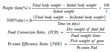
Growth performance and nutrient utilization of fish was determined following the methods of Jimoh, and Aroyehun, (2011) in term of Final Individual Weight, Survival (%), Specific Growth Rate (SGR %/ day), Feed Conversion Ratio, (FCR) and Protein Efficiency Ratio (PER), Net Protein Utilisation (NPU) responses was calculated as
Haematological Analysis
Blood (1-2ml) was collected from the vertebral caudal blood vessel according to Schmit et al., (1999), using disposable 2ml syringe and needle. The blood was emptied into the heparinized blood bottle treated with Ethyl Diamine Tetracetic Acid (EDTA). A blood sample was centrifuge (1500 rpm for 7mins) to obtain the blood plasma. Plasma samples was stored at (-200C) for the electrochemical and biochemical analysis. Computerized method employing System KX-2INTM Automated Hematology Analyzer was used in blood analysis, the KX-2IN is an ideal hematology analyzer for a clinical satellite laboratory or research testing. Spectrophotometric method was used for biochemical analysis as described by Svobodova et al. [15]. While the plasma electrolytes was determined using corning 400 flame photometer. Other metals were determined using (a back) Model 200A flame of the Atomic Absorption Spectrophotometer (AAS).
Histological Examination of Test Organ
At the end of the experiment, one fish per treatment, that is, three fish per concentration were sampled after 96hours of exposure for histological analysis, the test organism was killed with a blow on the head, using a mallet and was dissected to remove the vital organs (gill, liver, kidney and skin). The organs were fixed in 10% formalin for three days after which the tissue was dehydrated in periodic acid Schiff’s reagent (PAS) following the method of Hughes & Perry, [16], in graded levels of 50%, 70%, 90% and 100% alcohol for 3 days, to allow paraffin wax to penetrate the tissue during embedding. The organs were embedded in malted wax. The tissue was sectioned into thin sections (5-7µm), by means of a rotatory microtome and was dehydrated and stained with Harris haematoxyllin-eosin (H&E) stain, Bancroft & Cook [17], using a microtone and each section were cleared by placing in warm water (38oC), where it was picked with clean slide and oven-dried at 58oC for 30 minutes to melt the wax. The slide containing sectioned materials/tissue were cleared using xylene and graded levels of 50%, 70%, 90%, 95% and 100% alcohol for two minutes each.
The section was stained in haematoxyline eosin for ten minutes. The stained slide was observed under a light microscope at varying X100 magnification, sections were examined and photographed using an Olympus BH2 microscope fitted with photographic attachment (Olympus C35 AD4), a camera (Olympus C40 AB-4) and an automatic light exposure unit (Olympus PM CS5P).
Biochemical Composition (Proximate) Analysis
The biochemical composition of the carcass of the experimental fish was run to determine the Crude Protein (CP), crude Lipid (CL), Crude Fiber (CF), Moisture (M), Ash and Nitrogen Free Extract (NFE), using standard methods [18]. Nitrogen was determined by the micro-kjedahl method (Pearson, 1976) and the crude protein was taken as N% x 6.25 (constant factor) where N is equal to Nitrogen content per 100g sample. Total carbohydrate was determined using the phenol-sulphuric acid method. The crude fibre was obtained by dry ashing of the sample at 550oC dissolved in 10% HCl (25ml) and 5% Lanthanum Chloride (2ml) boiled, filtered and made up to standard volume with distilled water.
Water quality Analysis
The water quality parameters were recorded for temperature, dissolved oxygen (Do) content, pH and conductivity before and after the experiment. pH was determined using a digital pH meter (Mettler Toledo 320). DO and conductivity were measured using a digital dissolved oxygen meter (oxygen analyzer model JPB-607 portable) once in a day at 8.00am. The water quality was determined using the method of America Public Health Association (APHA, 2000) Model Number, E-9909 (pH, TDS, Salinity, EC, Temp.)
Method of Statistical Analysis
The data was collated and analysed using descriptive statistics, one way analysis of variance and Pearson’s correlation. The differences in the means between both values was assessed with Duncan multiple range test Using SPSS version 21 at P<0.05 significant level.
Results
The result of final total length of the Clarias gariepinus fed diets containing Tiny Moss (Bryum capillare) presented in Table 2, showed that there were significant (p<0.05) differences in the final total length during the period of the study. Though, the highest final total length was recorded among the T5, T3 and T1 respectively followed by T6 while T4 and T3 had similar records which were the least among the groups.
Proximate analysis of the Tiny Moss (Bryum capillare) in the diets
The result of the proximate composition of the diets containing Tiny Moss (Bryum capillare) used for feeding the Clarias gariepinus in the experiment is presented in table 3.
The result of proximate analysis of the fish diet containing Tiny Moss (Bryum capillare) in the diets as presented in the Table 3 showed varying levels of nutrients in diets containing Tiny Moss (Bryum capillare) for the experiment. The T6 diet had the highest percentage level of crude protein followed by T4 then T5 and T1, while T3 and T2 had the least crude protein. The crude fat was also highest in T6 followed by T4, T5, T1, T3 and T2 respectively. The crude fibre showed close similarities in the values such that they followed this trend; T2, T3, T1, T5, T4 and T6 respectively. The ash content, Moisture content Nitrogen Free Extract and Dry matter content equally showed little variations among the individual groups Performance of Clarias gariepinus Fed diets containing Tiny Moss (Bryum capillare).
The result of the productive performance of the Clarias gariepinus fed the diets containing Tiny Moss (Bryum capillare) used for feeding the fish in the experiment is shown in table 4.

Means with the same superscripts in the same column are not significantly different at P>0.05, while those with different superscripts in the same column are significantly different at same level.

%NFE =100 – (%CP+%CFAT+%CFIBRE+%ASH+%M); %DM = 100 - %M
Means with the same superscripts in the same column are not significantly different at P>0.05, while those with different superscripts in the same column are significantly different at same level.

The Total Dissolved Solid (TDS) of the pond containing graded levels of Tiny Moss (Bryum capillare) meal showed close relationship in body weights of the fish varying levels of Tiny Moss (Bryum capillare) at the start of the experiment. The Total Dissolved Solid (TDS) significantly increase (P<0.05) at T4 as the Tiny Moss (Bryum capillare) inclusion increased up to 4% among the individual treatment, followed by T1, T3, T5 and T6 that had similar values whereas T2 had the least Total Dissolved Solid (TDS) which the mean differences were statistically significant (p<0.05) among the individual treatments.
The productive performance of the Clarias gariepinus Fed diets containing Tiny Moss (Bryum capillare) as indicated in the Table 4, showed close relationship in body weights of the fish varying levels of Tiny Moss (Bryum capillare) at the start of the experiment. The final body weight and the daily weight gain decreased as the Tiny Moss (Bryum capillare) inclusion increased among the individual treatment, such that the T1 had highest final body weight and daily weight gain among those in control group (T1) followed by T2 then T3, but T5 and T6 had similar values whereas T4 had the least weight gain, which the mean differences were statistically significant (p<0.05) among the individual treatments. There were no significant (p>0.05) differences in the daily feed intake and the total feed intake during the period of the study. Though, the highest feed intake was recorded among the T3 while others had similar records of feed intake among the groups. The results of the feed conversion ratio were significantly (p <0.05) different among the groups, such that T1 and T2 had the best FCR followed by T5 and T6 which had similar values but the T4 had the least value. There were significant (p<0.05) differences in the survival rate during the period of the study such that T2 recorded the highest survival rate, followed by T3 and T6 while T4 and T5 had similar records and the least survival rate among the groups. Water Quality of the pond containing Clarias gariepinus fed diet with Tiny Moss (Bryum capillare). The result of the water quality of the pond containing clarias gariepinus fed diet with Tiny Moss (Bryum capillare) in the experiment is presented in table 5.

Means with the same superscripts in the same column are not significantly different at P>0.05, while those with different superscripts in the same column are significantly different at same level.
The result of the average water quality of the pond containing Clarias gariepinus fed diet with Tiny Moss (B capillare) in the experiment as presented in table 5, indicated that there were similarities in the pH level of the water such that the T3, T4, T5 and T6 had the same marginally higher pH followed by T2 whereas T1 had the least pH, which the mean differences were statistically not significant (p>0.05) among the individual treatments. The Total Dissolved Solid (TDS) of the pond containing graded levels of Tiny Moss (Bryum capillare) meal as indicated in the Table 5, showed close relationship in body weights of the fish varying levels of Tiny Moss (B capillare) at the start of the experiment. The Total Dissolved Solid (TDS) significantly increase (P<0.05) at T4 as the Tiny Moss (B capillare) inclusion increased up to 4% among the individual treatment, followed by T1, T3, T5 and T6 that had similar values whereas T2 had the least Total Dissolved Solid (TDS) which the mean differences were statistically significant (p<0.05) among the individual treatments.
The result of the water conductivity of the pond containing Clarias gariepinus fed diet with Tiny Moss (B capillare) in the experiment as presented in table 5, indicated that their significant differences (P<0.05) among the group such that T4 (4% inclusion of Tiny Moss (B capillare) meal) had the highest level of water conductivity followed by T1, T3 T5 and T6 that had similar conductivity while T2 (2% inclusion of Tiny Moss (B capillare) meal) had the least. The average water salinity of the pond containing graded levels of Tiny Moss (B capillare) meal as presented in the Table 5 were similar in their individual salinity of the water at varying levels of Tiny Moss (B capillare) during experiment. The average salinity is not significantly different (P>0.05) among the individual group as the Tiny Moss (B capillare) inclusion increased. Though there were marginal increases such that T4, had increased salinity followed by T2, then T3 and T6 whereas T5 had the least value which the mean differences were statistically not significant (p>0.05) among the groups.
The result of the average ammonia content of the water in the pond containing Clarias gariepinus fed diet with Tiny Moss (B capillare) meal in the experiment as presented in table 5. This indicated that there were significant differences (P<0.05) in the ammonia content of the water such that the T4, had the highest ammonia concentration followed by T3, T5 and T6 which had the same ammonia concentration, while T1 and T2 had ammonia concentration. The average Temperature (OoC), Nitrite (NO3), Nitrate (NO2), and Dissolved oxygen (DO) of the pond containing graded levels of Tiny Moss (Bryum capillare) meal as presented in the Table 5 were similar in their individual water at varying levels of Tiny Moss (Bryum capillare) inclusion during experiment their results indicated no significant differences among the groups.
Haematological parameters of Catfish Juveniles fed Tiny Moss (Bryum capillare) meal. The result of the average haematological parameters of catfish (Clarias gariepinus) juveniles fed diets containing Tiny Moss (Bryum capillare) is presented in table 6. The result of average haematological parameters of catfish (Clarias gariepinus) juveniles fed diet containing Tiny Moss (Bryum capillare) is presented in the Table 6. The study showed that the haematological parameters of the catfish (Clarias gariepinus) juveniles fed graded levels of diets containing Tiny Moss (Bryum capillare) were not significantly different (P>0.05). Though there were marginal variations among the individual group, there were still a lot of similarities among the values in each of the groups. Biochemical Characteristics of Catfish Juveniles fed Tiny Moss (Bryum capillare) meal. The result of the average biochemical characteristics of Catfish (Clarias gariepinus) juveniles fed diets containing Tiny Moss (Bryum capillare) is presented in table 7.


Means with the same superscripts in the same column are not significantly different at P>0.05, while those with different superscripts in the same column are significantly different at same level.
The result of average biochemical characteristics of catfish Clarias gariepinus juveniles fed diet containing Tiny Moss (Bryum capillare) is presented in the Table 8. The study showed that the protein content of the catfish (Clarias gariepinus) juveniles fed graded levels of diets containing Tiny Moss (Bryum capillare) were significantly different (P<0.05). The catfish fed T4 diet had the highest percentage level protein followed by T2, and T6 then T1, while T3 and T5 had the least protein. The average ether extract was also highest in T2 which were similar to those in T4, and T6, but were significantly different (P<0.05) to T1 and T3 which had the same protein level, whereas T5 had the least. The ash content, Nitrogen Free Extract, energy and glucose showed close similarities in the values such that they showed marginal variations which were not significantly different (P>0.05) among the individual groups.

Means with the same superscripts in the same column are not significantly different at P>0.05, while those with different superscripts in the same column are significantly different at same level.
The result of the effect of Tiny Moss (Bryum capillare) on Amino Acid Profile of Catfish Clarias gariepinus Juveniles fed diet containing Tiny Moss (Bryum capillare) is presented in the Table 9. The study showed that the Glutamic Acid, Aspartic Acid, Valine, Threonine, Serine, Phenylalanine, Proline and Methionine increased in value while Lysine, Leucine, Arginine, Alanine, Isoleucine, Glycine, Histidine and Tryptophan reduced in value and there was no significant change in cysteine (%) all were significantly at (P<0.05).

Means with the same superscripts in the same column are not significantly different at P>0.05, while those with different superscripts in the same column are significantly different at same level.
Figures 1-4 present the results of tissue analysis of fish from the respective treatment. Histological examinations of the test fish showed some pathological changes. The Tiny Moss (Bryum capillare) feed shows normal skin architecture with well outlined epithelia cell (EC) moderate effect on the skin layer with moderate necrosis (N) of the muscular region with the epithelia lining and superficial spreading of melanoma (M) restricted to the epidermis The heart shows normal cardiac tissue with cardiac cell (CC), cardiac fiber (CF) cardiac muscles (CM) shows moderate aggregate of myocardiac inflammation (AMI). The gill showed section of gill with ghost (G) appearance with severe aggregate of inflammatory cell (AIC). The liver cells revealed section of liver (X100) (H/E) shows severe effect on the hepatic tissue with severe I intra hepatic inflammation (IHI) and intra hepatic hemorrhage (IHIH). The damage done to these organs as the result of the feeds correlates with the concentrations of the feeds in each experimental tank.
Discussion
Non-conventional feed resources (NCFRs) are feeds that are not usually common in the markets and are not the traditional ingredients used for commercial fish feed production (Devendra, 1988; Madu et al., 2003). NCFRs are credited for being non-competitive in terms of human consumption, very cheap to purchase, by-products or waste products from agriculture, farm made feeds and processing industries and are able to serve as a form of waste management in enhancing good sanitation. The result of final total length of the Clarias gariepinus fed diets containing Tiny Moss (Bryum capillare) presented in Table 2 showed that there were significant (p<0.05) differences in the final total length during the period of the study. Though, the highest final total length was recorded among the T5, T3 and T1 respectively followed by T6 while T4 and T3 had similar records which were the least among the groups.
The result of proximate analysis of the fish diet containing Tiny Moss (Bryum capillare) in the diets as presented in the Table 3 showed varying levels of nutrients in diets containing Tiny Moss (Bryum capillare) for the experiment. The T6 diet had the highest percentage level of crude protein followed by T4 then T5 and T1, while T3 and T2 had the least crude protein. The crude fat was also highest in T6 followed by T4, T5, T1, T3 and T2 respectively. The crude fibre showed close similarities in the values such that they followed this trend; T2, T3, T1, T5, T4 and T6 respectively. The ash content, Moisture content Nitrogen Free Extract and Dry matter content equally showed little variations among the individual groups, this work is similar to the work of Elhassan et al., (2015) who reported a significant (p<0.05) increase in bambara groundnut protein (18.83±0.49), lipids (7.05±1.82), fiber (5.74±1.09), carbohydrate (63.37±2.57), moisture (12.59±1.14) and ash (3.52±0.22), which indicates that Bambara groundnut could be an excellent source of protein, lipid, carbohydrate and mineral elements in Clarias gariepinus Fed diets.
The relationship between Na and K as well as between Ca and P; are desirable with the respective ratios of Na/K (0.6) and Ca/P (1.2). They also contain high levels of carotenoids (30 to 41.5 mg/100g DW), vitamin C (137.5 to 197.5 mg/100g DW) [19].
Seventeen amino acids (isoleucine, leucine, lysine, methionine, cysteine, phenylalmine, tyrosine, threonine, valine, alanine, arginine, aspartic acid, glutamic acid, glycine, histidine, proline and serine) were detected. Their amino acid composition compare favourably with that of WHO/FAO protein standard indicating favourable nutritional balance except for lysine and methionine which appear marginal. The nutritional values of the phytochemicals were also assessed with a view of establishing and understanding their nutritional uses. The functional properties for the three vegetables were similar. Comparing the nutrient and chemical constituents with recommended dietary allowance (RDA) values, the results reveal that the leaves contain an appreciable amount of nutrients, minerals, vitamins, amino acids and phytochemicals and low levels of toxicants Essential or indispensable amino acids (EAAs) cannot be synthesised by fish and often remain inadequate but are needed for growth and tissue development [20,21]. The result of the effect of Tiny Moss (Bryum capillare) on Amino Acid Profile of Catfish Clarias Gariepinus Juveniles fed diet containing Tiny Moss (Bryum capillare) is presented in the Table 9. The study showed that the Glutamic Acid, Aspartic Acid, Valine, Threonine, Serine, Phenylalanine, Proline and Methionine increased in value while Lysine, Leucine, Arginine, Alanine Alanine, Isoleucine, Glycine, Histidine and Tryptophan reduced in value and there was no significant change in cysteine (%) all were significantly at (P<0.05).


Fishmeal is known to contain complete EAA profile that is needed to meet the protein requirement of most fish species. Since fishmeal is expensive as a feed ingredient, the use of non-conventional feedstuffs has been reported with good growth and better cost benefit values. The utilization of non-conventional feedstuffs of plant origin had been limited as a result of the presence of alkaloids, glycosides, oxalic acids, phytates, protease inhibitors, haematoglutinin, saponegin, momosine, cyanoglycosides, linamarin to mention a few despite their nutrient values and low-cost implications [22,23]. The report of this work is similar to the work of Sobowale et al. [19] who reported the nutritional components in three species of leafy vegetables using standard analytical methods. All the vegetables contained moisture (79.92 to 84.0%), crude protein (20.61 to 22.7%), crude fibre (10.7 to 22.44%), ash (6.8 to 10.44%), carbohydrate (55.86 to 68.22%) crude lipid (4.24 to 5.6%) and food energy (1507.19 to 1673.96 kJ/100g). The mineral element content were high with remarkable concentration of K (35.2 to 48.8mg/100g), Na (11.4 to 14.4 mg/100g), Ca (15.4 to 18.7mg/100g), Mg (12.2 to 18.7mg/100g), P (13.8 to 15.08mg/100g). These anti-nutritional factors negate growth and other physiological activities at higher inclusion levels [24].
The result of the essential amino acids indicated that these animal supplements could substitute fishmeal in fish feed (Table 2) since they all contain the required essential amino acids needed by fish for protein metabolism [25]. This result showed that earthworm meal is richer in methionine (Figure 1) than other animal protein sources studied which agreed with the observation of Finke (2003) when he compared the nutrient values in some invertebrates. Methionine has been credited as growth promoting essential amino acid, which is highly needed by cultured fish and limited in most plant and many animal supplements (Wilson, 2002).
Several reports have indicated that gill lesions do not only indicate possibilities of impaired respiratory functions but impaired osmo-regulatory functions as [26]. Even slight structural damage can render a fish vulnerable to osmo-regulatory as well as respiratory difficulties (Hughes and Morgan, 1973) thereby affecting the overall metabolism and survival of the fish. The histopathological alteration observed in the brain, gill, liver, intestine and muscle/flesh is an indication of the toxic effect of P. zeylanica extracts to fish. This agreed with Fafioye [27,28], observation when Clarias gariepinus and O. niloticus were exposed to lethal and sublethal concentrations of Parkia biglobosa and Raphia vinifera respectively. The gill lamellae play a significant role in regulating the exchange of gas, water and ions in fish. The role of the gill in excretion predisposes it in such a way that slight structural damage can render a fish very vulnerable to osmoregulation as well as respiratory difficulties.
Conclusion
Fish, like other organisms, required food (energy) in order to grow, survive and reproduce. The food items (source of energy) in aquatic habitat are in the form of plankton, periphyton, nueston, benthose, nekton and plants are available throughout the year. The success of intensive fish culture depends on the formulation of a fish feed that contains an optimum level of protein and energy necessary for the growth of fish and is also cheap. It is obviously necessary to formulate and manufacture fish feed from locally available feed ingredients. This type of feed prepared from such ingredients should serve as a source of essential amino acid, minerals, vitamins, growth promoting substances and energy [29-34].
Essential or indispensable amino acids (EAAs) cannot be synthesized by fish and often remain inadequate but are needed for growth and tissue development [21]. Fishmeal is known to contain complete EAA profile that is needed to meet the protein requirement of most fish species. Since fishmeal is expensive as a feed ingredient, the use of nonconventional feedstuffs has been reported with good growth and better cost benefit values. The utilization of nonconventional feedstuffs of plant origin had been limited as a result of the presence of alkaloids, glycosides, oxalic acids, phytates, protease inhibitors, haematoglutinin, saponegin, momosine, cyanoglycosides, linamarin to mention a few despite their nutrient values and low-cost implications [23].
To Know more about Oceanography & Fisheries Open Access Journal
Click here: https://juniperpublishers.com/ofoaj/index.php
To Know more about our Juniper Publishers
Click here: https://juniperpublishers.com/index.php


