Friday, July 30, 2021

Hypertrophic Megacolon in a 1-year-old child with Hirschsprung’s Disease: Case Report and Literature Review - Juniper Publishers

Open Access Journal of Surgery - Juniper Publishers  


Abstract

Hirschsprung’s disease (HSCR) is a common cause of paediatric intestinal obstruction. It is a functional colonic obstruction due to lack of ganglion cells in the affected segment of colon. Symptoms range from intestinal obstruction to chronic progressive constipation in older children. A 1-years-old infant was brought to Khartoum Teaching Hospital with abdominal distension, difficulty passing stool and chronic constipation treated by regular enema without improvement. On examination, patient looked ill and emaciated. Diagnosis of HSCR disease was established and patient was planned for operative management. The clinical presentation and management of chronic HSCR were briefly discussed in this report.

Keywords: Hirschsprung’s disease; Hypertrophic megacolon;1-year old, Child

Introduction

Hirschsprung's disease (HSCR) is caused by the failure of ganglion cells to migrate from the colon during gestation. The different lengths of the distal colon cannot relax, resulting in functional colonic obstruction. The disease usually appears in early infancy, although some patients experience severe and persistent constipation later in life. Symptoms include difficult defecation, poor feeding, poor weight gain, and Bloating. This disease can be familial or spontaneous, and multiple factors appear to play a causative role in HSCR disease [1].

Patients diagnosed later in life with chronic symptoms often have short-segment variant of the disease, and their clinical manifestations often include chronic constipation, bloating, vomiting and growth failure [2]. Older children who suffer from long term obstructive symptoms, are resistant to conventional treatment options and may require daily enema treatment.

Enterocolitis and ruptured colon are the most serious complications associated with the disease and the most common causes of mortality related to it [3]. Enterocolitis occurs in 17% to 50% of infants with HSCR disease, and the most common causes are intestinal obstruction and residual ganglion enteritis. For many years after corrective surgery, babies must continue to be closely monitored for enterocolitis, as infections have been reported up to 10 years later [1].

Case Report

A 1-years-old infant was brought to Khartoum Teaching Hospital with abdominal distension, difficulty passing stool and chronic constipation treated by regular enema without improvement. His condition started since birth when patient delivered at home by non-vaginal delivery, of full term pregnancy which passed uneventful, he cried immediately and started to breastfeed. Baby failed to pass meconium during first 48hrs after birth.

On examination; patient looked ill and emaciated. Digital rectal examination showed hard stool with gush of fluid upon finger removal. Plane abdominal X-ray showed dilated colon with fecal impaction. Contrast enema study showed huge dilated rectosigmoid with transitional zone and collapsed distal rectum. Interestingly, enema study also showed a thick hypertrophied colon with very big old stool that appeared to be hardened.

Patient was admitted, cannulated and given intravenous antibiotics, then rectal wash by saline 10-15ml/kg for one week was done. Thereafter, he was prepared for colostomy and biopsy. Histopathological report showed typical features of distal aganglionic bowel segment.

Patient was operated under general anesthesia and aseptic condition, left iliac fossa grid iron incision was made, and bowel explored. Three seromuscular biopsies was taken from the collapsed part of rectosigmoid colon, stoma site and the dilated part, then loop colostomy was created. Patient recovered well from anesthesia. Three day post-operative stoma were adequately functioning and patient was discharged home and planned for Swenson pull through operation after 6 months. The operation was done successfully and follow up went uneventfully (Figures 1-3).

Discussion

Aganglionic disease is the main etiology of HSCR. The full migration, proliferation, differentiation, survival and apoptosis of neural c cells all contribute to functional ENS. Any interference in these processes will cause HSCR disease phenotype. In 80-85% of HSCR cases, the aganglionic area is limited to the rectum and sigmoid colon (short-segment disease). Long-segment disease can occur in up to 20% of cases and has the characteristics of aganglionic disease that extends to the proximal end of the sigmoid colon. Total colonic aganglionic disease is rare, occurring in 3-8% of HSCR patients [3].

About 80% of cases present in the first months of life, with dysmotility, poor diet and progressive abdominal distention. Up to 90% of babies with HSCR disease do not pass meconium within the first 24 hours after birth [1]. Older children present with constipation that doesn’t respond to regular treatment, these children usually don’t develop normally and are often thin and malnourished.

A retrospective study was conducted on 64 patients of HSCR in Sudan, with male: female ratio 5:1, average age at first presentation was nine days and most patient were from central Sudan. Patients present commonly with delayed passage of meconium and constipation, moreover, emergency presentation was encountered in 21.9% of cases. Complications were reported most frequently on the 13th day following surgery with colostomy prolapse. The age of patients at Pull Through Procedure was ranging from seven to seventy two months and the mean body weight was around 12 kg, these factors were found to play and important role on surgical outcome and length of hospital stay [4].

The diagnosis of HSCR can be done in a variety of ways. However, the preferred diagnostic procedure of choice is a contrast enema. This will define the transition zone between the normal (dilated) bowel and the narrow aganglionic bowel. This transition zone can be seen in 70% to 90% of cases. Routine x-rays show dilation of the intestine, and anorectal manometry can also help the diagnosis [3]. In rectosegmoid HSCR the location of radiographic transitional zone correlate accurately with the level of aganglionosis in majority of cases, but fail tobe accurate in long-segment or total colonic disease [5]. The gold standard for diagnosing HSCR is rectal biopsy. Submucosal rectal aspiration biopsy can be performed without anesthesia. A biopsy sample is analyzed to look for the loss of ganglion cells and hypertrophic nerve trunks. Biopsy samples can be stained to increase the activity of acetylcholinesterase, thereby aid in the diagnosis. If aspiration biopsy does not provide an accurate diagnosis, a full-thickness biopsy should be performed [3]. Simple modified technique can improve the adequacy of rectal suction biopsy, and wall suction with Noblett forceps is more likely to produce an adequate specimen and decrease the need for repeated biopsy [6].

Until now, the only treatment for HSCR is surgery. Due to malnutrition or sepsis after bowel perforation, lack of surgical treatment for HSCR can be fatal. Although surgery is the conventional treatment for HSCR patients, the results of surgery vary widely and vary widely [3]. Traditional treatment by the three stage Pull Through Procedure carry disadvantages, such as stoma problems and might triple hospital stay time. Thus, one stage operation is more preferred option for neonates [7].

Surgical treatment aims to remove the ganglion intestine and anastomose the normal intestine to the anus, to maintain the function of the sphincter. The main technologies include total Transanorectal removal (TERPT) and laparoscopic assisted removal procedures. The modified TERPT procedure has generally better long-term outcomes than the classical approaches [8].

Conclusion

Early diagnosis and treatment of HSCR are essential to achieve better results. Initial diagnosis can be made by water-soluble contrast enema, but the gold standard for HSCR is rectal biopsy, and the mainstay of treatment is surgery. Chronic form of disease usually present with chronic constipation that doesn’t respond to regular enema, imaging may demonstrate hypertrophied colon with hard stool impaction. In emergencies, the age and weight at the time of removal surgery and the closing time of the colostomy can significantly affect the prognosis and extend the length of hospital stay.

To Know more about Open Access Journal of Surgery


To Know more about our Juniper Publishers

Click here: https://juniperpublishers.com/index.php    

Thursday, July 29, 2021

The Content of Natural Radionuclides in the Fish of Lake Issyk-Kul - Juniper Publishers

 Oceanography & Fisheries - Juniper Publishers

Abstract

This work is devoted to the study of the features of biogenic migration of natural radionuclides in the conditions of Lake Issyk-Kul located in the north-eastern part of Kyrgyzstan. Lake Issyk-Kul is a high-mountainous, thermal, drain less reservoir with brackish water with a high content of natural uranium. This is due to climatic conditions, the geological structure of river valleys, and the peculiarities of the composition of river waters capable of extracting uranium from rocks. Calculations using the Erica tool 1.3 software showed that the natural uranium content in the water of Lake Issyk-Kul (0.809 Bq/l) is safe for living organisms of the aquatic ecosystem, the level of absorbed dose, accumulation and risk factors vary within the normal range. According to the Erica tool 1.3 database, low radiation doses (0-50 μGy/h) can stimulate the growth of aquatic plants. The species composition of the ichthyofauna of Lake Issyk-Kul consists of 22 species of fish, including 12 aboriginal and 10 acclimatized. The content of natural radionuclides (226Ra, 228Th and 212Pb) in the bone and muscle tissue of some fish species of Lake Issyk-Kul was investigated. It has been established that radionuclides are predominantly found in bone tissue in comparison with muscles, their concentrations vary within the limits of natural parameters.

Keywords: Water; Natural radionuclides; Fish; Issyk-Kul Lake

Introduction

Lake Issyk-Kul is located in the northeastern part of Kyrgyzstan, between the ranges of the Northern Tien Shan: Kungey-Ala-Too and Terskey-Ala-Too, at an altitude of 1609 meters above sea level. The lake is closed, with about 80 relatively small rivers flowing into it. The largest of them are Tyup and Jergalan, flowing from the east. The water is brackish (water salinity 5.90 ‰), so it does not freeze in winter. The volume of water is 1738 km3, the area of the water surface is 6236 km2, the average depth is 278 m, the maximum depth is 668 m. The length of Issyk-Kul from west to east is 182 km, and from south to north - 58 km. The climate is temperate maritime, with the lake having a softening effect in the region. Average January temperature: -2° -6°, average July temperature: + 17° + 21°. In the western mountains bordering the lake, only 115 mm of precipitation falls, on the eastern coast - about 600 mm [1-3]. According to the research of prof. Kovalsky [4], the waters of the rivers in the regions of the Issyk-Kul depression are enriched with natural uranium. The increased content of uranium in the waters should be associated not only with the climatic conditions of the regions, but also with the geological structure of river valleys, as well as with the peculiarities of the chemical composition of river waters capable of well extracting uranium from rocks. So, for example, if in the northern rivers of Russia (Northern Dvina, Lena, Neva, Kama), the uranium content fluctuates within (2-13) ×10-7 g/l, while in the rivers flowing into Lake Issyk-Kul, its concentration increases to (58-71) ×10-7 g/l [4]. The average uranium content in the water of Lake Issyk-Kul varies within (62-65) ×10-6 g/l [5]. In modern literature, a number of studies are presented on the topic of pollution, migration, accumulation of radionuclides in the environment and their biological effect on living organisms. [6-12]. Based on the above, the aspect of the biological effect of natural concentrations of uranium and products of its decay on living organisms of the aquatic ecosystem of Lake Issyk-Kul is poorly studied.

Materials and Methods

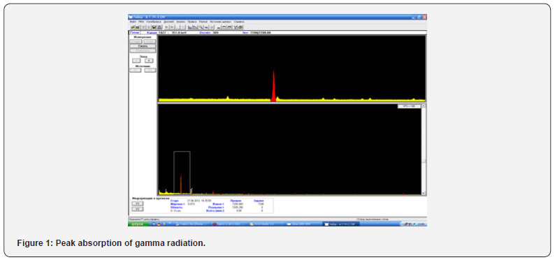
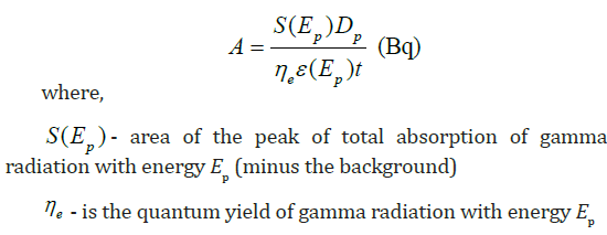
The content of natural radionuclides (226Ra, 228Th and 212Pb) in the bone and muscle tissue of some fish species of Lake Issyk-Kul has been investigated. 64 samples were prepared for gamma spectrometric analysis. The measurements were carried out on a GX4019 gamma spectrometer with Genie-2000 S 502, S501 RUS software. To obtain a stable equilibrium of radionuclides, the studied samples after their packing were stored from 25 to 180 days. The radionuclide activity in the sample on the day of spectrum measurement was calculated by the formula:

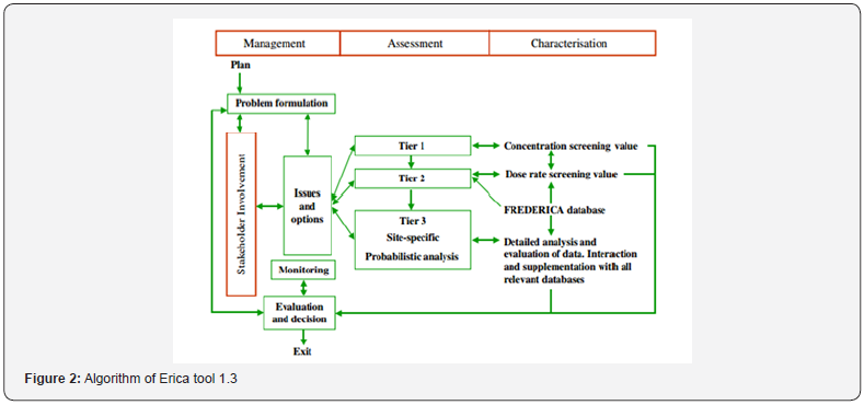
The Erica tool 1.3 application package is used for radioecological assessment of the state of terrestrial and aquatic ecosystems. Knowing the content of radioactive elements in soil or water, the program calculates the accumulation of radionuclides by living organisms, absorbed radiation doses, and an assessment of the radiation risk factor. The element of the radioecological assessment consists of three levels, at the first level the radiation risk factor is assessed, if its value is low, then level 1 can be limited (Figure 2). If the value of the radiation factor is increased, then the action is recommended to continue the assessment at level 2. To assess the statistical distribution of indicators, level 3 is used [13,14].

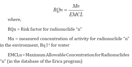
To calculate the radiation risk factor using the Erica tool 1.3 program, we entered the data on the specific activity of uranium in the water of Lake Issyk-Kul. If the calculated value of the risk coefficient is higher than 1, then there is a possibility of accumulation of radionuclides by living organisms. The risk factor (RQ) was determined using the following formula:

Results and Discussion

The species composition of the ichthyofauna of Lake Issyk- Kul is not rich and consists of 22 species of fish, including 12 aboriginal and 10 acclimatized [15,16]. We have investigated the distribution of natural radionuclides (226Ra, 228Th and 212Pb) in the bone and muscle tissue of some fish species of Lake Issyk-Kul. For the analyzes, fish species with wide distribution were selected, their habitat, type of food, commercial value were considered:

1. Pike perch (Lucioperca Lucioperca. 1958) is a predator, a widespread acclimatized species, the basis of its food is chironomids, mysids, amphipods, from about one year old it begins to switch to a predatory type of food. Forms large accumulations during the feeding period in shallow waters, and mainly in areas of the lake with a lower salinity of water.

2. Issyk-Kul trout (Salmo ischchan Issykogegarkuni Lushin. 1932) - an acclimatized species, distributed throughout the lake, with the formation of pre-spawning accumulations in the estuarine areas, near shallow waters with a spawning substrate. The diet of trout is predominantly benthic, but it can switch to a predatory type of diet.

3. Issyk-Kul Chebachok (Leuciscus bergi Kaschkarov. 1925) - an aboriginal and rare species, found in small quantities in the southwestern and southeastern parts of the lake.

4. The research results showed that natural radionuclides are predominantly found in bone tissue in comparison with muscles, especially 226Ra (Table 1). Concentrations of natural radionuclides vary within the background values, for example, the 226Ra content in the total mass of a two-year-old pike perch (Lucioperca Lucioperca) was 0.61 Bq/kg per wet weight, the absorbed dose level was 0.081 μGy/h, and the risk factor was 0.008. Lower concentrations are typical for 228Th and 212Pb.

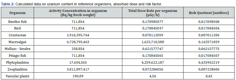
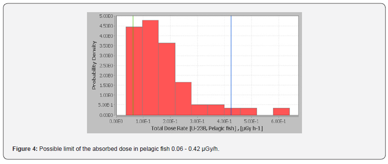
With the average uranium content in the water of Lake Issyk- Kul - 0.809 Bq/l, the following results were obtained (Table 2). Among the reference organisms, uranium is able to accumulate in aquatic plants (190.09 Bq/kg). The absorbed dose level was 4.56 μGy/h, it can vary in the range of 0.47 - 17.9 μGy/h. According to the Erica tool 1.3 database, absorbed doses in the range (0-50 μGy/h) do not have statistically significant negative biological effects on aquatic plants. At low doses of radiation, a slightly stimulating effect on growth is possible (by 1.2 times). For Lake Issyk-Kul, the greatest species diversity falls on the group of blue-green (Cyanophyta), diatoms (Bacillariophyta) and green (Chlorophyta) algae. The average value of the hazard coefficient for aquatic plants is (0.45). If the calculated value of the risk coefficient is higher than 1, then there is a probability of accumulation of radionuclides by living organisms (Figure 3). The estimated average uranium content in pelagic fish is 7.11 Bq/kg wet weight. The absorbed dose level was 0.17 μGy/h, it can vary in the range of 0.06 - 0.42 μGy/h (Figure 4).

According to the database of the Erica tool 1.3 software, no statistically significant negative biological effects on the fish organism are observed within these absorbed doses. It is known that uranium accumulates mainly in bone tissue, kidneys, gills, and then in the liver, and only a small part is retained in muscles. Since parts of fish organs: gills, liver and bones are usually not eaten by the population, the total daily dose for uranium established by the World Health Organization is no more than 50 Bq/kg [17,18].

Conclusion

Calculations using the Erica tool 1.3 software showed that the natural uranium content in the water of Lake Issyk-Kul (0.809 Bq/l) is safe for living organisms of the aquatic ecosystem, the level of absorbed dose, accumulation and risk factors vary within normal limits. According to the conclusions of the scientific of the UN Committee on the Effects of Atomic Radiation, the absorbed dose of 80 μGy/h is the threshold dose. The presented absorbed doses for reference organisms in Table 1 are much lower than this indicator. The content of uranium and other natural radionuclides (226Ra, 228Th and 212Pb) in certain fish species of Lake Issyk-Kul vary within the background values, mainly radionuclides are found in bone tissue as compared to muscles.

To Know more about Oceanography & Fisheries

Click herehttps://juniperpublishers.com/ofoaj/index.php


To Know more about our Juniper Publishers

Click here: https://juniperpublishers.com/index.php    


Wednesday, July 28, 2021

Noninvasive Continuous Blood Glucose Monitoring by Dielectric Spectroscopy - Juniper Publishers

 Current Research in Diabetes & Obesity - Juniper Publishers

Abstract

Aims: The Alergy noninvasive continuous blood glucose monitor (NICBGM) is a novel wristband device that reports glucose levels without entailing skin puncture. This study evaluated the performance of this device compared with an FDA-approved glucose meter in patients with type 2 diabetes.

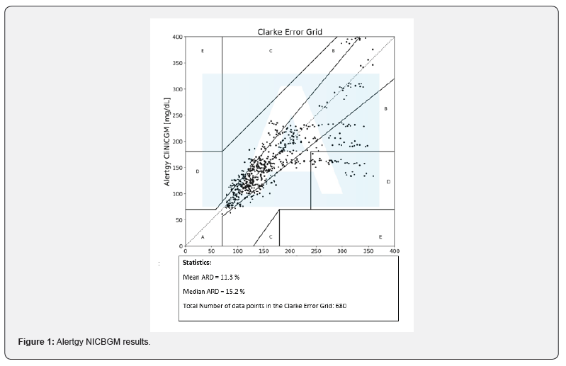
Methods: The Alertgy DeepGluco NICBGM device measures changes in the dielectric spectrum it collects specific to blood glucose levels three times a minute. This spectral data is analyzed by using neural network analysis, machine learning and then with a calibration process is used to generate algorithms to estimate blood glucose (BG) values once the system is calibrated to the individual. The Roche Accuchek Inform II glucometer was used as a reference technique for calibration and then to determine the accuracy of blood glucose determinations. 27 patients completed three or more 120-minute sessions. Mean absolute relative difference (MARD) was calculated on the data collected.

Results: MARD values were compiled for two or more days of data collection following the first day of calibration. The MARD for all measurements was found to be 15.3.

Conclusions: The resultant MARD suggest that this technique can achieve equivalent performance to that of existing CGM devices presently approved by the FDA when the same reference technique is utilized.

Keywords: Continuous glucose monitor; Glycemic control; Noninvasive glucose measurement; Diabetes

Abbreviations: NICBGM: Alertgy Noninvasive Continuous Blood Glucose Monitor; BG: Blood Glucose; DM: Diabetes Mellitus; FS: Fingerstick; T2DM: Type 2 Diabetes Mellitus, ESRD: End-Stage Renal Disease; POC: Point-of-Care; MARD: Mean Absolute Relative Difference; CGM: Continuous Glucose Monitor; GLP1: Glucagon-like Peptide-1; HbA1c: Hemoglobin A1c; ADHF: Acute Decompensated Heart Failure; SGLT2: Sodium Glucose Co-Transporter 2; DPP4: Dipeptidyl Peptidase-4; TZD: Thiazolidinedione

Introduction

Diabetes mellitus (DM) affects over 30 million people in the United States [1]. For most patients living with DM, frequent self-monitoring of blood glucose (BG) is needed for adequate outpatient glycemic control and has been associated with lower hemoglobin A1c [2]. The majority of patients with DM rely on fingerstick (FS) glucose measurements for self-monitoring, which necessitates lancing of the skin. FS testing is associated with pain and anxiety and can lead to nonadherence with home testing [3,4]. In recent years, the use of continuous glucose monitors (CGM) has become more widespread. CGMs significantly reduce the number of fingersticks needed for home monitoring; however, most devices still require the insertion of a new subcutaneous catheter every 10 to 14 days.

The Alertgy noninvasive continuous blood glucose monitor (NICBGM) is a novel device that does not entail skin penetration of the skin for continuous glucose monitoring. The device is worn on the wrist and measures BG levels through the use of dielectric spectroscopy. There have been several prior noninvasive BG monitors on the market; however, these have had limited success due to issues with inaccuracy and low reliability [5,6]. This study aimed to evaluate the performance of NICBGM retrospectively compared to an FDA-approved hospital-grade glucose meter in patients with T2DM.

Materials and Methods

Study population

This study was approved by the Complete Care Institutional Review Board. The study included 27 patients with T2DM recruited from the outpatient Complete Clinic. Inclusion criteria were known diagnosis of T2DM, current dietary or pharmacologic treatment for DM, recent hemoglobin A1c between 7.5 % (58 mmol/mol) and 10.0% (86 mmol/mol), and age between 18 and 75 years old. Exclusion criteria were prandial insulin use, fasting FS BG <70 or >250 mg/dL on the day of study sessions, pregnancy (tested at the start of each study session), end-stage renal disease (ESRD), decompensated heart failure, medications that might cause false readings (including acetaminophen, ascorbic acid, dopamine, maltodextrin, or mannitol), and any conditions that might limit the ANICGM device, such as lesions on the forearms.

Study protocol

The studies were performed at the Complete Care main clinic location in Melbourne Florida. Two sets of trials were conducted to study the effect of using difference amounts and types of sugar. The Complete Care Trial I dataset had 17 unique subjects and 14 of those subjects suffered from type II diabetes among other comorbidities. All subjects had their baseline blood glucose measured every 5 minutes for 20 minutes before 68 grams of sucrose were ingested. After ingestion, blood glucose was measured every 5 minutes for 2 hours. All glucose measurements were carried out using the Accu-chek Inform II glucometer. Each 2 hour set of data was considered as an individual test. There were 60 individual tests that had followed this protocol and all 60 tests were included for further analysis.

The CompleteCare Trial II dataset had 10 unique patients, each of which being a diagnosed diabetic. CompleteCare Trial II subjects ingested 75 grams to 100 grams of dextrose for each of their respective tests. The total number of individual tests available were 45. Patients participated in three or more visits, each with a 30-min warm-up period and a study period lasting 120 minutes, at least three days apart, and most within 14 days from Day 1. At each visit, study subjects were fitted with the Alertgy NICBGM device for a warm-up period of 30 minutes before the start of data collection, and the device was worn throughout the duration of the 120-minute session thereafter (Figure 1). A trained technician took FS BG measurements. Baseline FSBGs were obtained at -30 min, -15 min, and 0 min during the warm-up period. FSBG measurements were made before and during this warm-up period solely as health/safety checks to ensure that the patients were in adequate physiological condition to continue the study. These reference measurements were not paired with any NICBGM device estimates nor used within the machine learning training process.

Patients were instructed to fast for at least eight hours the night before. Patients were evaluated at the end of the study session to ensure that they were asymptomatic, and that their BG was in a safe range prior to discharge.

Device technology

FS BG was measured using the Accuchek Inform II glucose monitor to provide calibration values for the NICBGM. FS BG levels were entered into a secure database, and a proprietary calibration program was used to analyze the spectral data from the device during the training session on Day 1. These POC BG values were also used in calculations for the estimation of device accuracy. The NICBVGM uses a weak electromagnetic field generated by its wristband sensor to produce dielectric spectra. The core sensing technology used is dielectric spectroscopy, which has been shown in prior work to be capable of noninvasively measuring blood glucose in a laboratory environment [7].

The monitoring technology implemented in the study includes a sensor having a multi-portion dielectric composite. A microstrip transmission line is placed on one surface of the dielectric composite and includes an input trace, radiator portion, and an output trace. The dielectric material adjacent to the radiator portion is selected for its dielectric properties such that it is matched with that of the target anatomy (distal forearm/wrist region) of the tested subject. This dielectric matching between the forearm and the sensor allows the radiator portion to effectively respond as if it were embedded inside the target anatomy, removing substantial uncertainty from the measurement process. By then applying a plurality of signals to the sensor, the signal's reflected and transmitted components can be measured and used to determine the amounts of certain constituents (including blood glucose) present in the subject’s target anatomy. This study's dielectric sensor is capable of use in broadband sensing applications, ranging from 0 MHz (DC) to 2GHz. (United States Patent No. US20200217809A1, 2020).

The device sends and receives signals from the wrist area used to generate a dielectric spectrum once every twenty seconds. These spectral data are stored on the device and then downloaded wirelessly or through a USB port to a secure database. The spectral data are then inputted into a machine learning algorithm and used to create an estimate of the patient’s BG level.

Device algorithm

Measurements for the algorithm training procedures were collected on each of the test subject’s Day 1 trial. Blood glucose measurements were collected via the AccuCheck Inform II POC SMBG system with a 5-minute sampling period over a duration of 2 hours, resulting in 14 reference measurements per Day 1 session.

Data transfer

Alertgy received the complete FS BG dataset from Complete Care only for the first visit for each patient for calibration. For Days 2 and 3, Alertgy received only the -30 min FS BG data. The data were collected real-time. The data collected from the wristband were sent to the lab at Alertgy. Upon receipt in the lab, the data were processed from its original 'raw' form (consisting of many files of different types) and compiled into a format using internally-developed software modules.

Statistical methods

Categorical patient characteristics were summarized using frequencies and percentages, while continuous measures were described with means and standard deviations, after assessing that they met normality assumptions. Mean absolute relative difference (MARD) was chosen as the measure of agreement between NICBGM findings and FS BG levels [7]. MARD was computed by taking the arithmetic mean of the absolute relative differences between the NICBGM system measures and the reference standard FS BG, which serves as the denominator of the calculation. The MARD is expressed as a percentage, and a lower MARD signifies better concordance between the two measurements

Results

NICBGM results are shown in the Following Figure 1.

Conclusion

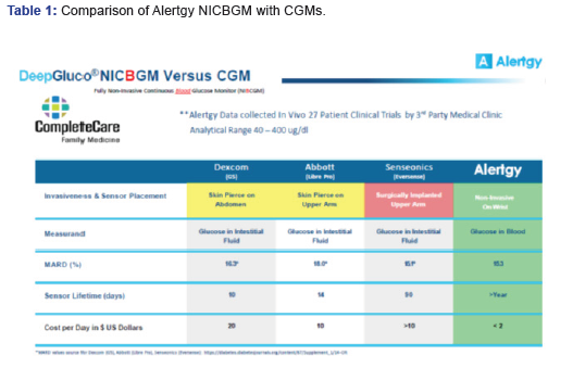
A comparative summary analysis of the results of the CompleteCare trial data generated by the Alertgy NICBGM are compared to other CGM devices in Table 1. Each listed device was evaluated against an SMBG system to develop the provided MARD values. The Alertgy NICBGM DeepGluco device provides equivalent accuracy. It also offers the advantages of no interstitial lag, non-invasive measurement, and a sensor life measured in years as opposed to days and is less costly to use.

To Know more about Current Research in Diabetes & Obesity


To Know more about our Juniper Publishers

Click here: https://juniperpublishers.com/index.php   


Tuesday, July 27, 2021

Evaluation of Mid-Lactation Holstein Cows During Heat Stress in Response to a Dietary Feed Additive - Juniper Publishers

 Dairy & Veterinary Sciences - Juniper Publishers


Abstract

Heat stress (HS) has detrimental effects on lactating cattle especially when they are in a negative energy balance. Feed additives have been shown to mitigate the effects of HS by improving metabolic and immune function. The objective of this study was to evaluate the effect of feeding a dietary supplement (Land O’Lakes Inc., Arden Hills, MN) on the HS response in multi-parturient dairy cows in mid lactation. Two pens of cows at a commercial dairy were fed either control (CON) or additive (YB) at approximately 56.5 kg/pen per d for two weeks prior to arrival. Study cows (n=12) were balanced in days in milk (DIM), milk production, and parity (111.91±4.85 d, 33.67±0.96 kg/d, and 2.25±0.18). On arrival, the cows (6 YB and 6 CON) were randomly selected and housed in environmentally controlled chambers for 18 d and fed appropriate diet. Cows were subjected to 7 d of thermoneutral (TN) conditions, 7 d of HS, and 4 d of recovery (REC) under TN conditions. Dry matter intake (DMI), milk production from AM and PM milkings, and milk composition were measured daily. Rectal temperature (RT) and respiration rate (RR) were measured at peak temperature daily. Blood samples were collected once daily at 0900 h following catheterization on d 5 of TN to d 4 of REC. Serum samples were analyzed for glucose, insulin, blood urea nitrogen (BUN), β-hydroxybutyrate (BHB), and non-esterified fatty acids (NEFA). Results were analyzed using repeated measures in the PROC MIXED of SAS. HS increased RT (P<0.0001), RR (P<0.0001), BUN (P<0.0001), BHB (P=0.0009), insulin (P=0.036), cortisol (P<0.0001), neutrophil (P=0.0272), and water intake (P<0.0001). HS decreased lymphocyte (P<0.0001), DMI (P<0.0001), and 4% fat corrected milk (FCM, P=0.02). YB increased the feed efficiency ratio (P=0.03). YB had no effect on blood parameters. There was a treatment interaction with cows fed YB having higher feed efficiency (P=0.05) during peak thermal loads than CON. Results of this study suggest that HS exposure had performance and metabolic impacts in mid lactation cows. Supplementation with YB alleviated some of the performance effects associated with HS.

Keywords: β-hydroxybutyrate; Altering cortisol; Rumen acidosis; Ketosis; Lameness; Mastitis; Metritis

Abbreviations: HS: Heat stress; DIM: Days In Milk; TN: Thermoneutral; REC: Recovery; DMI: Dry Matter Intake; RT: Rectal Temperature; RR: Respiration Rate; BUN: Blood Urea Nitrogen; BHB: β-Hydroxybutyrate; NEFA: Non-Esterified Fatty Acids; THI: Temperature-Humidity Index

Introduction

HS is a costly factor to the dairy industry [1] as it changes the livestock production system due to increasing climate variability [2]. Physiological responses occur when the temperature-humidity index (THI) exceeds 68 or when the ambient temperature exceeds 32.2˚C [3]. Measurable factors that are negatively affected by heat stress are milk production, dry matter intake, growth, and health disorders [4]. It has been shown that milk yield is partially affected by HS through independent mechanisms of reduced nutrient intake [5].

Increasing the energy density of the diet from carbohydrates sources to fatty acid sources are common methods for feeding heat stressed animals but increase the chances of rumen acidosis per Kadzere et al. [6]. Using supplemental dietary additives have been shown to stabilize the rumen and increase post-rumen nutrient flow [7] in non-stressed cows; however, Shwartz et al. [8] reported no effect of a yeast culture on mitigating the effects of heat stress on lactating cows. The feed additive used in this study was a proprietary phytogenic and specialty yeast blend (Land O’Lakes Inc., Arden Hills, MN). Proven feed additives have shown increased immune function in lactating dairy cows [9] by altering cortisol responses in supplemented cows [10].

We hypothesized that supplementing with the YB would increase energy availability and thus reduce the negative effect of the heat load on production measures. The study objectives were to evaluate the effects of the dietary YB on body, production, and metabolic parameters in heat stressed lactating cows.

Materials and Methods

This study was conducted at the Agricultural Research Complex (ARC) at the University of Arizona (Tucson, AZ). The protocol was reviewed and approved by the University of Arizona Institutional Animal Care and Use Committee. It occurred in two phases: on-farm and on-site.

The on-farm phase consisted of feeding one 500 cow pen and monitoring a control pen of the same number of cows at a dairy in Stanfield, AZ. Treatment cows were given the feed additive in the TMR at 56.5 kg/pen and received it for 2 weeks. Previous research has shown that there may be a cell-mediated response in the rumen and that requires time to see a response during heat stress [10]. After the 2-week period, 6 treatment cows and 6 control cows were randomly selected and shipped to the ARC for the on-site phase with no health issues (rumen acidosis, ketosis, lameness, mastitis, metritis).

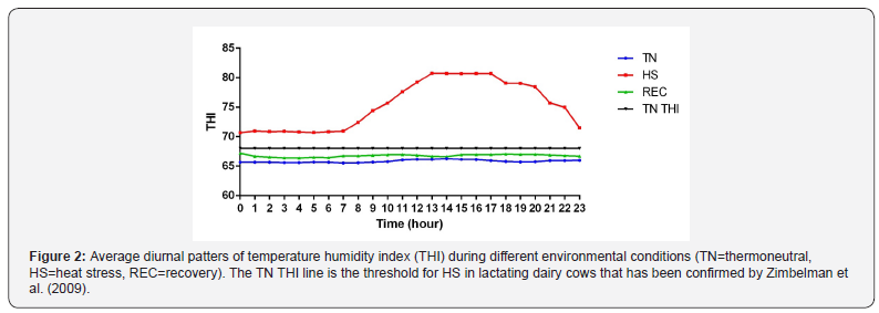
Upon arrival, twelve multiparous Holstein cows producing 33.67 ± 0.96 kg of milk/d, lactation (2.25 ± 0.18), and stage of lactation (111.92 ± 4.85 DIM) were weighed and randomly assigned to individual tie stalls in one of two environmental chambers. Both chambers housed 6 cows (3 control and 3 treatment). Chambers operated on the same environment throughout the study. There were 4 periods: acclimation (4 days), thermal neutral (TN, 7 days), heat stress (HS,7 days), and recovery period (4 days) (Figure 1). The acclimation period and TN period were set on the same environmental cycle. The TN environment was set at a minimum temperature of 21.16 ̊C, a maximum of 21.48 ̊C, and an average of 21.39 ̊C. The relative humidity (RH) had a minimum of 28.32 %, a maximum of 36.73 %, and an average of 32.30 %. The temperature-humidity index (THI) had a minimum of 65.54, a maximum of 66.16, and an average of 65.84 (Figure 2). The HS period had a minimum temperature of 25.40 oC, a maximum of 34.89 oC, and an average of 30.09 oC. The RH had a minimum of 23.40 %, a maximum of 36.48 %, and an average of 30.88 %. The THI had a minimum of 70.68, a maximum of 80.70, and an average of 75.46 (Figure 2). The Recovery period had a minimum temperature of 21.11 oC, a maximum of 21.43 oC, and an average of 21.38 oC. The RH had a minimum of 40.46 %, a maximum of 50.93 %, and an average of 45.93 %. The THI had a minimum of 66.39, a maximum of 66.99, and an average of 66.78 (Figure 2). The THI of 68 identifies as the threshold for HS in lactating dairy cows [11].

Journal of Dairy & Veterinary Sciences

THI values were calculated using the average temperature and relative humidity obtained from the data logger. THI was defined by the formula [12]:

Where: db T = dry bulb temperature (̊oC), RH = relative humidity percentage.

The diet was an alfalfa based TMR that was balanced to be consistent with the green chop based TMR on the commercial dairy. Grab samples were collected once every 3 days when a new batch of feed was mixed. Alfalfa was added to the TMR pre-mix and was stored at 4 oC. Samples were analyzed by Dairy Nutrition Services INC (Chandler, AZ) by wet chemistry

Feeding and milking were done twice daily at 0530 and 1730 h. The CON cows were fed the base TMR. The YB cows were fed the TMR plus 113.4 g of YB mixed. Orts were removed and weighed in the morning prior to feeding. The feed efficiency was determined by using 4% fat corrected milk (FCM) per kilogram of DM consumed for each cow in each treatment group. Water consumption was recorded daily from the water meters after the AM feeding. Milk weights were recorded daily and a daily milk sample was taken from the AM milk. Milk samples were individually refrigerated with a preservative (Bronopol tablet, D&F CON Systems, San Ramon, CA) at 4 oC. Samples were analyzed for fat, lactose, protein, somatic cell count (SCC), and solids-not-fats (SNF) by Arizona DHIA (Tempe, AZ). Body weights were recorded twice during the study: once during HS and at the beginning of REC.

Physiological measurements such as respiration rate and rectal temperature were recorded for each cow once daily at 1400 h. Respiration rate was recorded as breaths per minute. It was calculated by counting the flank movements for 15s and multiplying by 4. Rectal temperature was measured using a Cooper TM99A thermometer (Cooper-Atkins, Middlefield, CT). Cows that exceeded 40.5oC were removed from the chamber and were cooled with water until a rectal temperature of 38.3oC. Vaginal temperatures were recorded with HOBO® U12 stainless steel temperature data loggers (Onset Computer Corp., Bourne, MA) in 5 minutes increments. Blank controlled internal drug releasing devices (Eazi-BreedTM CIDR®; Zoetis, Parsippany-Troy Hills, NJ) were used to hold the HOBO in the vagina. The cows were fit with this apparatus on d 1 for the duration of the study. CIDR placement was monitored and were refitted if they were expelled from the vagina.

Blood was collected via indwelling jugular catheters that were surgically inserted into cows on day 4 and 5. Blood was collected at 0900 h from day 5 to 18. Catheters were flushed before sampling and once at 1500 h with heparinized saline (100 USP/mL). The first 6 mL of fluid was discarded to eliminate heparin. Two 12 mL syringes with a 22-gauge needle were used to draw blood and were transferred to BD Vacutainer tubes (BD, Franklin Lakes, NJ): sodium heparin and blank. Samples were chilled for one hour at 4 oC. Serum and plasma were collected after centrifuging samples at 1,500 x g for 15 min at 4 oC and stored at -80 oC until analysis. Additional blood samples for white blood cell differentials were collected in BD Vacutainer tubes with EDTA. Differentials were run by Antech Diagnostics (Phoenix, AZ).

Serum β-hydroxybutyrate (BHB) was determined by a colorimetric kit (β-hydroxybutyrate (Ketone Body); Cayman Chemical, Ann Arbor,MI). Serum blood urea nitrogen (BUN) was determined using a colorimetric kit (DetectX Urea Nitrogen; Arbor Assays, Ann Arbor, MI). Serum glucose was measured using a colorimetric assay (Glucose Oxidase; Pointe Scientific Inc., Canton, MI). Serum non-esterified fatty acid (NEFA) was determined enzymatically through a commercial kit (Wako NEFA-HR(2); Wako Chemicals USA, Richmond, VA). Serum insulin was determined using an enzyme linked immunosorbent assay (ELISA) kit (Bovine Insulin, ELISA kit; ALPCO, Salem, NH).

Plasma cortisol was assessed using an enzyme immunoassay (EIA) kit (Cortisol Parameter Assay; R&D Systems Inc., Minneapolis, MN) with an intra- and interassay CV <5 %.

Statistical Analysis

Physiological and blood data were analyzed using a PROC MIXED model as a 2 x 2 replicated factorial design with the LSMEANS and PDIFF options within each environmental period as the REPEATED option with day within period as the repeated measure, and effect of the cow was nested within treatment, using a covariant structure. Rectal and vaginal temperatures were averaged per hour by day of trial. Environment (ENV), treatment (TRT), ENV x TRT, day(ENV), and treatment x day(ENV) effects were tested by means of the PDIFF option and were declared significant at P < 0.05. One YB cow was removed because of adaptation complications: severe hypophagia and constant standing.

Results and Discussion

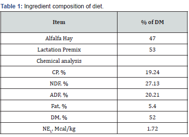
Environmental conditions prior to arrival to ARC were not documented. All cows of the same treatment were housed in the same Saudi style barn cooled using Koral Kool systems and were kept below 72 THI. It isn’t known whether conditions were over 68 THI. The recorded environmental conditions for the TN, HS, and REC periods of the study are shown (Figure 2). As cows are exposed to conditions above 68 THI, RR exceeds 60 [13] and RT elevates above 38.5oC [3]. Due to these differences in parameters, changes in physiology and performance measures are prevalent between environmental groups Table 1.
Journal of Dairy & Veterinary Sciences

Mean RR and RT in CON and YB fed cows are shown in Table 2. There was no difference in treatment for RR and RT. There is conflicting data on the effect of treatment on RT. Baumgard et al. [14] reported an increase in RT when cows were fed a feed additive during HS. Other studies reported a reduction in RT with supplementation during hyperthermia [9,15]. There was an effect of environment (P<0.0001) for both these variables as cows exposed to a THI above the threshold increases these parameters. HS cows increased RR (42 breaths/min) and RT (1.32 oC) compared to TN cows which indicates heat stress was achieved.

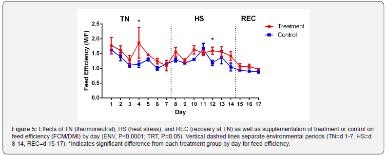
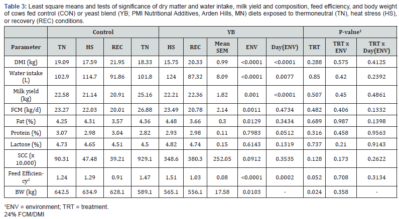
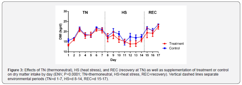
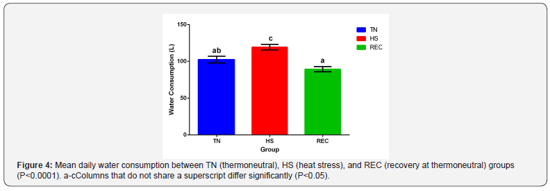
Feed intake, water intake, milk yield, and components are reported in Table 3. Dry matter intake (DMI) increased in both groups as cows adjusted to the facility during the TN period but experienced a slight decrease due to feed heating during 2 days of storage during TN period (Figure 3). DMI differed between TN and HS (P=0.0007). Thermal stress has been well documented in causing a decrease in DMI and how that drop in DMI may impact milk yield. Previous heat stress research has shown an altered endocrine profile that differs from nutrient restricted cows which affect precursors of milk components, such as glucose [14,16,5]. There was no effect of TRT and TRT x ENV for DMI. Schingoethe et al. [17] had similar findings in lactating Holsteins cows that were fed supplementation of a yeast culture had no effect on DMI during the summer. On the contrary, Wiedmeier et al. [18] suggested that during periods of stress, fungal supplemental products could be effective with increasing digestibility of structural carbohydrates. This is supported by Moallem et al. [19] using a live yeast supplementation which saw an increase in feed intake and efficiency during the hot season. There was no difference in water intake between treatment groups for each environmental phase (Figure 4). Water intake increased during HS from TN (P=0.0005) and decreased during REC from HS (P<0.0001). Murphy et al. [20] reported similar water intake values in lactating Holstein cows. Cows fed YB had a higher feed efficiency (1.34±0.08 vs. 1.15±0.08, P=0.05) compared to CON (Figure 5). There was also an effect of ENV (P<0.0001) with feed efficiency increasing during HS and decreasing during REC. Improvement in feed efficiency has been reported in hyperthermic conditions with supplementation of feed additives that contained live yeast and yeast culture [19,17]. YB increased feed efficiency in general even though there was no ENV effect. BW differed between TRT (P=0.0235) with YB cows having lower BW than CON. There was an effect of ENV (P=0.0103) with BW decreasing during periods of HS. The interaction between ENV x TRT was not significant for BW.
Journal of Dairy & Veterinary Sciences

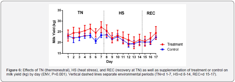
Milk and FCM yield differences weren’t detected between CON and YB groups (Figure 6 and Table 3). These results are similar to previous studies using feed additives containing yeast cultures [17,21]. Other studies have shown an increase in milk yield with yeast supplementation but it is correlated with greater DMI for ruminants [22,23]. Milk yield declined during HS (P=0.002). This decline in milk yield is only partially due to a decrease in DMI which has been shown by Rhoads et al. [16]. Differences between environments were found in FCM with TN being more elevated than HS (P=0.02). There was no significant difference in the following milk components: protein percentage, lactose percentage, and SCC. Previous work by Hall et al. [10] with HS has shown an increase in SCC during the REC but there was no significance observed in this study. The hypothesis is that during heat stress, mammary epithelial cells sluff post HS and contribute to the rise in SCC. Fat percentage had an effect of ENV (P=0.0129). Milk fat percentage had no difference between TN and HS which is surprising while there was fat depression observed during the REC. Rhoads et al. [16] reported that climate-controlled HS cows don’t have reduced milk fat percentages, unlike commercial dairies that frequently experience fat depression during the summer months due to supplementation with high concentrate diets [6]. Not much work has been done during the REC and further research needs to evaluate this in climate controlled cows.
Journal of Dairy & Veterinary Sciences

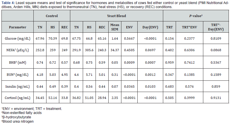
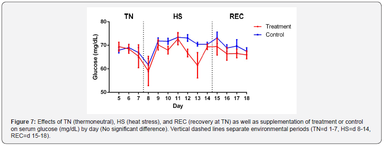
Hormone and metabolite data are presented in Table 4. No differences were detected in serum glucose due to TRT, ENV or TRT x ENV (Figure 7). This is contrary to other findings [16,8]. Cowley et al. [24] had similar results and suggests that the glucose homeostatic regulatory system is able to compensate for the reduction of DMI. No significant differences in serum NEFA for TRT, ENV or TRT x ENV were detected. Serum BHB did not have any significant differences in TRT or TRT x ENV but an effect of ENV was observed (P=0.0009). NEFA and BHB are a significant source of energy for cows in a negative energy balance. BHB decreases during HS according to Dale and Brody [25] but a decrease during REC which has TN conditions is surprising. This suggests a potential shift in postabsorptive changes in lipid metabolism since ketones are a by-product of fatty acid oxidation and the cow now favors glucose to lower heat production.
Journal of Dairy & Veterinary Sciences

There was a difference in ENV for BUN (P<0.0001) and there were no differences in effect for TRT or TRT x ENV. The increase of BUN during HS is similar to previous studies [8,5]. BUN originates from two sources: inefficient rumen ammonia incorporation or deamination of amino acids as glucose precursors. Erasmus et al. [7] found that yeast supplemented groups during HS had improved rumen nitrogen balance which supports the increase in urea-nitrogen in circulation. During periods of inadequate nutrition, cows will undergo proteolysis in skeletal muscle to support lactation [26]. BUN source could have derived from muscle catabolism; however, BUN isn’t a good marker and either 3-methyl-histidine or creatine should be used which increase in HS lactating cows [27,28].

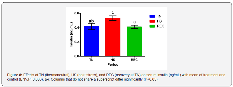
No difference was detected in serum insulin between TRT groups and TRT x ENV. There was an effect of ENV (Figure 8) with insulin increasing during the HS period (P=0.036). Elevated insulin levels have been reported likewise during HS periods [29,5]. This helps explain why there is no NEFA response due to insulin’s antilipolytic properties [30]. Insulin also stimulates protein synthesis and serves as an antiproteolyic hormone [31] which supports the decrease in BUN.
Journal of Dairy & Veterinary Sciences

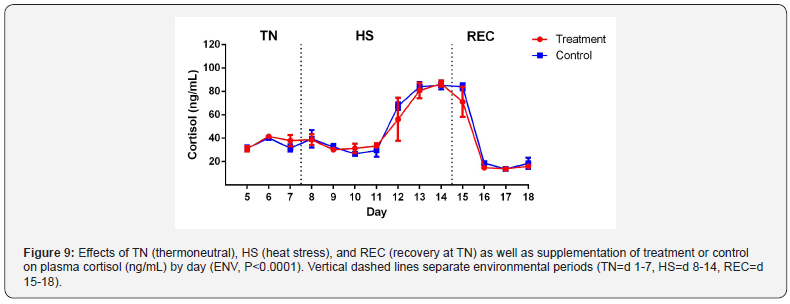
Plasma cortisol had no effect of TRT and TRT x ENV. As expected, there was a significant impact of ENV on plasma cortisol (Figure 9; P<0.0001). Plasma cortisol has been noted to have an increase during acute but not chronic HS [32]. Interestingly, we found no response during the acute phase for acclimation to HS and an elevation during the chronic phase. Alvarez and Johnson [33] reported cortisol spikes that ranged from minutes to hours after an immediate insult of heat stress which would be on d 8. During acute heat stress, cows adjust cellular pathways to mitigate the effects of heat stress and maintain homeostasis. This is no longer needed once the acclimated phenotype and cows have reached long term heat acclimation [34]. This supports the decrease of cortisol during the acclimation period. One possibility is the collection of blood samples in the AM may have missed the spike in cortisol since cortisol levels peak in the afternoon. Another possibility is that although similar THI was used to previous studies conducted on HS, a difference in temperature and humidity could have caused this since there can be the same THI but different values for temperature and humidity
Journal of Dairy & Veterinary Sciences

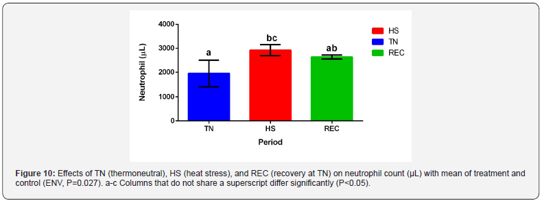
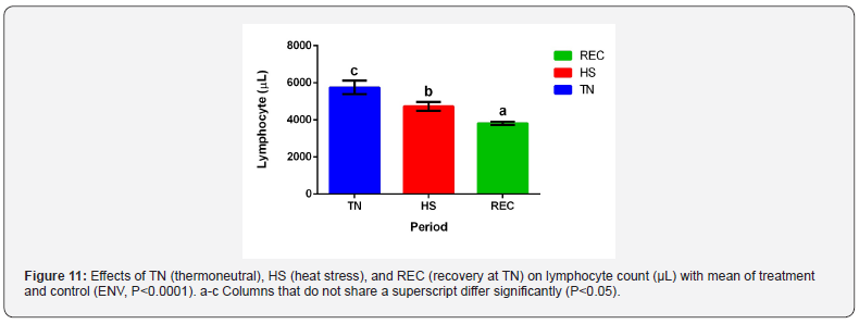
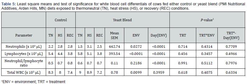
White blood cell differentials are shown in Table 5. Neutrophil count increased during HS (Figure 10) with an observable difference with an effect for ENV (P=0.02) but not for TRT and TRT x ENV. Lymphocytes decreased during HS and REC (Figure 11; P<0.0001) while having no other differences in TRT and TRT x ENV. Interestingly, there was no difference in the neutrophil to lymphocyte ratio. Total white blood cell count was more elevated during TN and decreased throughout the thermal periods respectively (ENV, P=0.009). The results agree with stress induces neutrophilia and lymphopenia [35,36-38,30].
Journal of Dairy & Veterinary Sciences

In conclusion, supplementing mid-lactation Holstein cows with YB beginning 14 days before and during exposure to moderate HS showed an increase in some production responses measured by feed efficiency. Feeding YB; however, did not result in the mitigation of negative physiological responses associated with HS. The mechanism behind the increase in feed efficiency is unknown due to there being no glucose or DMI response to treatment and further research should focus on it.

To Know more about Dairy & Veterinary Sciences


To Know more about our Juniper Publishers

Click here: https://juniperpublishers.com/index.php    

Monday, July 26, 2021

Monitoring Processes and Improvement of Wine Quality by Determining of Catechin and Phenolic Compounds in Slovakian Wines - Juniper Publishers

Biotechnology & Microbiology - Juniper Publishers 

Abstract

Moderate consumption of wine seems to be beneficial to health, because phenolic compounds and catechin as antioxidants are considered to be important bioactive compounds. Interest in wine polyphenols, their interactions has increased among food research fields. In this study, Analytical properties of caftaric acid, (+)-catechin, caffeic acid, 4-OH-cinamic acid, vanillic and gallic acid were studied in five Slovakian protected white wines from Južnoslovenská winegrowing region. (+) Catechin was the major individual phenolic compound in PDO (protected designation of origin) wines obtained from this region harvested in vintage 2016-2018. The content of (+) catechin varied from 16.58 mg/L in the year 2016 to 26.29 mg/L in the year 2018. Much more varied the content of caffeic acid in the vitage 2016 2.30 mg/L to 11.38 mg/L in vintage 2018, which could be responsible for oxidation levels that the wine is undergone and/or grape varieties. The purpose of this study was also to discuss different styles of Slovakian wine varieties: Traminer rose, Pinot gris and Irsai Oliver.

Keywords: Wine phenolic compounds; (+) Catechin in wine and wine polyphenols

Introduction

There has been an explosion in diverse types of wines including petillant naturel, oxidised and ‘whole bunch’ wines. The white wines, oak-driven white wines, full-flavoured, high alcohol, broad-shouldered wines and also aromatic white wines made from Sauvignon or Riesling are preferred. There is no doubt that this explosion in diversity comes to a revolution in the way that a new generation of wine drinkers who are more educated and more discerning about value of aromas than ever before [1]. Consumers are demanding more information about phenolic acids in wine products. Wine typicity and terroir is that the geographical origin and oenological practices, along with the grape variety, make an important contribution to the final expression of the finished wine. It means that protected designation of origin (PDO) or Protected Geographical Indication (PGI) in more european wines carry more of the variety and region’s signature taste, texture, flavour and aroma. In the end of clarification process, total phenolic acids of the wines was higher than in the must. Polyphenols are one of the most important secondary metabolites in grapes and wine [2] It was found out that geographic location could affect how wine experts and degustators rate quality of wine like balance and acidity of wine. Therefore, it is very useful to have an analytical tool for identification of odor and polyphenols in wine [3]. (+) Catechin is a major individual flavonoid phenolic compound in the wine. The differences in catechin and caffeic acid content of wines in Turkey were found to be significant, the differences in epicatechin, ferulic acid and vanillic acid contents were not found to be significant [4, 5]. In Portugal wine was found out the role of gallic and caffeic acid in white wine preservation. Moreover, the wine with gallic acid obtained the highest scores according to the sensory evaluation and wines demonstrated a rich aromatic profile [6]. Slovakian legislation prescribes strict controls before introducing the wine into the market. The aim of this study was to evaluate the differences between phenolic compounds (flavonoid and non-flavonoid) in Slovakian wine varieties Traminer rose, Pinot gris and Irsai Olliver which are PDO wines from Južnoslovenská winegrowing region. It was found, that vintage year plays role in content of polyphenols and also grape variety. The article documents that wine from the same vineyard with the same PDO Južnoslovenská could have similar climatic conditions and similar technology, but do not possess the same aromatic characteristics, which is in agreement with literature [7].

Materials and Methods

Two different vineyards from the same PDO area were chosen. In total 5 samples of wine (Traminer rose 2016, Traminer rose 2017, Irsai Oliver 2018, Pinot gris 2016, Pinot gris 2017) were tested. The HPLC method with fluorescence detection was used for the determination of phenolic compounds in samples. The samples were filtered through a 0,45 μm membrane and direct injection of 20 μl was used. Agilent Technologies system was used with an injection valve (Rheodyne), thermostat, diode array detector and fluorescent detector (Agilant Technologies, Santa Clara, USA). The chromatographic column was LiChrospher 100RP-18 (5um), the mobil phase for gradient elution was a mixture of methanol: water: formic acid (70:28:2) The column temperature was kept at 40°C, Spectrophotometric detector was operated at 280 nm. The Fluorimetric detector was operated at λ(ex)=278 nm and λ(em)=360 nm (gallic acid, vanillic acid) , λ(ex)=278 nm and λ(em)=454 nm (p-coumaric, caffeic acid and (+)catechin) The identities of the different chromatographic peaks were confirmed by comparing their spectral characteristics to standards and retention times. Each sample was measured 3 times and average was taken in all cases. The white wines were obtained from famous Slovak wine producers in Južnoslovenská winegrowing region Dufrex s.r.o., Hurbanovo (Traminer rose and Pinot gris) and Vinohradnícko-vinárske družstvo, Dvory nad Žitavou (Irsai Oliver). OIV methods were used for the determination of basic parameters in all wines (alcohol, volatile acids, total acids, extract, reducing sugars, total SO2). Analysis were carried out in accredited laboratory in Bratislava, under ISO 17025.

Results and Discussion

Phenolic compounds are divided into flavonoid and nonflavonoid compound. Flavonoids are flavan 3-ol (anthocyanin, catechin, epicatechin) and Nonflavonoids are hydroxycinnamate acid, hydroxybenzoic acid. Caftaric acid is a non-flavonoid compound in wine, it is an esterified phenolic acid, composed of caffeic acid, a hydroxycinnamate produced by plants, and tartaric acid, the principal organic acid found in grapes berries. Winemakers often measure caftaric acid levels as their primary method to estimate the oxidation levels of wine. The content of caftaric acid was obtained 11,78 mg/l in Traminer rose 2016 and 12,78 mg/l in Traminer rose 2017 which shows that oxidation level is good and wine variety is suitable for aging. The highest content 26,29 mg/l of (+) Catechin was found in Irsai Oliver variety in vintage 2018 and the lower content 16,58 mg/l was in the Traminer rose 2016 which shows, that aroma profile is much more higher in Irsai Oliver variety and also vintage plays role For example caffeic acid varied from 2,3mg/l in vintage 2016 to 11,38mg/l in 2018. 4-OH cinamic acid varied from 3,28 mg/l in 2016 to 4,13mg/l in 2018. Slovakian wines with PDO Južnoslovenská had to have minimum content of actual alcohol 10,5%obj., total content of acids is minimal 3,5g/l, sugarfree extrakt is minimum 16g/l and volatile acids maximum 1,1g/l, which all these wines achieved. See Table 1 & 2.

Material Science
Material Science

Global warming could affect the aroma of wine and harvest time of later harvested varieties (Pinot gris and Traminer rose) is moving from the end of September to the end of August, so harvesting in earlier time (on the north hemisphere) shows lower acids and this affect aroma [8]. This changes grapes’ sugar and acid levels, leading to lower-quality wines with higher alcohol content. The last six weeks of ripening are critical for flavour and colour development and sugar-acid balance, so we don’t want to have too much heat at the end of the season before harvest. International research published in the prestigious journals is looking for the different grape varieties that may be able to better withstand climate change. In this work we confirm that the earlier variety IO is by content of phenolic compounds very near and comparable with later varieties Traminer and Pinot gris, which is in agreement with literature [7,9].

Conclusion

Polyphenols are organic substances, which have got in the structure benzene ring and HPLC is the best method to determine organic substances because of very fast separation and retention times, separation recovery, reproducibility and accurate data [10,11]. For the determination are used FID detector, UV-detector and/or Fluorescence detector (LFD). We used HPLC (LFD) detector with very good precision, peaks of samples were compared with peaks of standards and retention times. Wines were produced by OIV practices, no aroma faults were found by sensory analysis. The basic analytical parameters were in limits. The aim of study was to determine acids and phenolic compounds to compare the contents in the five Slovakian wines Traminer rose, Pinot gris and Irsai Oliver, to show their typicity and vintage differences. Our opinion is that the oenological practices in addition to the grape variety, vintage and the geographical origin contribute to the expression in the wine. Wines of these monitored varieties have always been considered rich in content and long-lasting. The typical aroma of Pinot gris is described with pear, apple, banana, apricot, caramel and citrus notes. For mature wines, bread flavours can be recognized [12,13]. Phenolic acids found in Slovakian wines are significant as in Czech Republic or Austria wines and could be compared.

To Know more about Biotechnology & Microbiology

Click herehttps://juniperpublishers.com/aibm/index.php


To Know more about our Juniper Publishers

Click here: https://juniperpublishers.com/index.php   


Friday, July 23, 2021

Compound Odontoma of the Anterior Maxilla in a 4-Year-Old Patient: A Case Report - Juniper Publishers

Dentistry & Oral Health - Juniper Publishers


Abstract

Background: Odontomas are an odontogenic tumor containing all the components of the odontogenic tissue. They are divided into compound and complex odontomas depending on their macroscopic appearance and organization. Odontomas are frequent in the first two decades of life and generally present as asymptomatic entities in most patients. However, they can hamper normal eruption of permanent teeth or cause swelling.

Material and Methods: We present the case of a 4-year-old boy who presented with progressive unpainful swelling of the anterior maxilla and absence of the upper right temporal central incisor. An orthopantomography was taken showing a tooth-like mass in the region of the permanent upper right incisors surrounded by a radiolucent halo. The suspicion diagnosis of odontoma was stablished and surgical excision was decided to avoid impairment of the eruption of the permanent incisor. The histopathological analysis confirmed the diagnosis of compound odontoma.

Conclusion: Odontomas are benign odontogenic tumors that can produce alterations of the eruption of teeth and progressive swelling with associated facial deformity. Surgical excision is safe, and no recurrences have been described. However, need for orthodontic treatment to aid in the eruption of the retained teeth should be bear in mind when treating odontomas.

Keywords: Odontogenic epithelium and Ectomesenchyme; Odonto-ameloblastoma; Radio dense lesion; Odontogenic cyst; Calcifying odontogenic tumour; Fibro-osseous lesion; Ameloblastic fibro-odontoma

Introduction

Odontomas are benign entities that have been classified as odontogenic tumors by the World Health Organization (WHO). They are commonly found in the first two decades of life. They are considered a type of hamartomas composed of odontogenic epithelium and ectomesenchyme. They consist of enamel, dentine, cementum and pulpal tissue. Their etiopathology remains unknown, but different theories have been proposed including trauma, infection, family history and genetic mutation. They can be classified into compound and complex odontomas attending to their macroscopic appearance and organization. The compound odontoma appears as a mass containing numerous small tooth-like structures. The complex odontoma, on the contrary, presents as a disorganized mass of enamel and dentin showing a white-yellow hard tissue appearance. Complex odontomas are more frequent in the posterior mandible, whilst compound odontomas are generally found in the anterior maxilla.

These tumors are generally asymptomatic and are usually detected on routine dental imaging [1]. However, they may cause progressive bone swelling, facial asymmetry, pain or, occasionally, they might alter normal eruption of primary or permanent teeth. Radiographically, the compound odontoma appears as a collection of tooth-like structures surrounded by a narrow radiolucent zone, hence, it can seldom be confused with any other entity such as supernumerary teeth [2]. Complex odontomas, however, appear as a radio dense lesion surrounded as well by a narrow radiolucent halo. Odontomas must be surgically removed in order to prevent cyst formation, possible conversion to odonto-ameloblastoma [3] and allow teeth eruption. Excision of the odontoma with the associated soft tissue is needed. Recurrences have not been reported.

Case report

We present the case of a 4-year-old boy who presented with a slowly progressive swelling on the anterior maxilla. On physical examination, a hard-bony swelling was found on the right anterior maxillary anterior region, in the area of the upper right incisors, and absence of the upper right temporal central incisor (Figure 1). An orthopantomography showed a collection of multiple radio opaque tooth-like structures with a narrow radiolucent rim in the region of the lateral and central permanent incisors in the right maxilla (Figure 2). A provisional diagnosis of compound odontoma was made and surgical excision was decided. A CT was performed preoperatively in order to confirm the diagnosis and stablish a surgical plan. A mucoperiosteal flap was raised from medial to the upper left central incisor to distal to the upper right canine. The thin shell-like bone covering the odontoma was removed using a needle holder. The odontoma was exposed and was carefully removed using a scoop, paying special attention to avoid damage to the permanent central incisor (Figure 3). The macroscopic appearance consisted of several malformed tooth-like structures with surrounding soft tissue (Figure 4). A cellulose hemostatic agent was placed and closure with absorbable sutures was performed. The patient was discharged the same day. The histopathological examination confirmed the diagnosis of a compound odontoma. During the follow-up the patient had an uneventful healing with no signs of infection or dehiscence. No orthodontic traction of the permanent incisor was decided at the moment given the age of the patient, in order to wait for spontaneous eruption.

Discussion

Odontomas are the most frequently occurring odontogenic tumors in the oral cavity and are considered to be hamartomas rather than true neoplasms [4]. The mean age of detection on an average is 14.8 years, with the prevalent age being the second decade of life [2]. These odontogenic tumors appear more frequently in the maxilla than the mandible. They are most often seen in association with permanent dentition and are rarely associated with primary teeth [5]. Odontomas are composed of various tooth tissues such as enamel, dentin, pulp, and cementum and representing the second most common odontogenic tumor of the jaw bones [6]. The World Health Organization (WHO) classified odontomas as a “benign odontogenic tumor composed of odontogenic epithelium and odontogenic ectomesenchyme with dental hard tissue formation”. The tissues may either form disorganized conglomerates of dental tissues, known as complex odontomas or appear as multiple, rudimentary tooth-like structures, which are known as compound odontomas. Compound odontomas are frequently found in the anterior region of the maxilla, whilst complex odontomas are usually located in the posterior region of the mandible [1].

It can present as a slowly expanding lesion of the bone which is nonaggressive and usually causes disturbances in the eruption of permanent teeth [5]. Occasionally, they can produce facial asymmetry. However, in most cases, odontomas are asymptomatic and are diagnosed incidentally on routine dental imaging. An orthopantomography is needed to stablish the initial diagnosis. Both complex and compound odontomas display a radiolucent rim, representing dental follicular tissue. CBCT or CT can be performed for further analysis and to establish a surgical plan. Radiographic differential diagnosis of compound odontomas are limited, as they appear as a collection of small teeth like structures. Nonetheless, in complex odontomas a wider spectrum should be considered because they appear as a radiodense mass of hard tissues, including calcifying odontogenic cyst, calcifying odontogenic tumour, fibro-osseous lesion, ameloblastic fibro-odontoma and osteoblastoma. Ossifying fibroma may mimic an odontoma but can be differentiated from it by the fact that it is well circumscribed and usually separates easily from its bony bed [7].

Surgical treatment of odontomas consists of complete removal with any associated soft tissues. Spontaneous eruption of impacted or retained teeth can sometimes be expected. Nevertheless, further malocclusion or any misalignment of teeth can be corrected by orthodontic treatment [8]. Recurrences have not been reported. In a study performed by Isola et al. [9] in 45 patients with 29 complex odontomas and 16 compound odontomas, 25 of these patients presented with delayed eruption of permanent teeth, 6 presented with pain, 4 with swellings and 10 were asymptomatic. Of the non-extracted teeth, 33 teeth were displaced and retained. Of those, 29 teeth were aligned through orthodontic-surgical approach and 4 teeth erupted spontaneously after surgery during the follow-up. During a maximum follow-up of 15 years (0.3–15), 2/45 teeth were lost due to periodontal disease, 1/45 due to endodontic reasons, and 1/45 due to trauma and in all of the patients, no recurrence was seen.

In this article, we report a rare clinical case of a 4-year-old boy who presented with an indolent and slowly progressive bony swelling in the anterior maxilla and an absent upper right temporal central incisor. We believe the absence of this deciduous tooth was explained by the odontoma and was probably part of it. Radiographic examination showed a “multi tooth-like” structure with a radiolucent halo, compatible with a compound odontoma. Surgical removal was decided, and the tumor was successfully removed, preserving the permanent central incisor. Histopathological examination confirmed the diagnosis of compound odontoma. It is infrequent that odontomas affect primary dentition and are more exceptional in younger patients, such as this clinical case. During the follow up, the wound healed well with no sequelae or complications during the follow-up. In this case, orthodontic traction of the central incisor was not decided due to the age of the patient and in order to expect for spontaneous eruption.

Conclusion

In conclusion, odontomas are frequent odontogenic tumors that must be considered in the differential diagnosis of a bone-expanding mass hampering normal eruption of teeth in patients in the first two decades of life. However, many are diagnosed as an incidental finding on routine dental imaging. Surgical removal is safe and predictable, with no recurrences. However, spontaneous eruption of permanent teeth may not be achieved, and orthodontic traction may be needed.

To Know more about Dentistry & Oral Health


To Know more about our Juniper Publishers

Click here: https://juniperpublishers.com/index.php   

Artificial Intelligence System for Value Added Tax Collection via Self Organizing Map (SOM)- Juniper Publishers

  Forensic Sciences & Criminal Investigation - Juniper Publishers Abstract Findings:  Based on our experiments, our approach is an effec...